

 PDF
PDF ePub
ePub Citation
 Print
Print


Dear Editor:
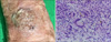
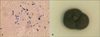
An 89-year-old female presented with a tender, dark gray, scaly, verrucous plaque on the right wrist for several months (Fig. 1A). The plaque measured 3 cm in diameter. This lesion developed after traumatic injury. She had a history of diabetes mellitus and human immunodeficiency virus infection. She was diagnosed with phaeohyphomycosis caused by Exophiala spinifera by KOH examination, fungal culture, lactophenol cotton blue stain, biopsy, and ribosomal DNA (rDNA) sequencing. Yeast-like fungal elements with pseudohyphae were observed on KOH preparation (Fig. 2A). Fungal culture on potato dextrose agar and corn meal agar, with peptone and tween 80, showed dark black, velvety colonies (Fig. 2B). Long annellophores and annelloconidia were observed on lactophenol cotton blue staining. Histopathology highlighted dark brown, yeast-like fungal elements and showed mixed inflammatory and granulomatous infiltrate in the dermis (Fig. 1B). Sequencing analysis of the internal transcribed spacer (ITS) region of the rDNA identified E. spinifera using the GenBank Basic Local Alignment Search Tool (BLAST). The GenBank BLAST search revealed 98% (530/539 bp) similarity with accession number NR111131 and 99% (537/540 bp) similarity with accession number KP132127. The lesions are resolving with oral antifungal medication (fluconazole 50 mg once a day for 3 months).
Cutaneous, subcutaneous, and systemic infections can be caused by Exophiala, which is characterized by annellidic conidiogenesis1. Identification of Exophiala has been based upon recognition of macroscopic and microscopic appearance1. On the basis of morphological examination, the most common species of Exophiala are E. jeanselmei and E. dermatitidis1. However, morphological examination is unreliable in differentiation of the genus Exophiala. The advent of gene sequencing technique has permitted more accurate differentiation of Exophiala2. Heterogeneous species of E. jeanselmei have been reclassified as several species, including E. heteromorpha, E. lecanii-corni, E. oligosperma, E. xenobiotica, and E. jeanselmei, using data sequence analysis of the rDNA ITS region and ITS-restriction fragment length polymorphism analysis3. Although E. dermatitidis is very homogeneous, it was reclassified into five subgroups. A recent study on the order of frequency of Exophiala infection in the USA showed the following: E. dermatitidis (29.3%), E. xenobiotica (19.7%), E. oligosperma (18.6%), E. lecanii-corni (6.9%), E. jeanselmei (3.7%) and E. spinifera (2.7%)1. Although a few cases of E. spinifera infection identified by molecular techniques and morphological features have been reported, none has been observed in Korea4. Our case was identified with KOH, fungal culture, and biopsy. In addition, sequencing analysis of the ITS region of rDNA verified the isolate as E. spinifera. A few reported cases of phaeohyphomycosis caused by E. spinifera came from tropical and subtropical areas5. E. spinifera is distributed in soil, plants, water and decaying wood material. Cutaneous E. spinifera infections show various clinical presentations: erythematous papules, verrucous plaques, and deep subcutaneous abscesses. The distribution and course of disease are variable according to the age and immune competency of the patient. Although susceptibility of E. spinifera to antifungal agents has been variable, our patient was successfully treated with oral fluconazole. We report a case of phaeohyphomycosis caused by E. spinifera, identified newly in Korea.
 XML Download