

 PDF
PDF ePub
ePub Citation
 Print
Print


INTRODUCTION
Connective tissue growth factor (CTGF, also known as CCN2) is a member of the CCN family of proteins that contribute to a variety of cellular processes, including angiogenesis, chondrogenesis, wound healing, and organ fibrosis1. CTGF is known to be a full-length molecule consisting of four conserved domains, i.e., insulin-like growth factor-binding protein domain (module 1: M1), von Willebrand factor type C domain (module 2: M2), thrombospondin type I repeat domain (module 3: M3) and C-terminal cysteine-knot I repeat domain (module 4: M4). CTGF overexpression is induced by transforming growth factor-β in many pro-fibrotic conditions such as scleroderma12. In articular tissue, CTGF is produced by chondrocytes and maintains cartilage tissue homeostasis via an autocrine process3. We previously demonstrated that tumor necrosis factor (TNF)-α inhibited CTGF production in human chondrocytes, whereas TNF-α induced CTGF in human synovial cells4. Based on those results, we found that a neutralizing anti-CTGF monoclonal antibody significantly ameliorated arthritis in collagen-induced mice5.
Psoriasis is a chronic inflammatory skin disease characterized by epidermal hyperplasia and dermal infiltration of immune cells. Cytokines, such as interleukin-17A and TNF-α, produced by T helper cells are implicated in disease pathogenesis6. Since the development of biological agents, anti-TNF-α antibodies have been used for the treatment of psoriasis7. Anti-TNF-α antibody therapy also improved the treatment of rheumatoid arthritis (RA). Psoriasis and RA have a commonality in terms of pathogenesis and treatment8. However, the function and role of CTGF are unknown in psoriasis.
In this study, we investigated the function of CTGF in psoriasis using the established imiquimod (IMQ)-induced psoriasis murine model9 and samples from psoriasis patients.
MATERIALS AND METHODS
Experimental conditions
BALB/c mice were obtained from Charles River Japan (Yokohama, Japan). Experiments were performed in compliance with institutional guidelines and were approved by the Institutional Animal Care and Use Committee (IACUC) of Juntendo University (Approval no. 2900053). At eight weeks of age, mice were shaved on the back skin and left ear, and 62.5 µl and 3.125 µl of 5% IMQ cream (Beselna Cream; Mochida Pharmaceutical, Tokyo, Japan) were respectively applied to these areas for six days. One day before IMQ application, 200 µg of anti-CTGF monoclonal antibody (Nosan Corporation, Yokohama, Japan) was intraperitoneally injected. An objective scoring system called the psoriasis severity index, which considers erythema, scaling, and thickness, was used, and skin lesions were scored from 0 to 4. The cumulative score (erythema plus scaling plus thickening) was also employed for measuring severity (scored from 0 to 12)9.
Patients and samples
The human subject research component was approved by the Juntendo University Urayasu Hospital Institutional Review Board (IRB no. 2009-009). After informed consent, 4-mm punch biopsies were obtained from both psoriasis patients (n=6) and other disease patients with normal condition of skin (n=4). The diagnosis was confirmed by both pathologists and dermatologists. Plasma was also collected from patients with psoriasis (n=28) and healthy control (n=8).
Pathological analysis
Mouse and human skin samples were fixed in a 20% formalin solution and embedded in paraffin, sectioned, and subjected to hematoxylin-eosin (H&E) staining, immunofluorescence, and immunohistochemistry. Epidermal and dermal thickness were determined at five positions per section by measuring the average distance from the basal lamina to the bottom of the stratum corneum using a BZ-II Analyzer ver. 1.1 (Keyence, Osaka, Japan). For immunofluorescence staining, back skin samples were blocked and stained using goat anti-CTGF antibody (Santa Cruz Biotechnology, Santa Cruz, CA, USA), rat anti-Ki-67 antibody (eBioscience, Sun Diego, CA, USA), rat anti-CD3 antibody (Abcam plc, Cambridge, UK), and biotin conjugated Armenian hamster anti-Vγ3 antibody (Leinco Technologies Inc., St. Louis, MO, USA). Sections were further incubated with Alexa Fluor 488 conjugated anti-goat IgG (Invitrogen, Carlsbad, CA, USA) or anti-rat IgG (Invitrogen), and Alexa Fluor 594 conjugated streptavidin (BioLegend, Sun Diego, CA, USA). Nuclei were stained with 4′,6-diamidino-2-phenylindole. For immunohistochemical staining, sections were incubated with primary rat anti-F4/80 antibody (Abcam plc, Cambridge, UK), rat anti-Ly-6G antibody (BD Biosciences, San Diego, CA, USA), Armenian hamster anti-CD11c antibody (BioLegend), and rat anti-PDCA1 antibody (DENDRITICS, Lyon, France). Sections were further incubated with horseradish peroxidase-conjugated anti-rat IgG (Dako, Glostrup, Denmark) and anti-Armenian hamster IgG secondary antibody (Jackson Immunoresearch Laboratories, West Grove, PA, USA). Immunoreactivity was detected by incubating with a DAB+ Liquid kit (Dako). Sections were then counterstained with hematoxylin. For evaluation of leukocyte infiltration in the skin, three researchers quantified the staining of three sections from all mice. The numbers of cells positive for F4/80, Ly-6G, PDCA-1, CD3, and Vγ3 were quantified in each section. Photomicrographs were taken using a BZ-9000 microscope (Keyence).
Immunoblotting analysis
Skin tissues were collected and homogenized. Ten micrograms of total protein was loaded in each well. The primary antibodies used were anti-CTGF antibody (Santa Cruz) and anti-GAPDH antibody (Millipore, Billerica, MA, USA). Densitometric analysis was performed using ImageJ software (National Institute of Health, Bethesda, MD, USA).
Sandwich enzyme-linked immunosorbent assay analysis
Human CTGF enzyme-linked immunosorbent assay (ELISA) analysis was performed as described before1011. Briefly, plasma samples were added to 96 well-plates (ThemoFisher Scientific, Waltham, MA, USA) that were coated with a monoclonal antibody against human CTGF modules. A biotin–labeled anti-human CTGF monoclonal antibody and a peroxidase-conjugated streptavidin (Jackson Immuno Research, West Grove, PA, USA) were added, Substrate and stop solution were sequentially added. The optical density at 450 nm was read to microplate reader (ARVO SX; Perkin Elmer, MA, USA), and measure the CTGF concentration.
Statistical analysis
Data were expressed as the mean±standard deviation and analyzed using GraphPad Prism 6 software (La Jolla, CA, USA). Statistical significance was determined by the Mann-Whitney U test, 2-way ANOVA, and unpaired t-test with Welch's correction. A p-value <0.05 was considered statistically significant.
RESULTS
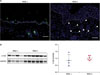
To identify CTGF expression in IMQ-induced psoriatic mice, we used immunohistological analyses and Western blotting (Fig. 1). Immunofluorescence tissue staining revealed that CTGF expression was increased in IMQ-induced psoriatic back dermal lesions compared with control IMQ untreated mice (Fig. 1A). To confirm CTGF protein expression levels, we performed a Western blot analysis using back skin samples. CTGF protein expression showed a tendency to increase in IMQ-induced psoriatic mice relative to untreated mice (Fig. 1B, left). However, the increased expression of CTGF was not statistically significant using this method (Fig. 1B, right).
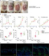
To confirm the involvement of CTGF in this disease model, we neutralized CTGF using an anti-CTGF antibody. After starting the IMQ application, the scoring of erythema, scaling, and thickening was performed using the back skin of mice. Anti-CTGF administration did not affect the systemic immune system; for example, IMQ-induced splenomegaly was not affected (data not shown). Representative skin condition of anti-CTGF treated and untreated mice are exhibited in Fig. 2A. Anti-CTGF treated mice demonstrated declining trend comparing with untreated mice. The independent skin scores in a representative experiment are shown in Fig. 2B. Skin scores were slightly but significantly increased in CTGF-blocked mice several days after IMQ application. Pathological analysis demonstrated that the epidermal and dermal thickness of back skin (Fig. 2C) and ears (Fig. 2D) were not changed by CTGF blocking. However, more Ki-67 positive cells were detected at the epidermis boundary in dermis of anti-CTGF antibody-treated IMQ-induced psoriatic mice (Fig. 2E) and the difference between anti-CTGF treated and non-treated mice was statistically significant (Fig. 2F). Thus, these data based on the effect of anti-CTGF suggest CTGF slightly suppressed the proliferation of suprabasal cells.
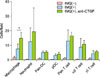
Next, we evaluated the infiltration of immune cells in IMQ-induced psoriatic skin lesions. The numbers of macrophages, neutrophils, pan-dendritic cells (DCs), plasmacytoid DCs, pan-T cells, Vγ3− T cells (most of αβ T cells), and Vγ3+ T cells (γδ T cells) were counted. Macrophages were tended to increase in IMQ induced mice and showed significant increase in only CTGF-blocked mice (Fig. 3).
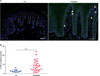
CTGF expression also increased in skin lesions from psoriasis patients comparing with healthy control (Fig. 4A). We previously reported that plasma CTGF levels from RA patients were significantly higher from healthy control4. The same analyzing methods demonstrated that CTGF levels from psoriasis patients were significantly higher from healthy control (Fig. 4B).
Overall, our results indicated that CTGF is involved in IMQ-induced psoriatic dermatitis and the neutralizing CTGF by anti-CTGF treatment could worsen this condition. CTGF expression in skin lesions showed similarity between IMQ-induced psoriatic mice and psoriasis patients. In addition, plasma CTGF levels increased in psoriasis patients comparing with healthy control.
DISCUSSION
The role of CTGF in psoriasis has not been previously reported. Genome-wide expression analysis has shown that IMQ induced-psoriasis does not fully mimic human psoriasis12. In this sense, the analysis of our study resulted in reflecting the limited condition in psoriasis. However, IMQ induced mice is the representative model of psoriasis about clinical and immunological status in the dermatitis. In the current study, we described novel functions of CTGF in psoriatic dermatitis using IMQ-treated mice and found that CTGF tended to increase in the dermis (Fig. 1). It is likely that the enhanced CTGF expression observed with IMQ treatment was associated with dermal fibroblasts1. As a result of neutralizing CTGF by anti-CTGF in IMQ-treated mice, the psoriatic score worsened (Fig. 2B). In skin lesions, suprabasal cells proliferated (Fig. 2E, F), and infiltration of immune cells, notably macrophages, significantly increased (Fig. 3).
Macrophages upregulate healing through their inflammatory and reparative phenotypes and the balance between inflammatory and repair phenotype of macrophages is crucial for successful wound healing13. The application of macrophages or stem cells onto diabetic rodent wounds has been shown to accelerate wound healing and epithelial closure14. In the current study, macrophages were tended to increase in IMQ dermatitis induced mice and showed significant increase in only CTGF-blocked mice using anti-CTGF (Fig. 3). This result suggested that CTGF inhibition may delay the process of wound healing and CTGF could induce repair phenotype of macrophages to improve skin lesions in psoriasis. Recent reports showed CTGF contribute wound repair,1516 and both CTGF and macrophages are involved in tissue remodeling17. However, another reports showed CTGF function as chemotactic factor for some lymphocytes18. In vivo CTGF function is still unclear. At least, our results suggested that reduction of CTGF by anti-CTGF could have an effect on worsening skin condition in psoriasis.
We demonstrated CTGF expression increased in skin lesions and plasma from psoriasis patients comparing with healthy control (Fig. 4). Taken together, CTGF could contribute to improve psoriatic skin lesions through inducing repair phenotype of macrophage in both IMQ-induced psoriatic mice and psoriasis patient.
In summary, the present data based on the effect of anti-CTGF treatment demonstrated that CTGF suppressed the progression of IMQ-induced psoriatic dermatitis. Additionally, CTGF expression was increased in skin lesions and plasma from psoriasis patients. Our findings suggest that CTGF could contribute to the healing rather than the worsening of psoriasis skin lesions including the induction of reparative macrophage.
 XML Download