

 PDF
PDF ePub
ePub Citation
 Print
Print


INTRODUCTION
Laparoscopy-assisted distal gastrectomy (LADG) is the most commonly performed procedure for the treatment of early gastric cancer, involving laparoscopic mobilization of the stomach and systematic lymph node dissection. Billroth-I (B-I) gastroduodenostomy is widely performed after LADG [12]. Several methods have been developed for the ligation of vascular structures during laparoscopic surgery, with each technique having advantages and disadvantages. For example, application of end loops requires dexterity and training, whereas titanium clips can slip from their primary position [34].
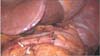
The Hem-o-Lok clip (Weck Closure Systems, Research Triangle Park, NC, USA), first introduced in 1999 (Fig. 1), is a nonabsorbable polymer clip with a lock engagement feature and teeth in the jaws that provide good security. These clips are frequently used to ligate the renal hilum vessels during minimally invasive nephrectomy, as well as for other laparoscopic procedures [56]. However, complications related to Hem-o-Lok clips have been reported in various settings. In particular, complications after upper abdominal surgery have been reported; most of these complications involve a duodenal ulcer after laparoscopic cholecystectomy [78]. This report describes a very rare complication of Hem-o-Lok clip migration into an anastomosis site after LADG with B-I and management.
CASE REPORT
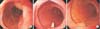
A 58-year-old man with early gastric cancer underwent LADG, followed by B-I gastroduodenostomy using a circular stapler through a small incision made in the epigastrium. During the operation, the perigastric vessels were ligated with Hem-o-Lok clips and metal clips (Fig. 2). The patient recovered normally after the operation and was followed up every 6 months as an outpatient. Six months after LADG, the patient underwent a routine follow-up esophagogastroduodenoscopy (EGD), which demonstrated a fungating mass with ulcer at the anastomosis site (Fig. 3A). An endoscopic biopsy of the area around the ulcer revealed a benign ulcer and the patient was treated with an oral proton pump inhibitor for 14 days. One year after LADG, the patient underwent routine follow-up EGD and an abdominal CT scan. The CT scan showed nonspecific findings, whereas the EGD showed a Hem-o-Lok clip at the site of the fungating mass (Fig. 3B). Initially, we considered laparoscopic exploration or endoscopic removal. Although laparoscopic surgery usually results in fewer adhesions around the operation field, the precise condition of the intra-abdominal region would have been difficult to predict. In addition, endoscopic removal of the clip would have been difficult due to the presence of severe inflammation and possible staple line or suture material entrapment by the clip, thereby increasing the risk of perforation or leakage. Above all, the patient was asymptomatic, with no major abnormalities being detected on clinical examination. Therefore, no attempt was made to remove the clip. The patient remained well after the exposed Hem-o-Lok clip was identified. A repeat EGD 6 months later showed that the clip had disappeared from the anastomosis site, and that the anastomosis site was covered by normal mucosa surrounding the scar (Fig. 3C). The patient was asymptomatic, suggesting that the clip had passed naturally.
DISCUSSION
Hem-o-Lok clips are routinely used during laparoscopic surgery as substitute ligation materials. These nonabsorbable polymer locking clips are inert, nonconductive, compatible with CT scan and MRI, and safe for patient use [345]. In addition, the lock engagement feature and the presence of teeth in the jaws provide good security.
Several reports, however, describe complications related to Hem-o-Lok clips, especially clip migration, after laparoscopic cholecystectomy and urologic surgery procedures, such as prostatectomy and nephrectomy [78]. In almost all previous cases, the clips were removed endoscopically or surgically. Although one study recommended clip removal immediately after diagnosis of a duodenal ulcer secondary to a migrated clip [8], another study recommended control of mucosal injury with an oral proton pump inhibitor when there are no symptoms or warning signs meriting further intervention [7]. The patient in the present report was asymptomatic despite having a benign ulcer. As the discovery of the migrated Hem-o-Lok clip was incidental, no effort was made to remove it. The patient remained well in the absence of treatment, and the Hem-o-Lok clip disappeared spontaneously.
Several mechanisms of clip migration have been suggested. The anatomic proximity of the ligation site to the bowel may provoke a rejection response mechanism, leading to fistula formation around the clip and extending into the bowel. Alternatively, the clip may adhere to an undiagnosed, preexisting ulcer [9]. Another possibility is that a difficult dissection and clip-induced inflammation may result in the clip adhering to the bowel [7]. Finally, inflammation around the anastomosis site may involve the clip, resulting in clip erosion of the bowel wall and eventual migration into the bowel [10]. In our patient, the mechanism of clip migration is unclear, although it was likely due to clip involvement in inflammation around the anastomosis site.
To our knowledge, Hem-o-Lok clip migration into the anastomosis site after LADG with B-I has not been previously reported. Efforts that may prevent this complication include meticulous dissection, removal of misplaced and wandering clips, and use of suture materials or absorbable clips for ligation of vessels. Properly applied Hem-o-Lok clips during LADG are safe and effective for the ligation of involved tissues. In our patient, the Hem-o-Lok clip disappeared spontaneously without endoscopic or surgical treatment. These findings suggest that, in the absence of migrated clip-related complications, these clips can be left undisturbed with passive monitoring. Although Hem-O-Lok clips are useful and safe, surgeons should consider minimizing the use of clips on tissue immediately adjacent to an anastomosis during LADG.

 XML Download