Abstract
Purpose
We developed a technique of totally-robotic right colectomy with D3 lymphadenectomy and intracorporeal anastomosis via a suprapubic transverse linear port. This article aimed to introduce our novel robotic surgical technique and assess the short-term outcomes in a series of five patients.
Methods
All colectomies were performed using the da Vinci Xi system. Four robot trocars were placed transversely in the supra pubic area. Totally-robotic right colectomy was performed, including colonic mobilization, D3 lymphadenectomy, and intra corporeal stapled functional anastomosis. The 2 middle suprapubic trocar incisions were then extended to retrieve the specimen.
Results
Five robotic right colectomies via the suprapubic approach were performed between August 2015 and February 2016. The mean operation time was 183 ± 29.37 minutes, and the mean estimated blood loss was 27 ± 9.75 mL. The time to clear liquid intake was 3 days in all patients, and the mean length of stay after surgery was 6.2 ± 0.55 days. No patient required conversion to conventional laparoscopic surgery. There were no perioperative complications. According to the pathology report, the mean number of harvested lymph nodes was 36.6 ± 4.45. Four patients were stage III, and 1 patient was stage II according to the 7th edition of the American Joint Committee on Cancer system.
Currently, laparoscopic right colectomy is the standard treatment method for patients with right-sided colon cancer. A large number of studies revealed that laparoscopic right colectomy has better, or at least similar, oncological and surgical outcomes compared to conventional open surgery [123]. Furthermore, several studies have demonstrated that robotic right colectomy has similar surgical and oncological outcomes to laparoscopy [456].
Previously, we reported a randomized clinical trial evaluating robotic versus laparoscopic-assisted colectomy with lymphadenectomy for right-sided colon cancer [7]. We concluded that robotic right colectomy with lymphadenectomy was safe and feasible but not justified for routine use because of its higher cost and lack of clinical benefits [78].
Recently, the da Vinci Xi system was introduced, demonstrating several improvements compared to its prior versions. One of the improvements of the da Vinci Xi system is a new overhead arrangement of the arm with a greater degree of freedom, which allows access to a greater area of the body without repositioning or redocking [9]. The mechanical merit of the da Vinci Xi system inspired us to perform oncologic right hemicolectomy with intracorporeal anastomosis with horizontal linear placement of ports in the suprapubic area, which may give better results clinically and cosmetically. Herein, we describe our novel technique with an assessment of short-term outcomes in a series of the first 5 patients to undergo this surgical approach at our institution.
From August 2015 to February 2016, 5 patients underwent right colectomy using the da Vinci Xi system by a single surgeon. All 5 patients were preoperatively diagnosed with resectable colon malignancy without distant metastasis. All data, including baseline patient characteristics and perioperative results, were collected prospectively. All patients were provided informed consent and this study was approved by the Institutional Review Board at Kyungpook National University Chilgok Hospital approved this study (KNUCH-16-05).
Under general anesthesia, the patient is placed in a 10- to 15-degree Trendelenburg position, and the table is then rolled to the left 10–15 degrees, with both arms placed alongside the body to reduce the risk of shoulder injury and to prepare space for docking the robot. Four robot trocars (8 mm) are placed in the suprapubic area transversely. An additional 5-mm trocar for the assistant is placed in the left upper quadrant (Fig. 1).
During the robotic operation, we use a double-fenestrated grasper, vessel sealer, 0-degree endoscope, and a hot shear on the first to the fourth arms, respectively (Supplementary material). After the small bowel is moved to the left upper abdomen, we incise the peritoneal sulcus along the ileocecal mesentery, continuing dissection through the avascular plane of Toldt's fascia. In detail, the appendix or terminal ileum is pulled up and medially using a grasper while the assistant pushes the mesentery of the ileum superiorly. We dissect an avascular plane between the ileocecum and retroperitoneum, creating a tunnel, until the duodenum and the head of pancreas are identified.
When the inferior dissection is complete, we return the bowels to the normal anatomic position. Then, we are ready to perform lymphadenectomy and vessel division. To create clear exposure of the mesenteric axis, the ileocolic vessels are lifted up by a double fenestrated grasper while the assistant pushes the middle colic pedicle superiorly. We create a mesenteric window inferior to the ileocolic vessels and dissect the lymph nodes upward. Once the ileocolic vessels are divided at their origins, dissection continues up to the origin of the middle colic artery. During lymphadenectomy, we divide the right colic artery if present. Depending on the tumor location, we may divide the right branch of the middle colic artery rather than the root of the middle colic artery, but in all cases we clear the lymphoareolar tissue. All the vessels can be securely divided using the vessel sealer without clips.
After completion of lymph node dissection and vessel ligation, the mesentery on the ileum and transverse colon is trimmed accordingly. Finally, the remaining peritoneal attachment is freed to fully mobilize the right colon. To prepare intra corporeal anastomosis, we exchanged the second robot port with a 12-mm cannula for the robotic stapler, and the trans verse colon and the terminal ileum are divided by linear staplers. After making enterotomies in the transverse colon and the ileum using monopolar curved scissors, a side-to-side functional anastomosis is created using a linear stapler. Subsequently, the enterotomy is closed manually with an absorbable suture using a robotic needle driver.
The specimen is wrapped in a plastic bag to reduce the risk of cell spillage during extraction and is retrieved through a transverse incision connecting the second and third trocars, double-protected by a plastic wound protector (Alexis wound retractor, Applied Medical, Rancho Santa Margarita, CA, USA). Once the specimen is removed, we cover the wound protector with a glove and reestablish insufflation to check for bleeding and orientation of the anastomosed bowel. The suprapubic incision is closed with absorbable sutures layer by layer. The cannula sites are closed with 2-0 absorbable sutures at the fascia level. Skin closure is performed using a skin stapler.
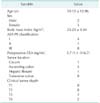
Table 1 shows the patients' preoperative baseline characteristics. There were 2 men and 3 women. The mean patient age was 59.15 ± 12.96 years, and the mean body mass index was 24.23 ± 3.54 kg/m2.
The mean operation time was 183 ± 29.37 minutes (range, 150–230 minutes), and the mean estimated blood loss was 27 ± 9.75 mL (Table 2). The time to clear liquid intake was 3 days after operation in all patients. The mean length of hospital stay after surgery was 6.2 ± 0.55 days. No patient required conversion to conventional laparoscopic surgery. There were no perioperative complications or mortality.
According to the pathology report, the mean tumor size was 5.0 ± 1.51 cm. The mean number of harvested lymph nodes and positive lymph nodes was 36.6 ± 4.45 and 6.6 ± 3.67, respectively. Four patients were stage III, and one patient was stage II according to the 7th edition of the American Joint Committee on Cancer system.
Despite several merits of using surgical robots, such as endowrist, stable traction, and counter-traction, and simultaneous control of the instruments and the scope, the previous version of the da Vinci system had inherent drawbacks because of frequent collisions of the robotic arms and instruments during surgery [81011]. This mechanical limitation led us to place the ports in unusual sites compared to conventional laparoscopy. Consequently, a 12-mm port for the camera is often placed away from the umbilicus, which is the most commonly used site for camera insertion, anastomosis, and specimen delivery in conventional laparoscopic surgery. The latest da Vinci Xi system has addressed some of these issues so that more compact, linear placement of the ports can be accomplished in order to perform multi-quadrant organ exposure with single docking of the system.
Therefore, we developed a novel port set-up on the suprapubic horizontal line to maximize the advantages of minimally invasive surgery with multiple rationales. First, multiple small incisions in a horizontal line in the suprapubic area are easily concealed by skin creases. Second, a transverse incision connecting the middle 2 trocar incisions might reduce the rate of incisional hernia compared to vertical or midline incisions [12131415], and could reduce the size of the incisions because the newest da Vinci Xi system has a smaller minimum working distance between the trocars to avoid collision. Third, placing all incisions on the lower abdomen, except one 5-mm port in the left upper quadrant, may result in reduced postoperative pain.
Technically, we selected the fewest possible number of instruments that performed multiple functions and ultimately chose five instruments, which are as follows: (1) a tip-up double fenestrated grasper for grabbing the tissue and wider retraction with jaws opened, (2) a vessel sealer for division of the vessels and trimming the mesentery and omentum, as well as assisting when suturing, (3) a hot shear for dissection of the avascular plane, precise lymph node dissection, and enterotomies, (4) robotic linear staplers for division of the bowel and creation of an anastomosis, and (5) a needle drive for suturing to close the enterotomy.
This robotic right colectomy via the suprapubic approach was performed safely and successfully with satisfying short-term outcomes in our experience. The mean operation time and estimated blood loss were 183 ± 29.37 minutes and 27 mL, respectively, which are comparable to results from our previous study in 2012 (201.4 minutes and 41.7 mL, respectively) [7]. No patient required conversion to conventional laparoscopic or open surgery. In addition, there were no perioperative complications or mortality in this series. The number of retrieved lymph nodes in the specimens was acceptable and comparable to other studies [78].
After retrieval of the specimen, we closed the transverse mini-laparotomy layer by layer. In this series, we did not experience any complications related to the mini-laparotomy. As briefly discussed, several comparative studies showed the superiority of a transverse incision in the lower abdomen compared to a midline incision in terms of cosmetic satisfaction, wound pain, risk of incisional hernia, and adhesions [12131415]. Although our data have a definite limitation due to the small number of cases and short duration of follow-up, all patients were satisfied regarding their wounds and postoperative recovery.
There are still some obstacles for robotic right colectomy. First, in most of areas in the world, robotic surgery costs significantly more than laparoscopic surgery. Nonetheless, affordability may differ from country to country, and when it is available, our technique can be easily used for right colon cancer patients. Second, the operative time for robotic right colectomy is longer, although still acceptable, compared to conventional laparoscopically-assisted right colectomy with extracorporeal anastomosis. This may be due to the intracorporeal anastomosis method as well the nature of the robot system itself. Lastly, although there was no conversion to laparoscopic or open surgery in this series, it may be difficult to share the same port in cases of a “converted” laparoscopic approach. Therefore, careful patient selection is needed.
In conclusion, totally-robotic right colectomy via the suprapubic approach can be performed successfully with satisfying short-term outcomes in selected patients. Further comparative studies are required to verify the clinical advantages of our technique over conventional robotic surgery.
Figures and Tables
Fig. 1
Suprapubic approach. (A) Trocar positioning of da Vinci Si system. (B) Novel trocar positioning of da Vinci Xi system. (C) A postoperative scar after suprapubic approach. SUL, spinoumbilicus line; MCL, midclavicular line.

References
1. Lacy AM, Garcia-Valdecasas JC, Delgado S, Castells A, Taura P, Pique JM, et al. Laparoscopy-assisted colectomy versus open colectomy for treatment of non-metastatic colon cancer: a randomised trial. Lancet. 2002; 359:2224–2229.

2. Clinical Outcomes of Surgical Therapy Study Group. Nelson H, Sargent DJ, Wieand HS, Fleshman J, Anvari M, et al. A comparison of laparoscopically assisted and open colectomy for colon cancer. N Engl J Med. 2004; 350:2050–2059.

3. Fleshman J, Sargent DJ, Green E, Anvari M, Stryker SJ, Beart RW Jr, et al. Laparoscopic colectomy for cancer is not inferior to open surgery based on 5-year data from the COST Study Group trial. Ann Surg. 2007; 246:655–662.

4. Park JS, Choi GS, Lim KH, Jang YS, Jun SH. Robotic-assisted versus laparoscopic surgery for low rectal cancer: case-matched analysis of short-term outcomes. Ann Surg Oncol. 2010; 17:3195–3202.

5. Xu H, Li J, Sun Y, Li Z, Zhen Y, Wang B, et al. Robotic versus laparoscopic right colectomy: a meta-analysis. World J Surg Oncol. 2014; 12:274.

6. Trastulli S, Coratti A, Guarino S, Piagnerelli R, Annecchiarico M, Coratti F, et al. Robotic right colectomy with intracorporeal anastomosis compared with laparoscopic right colectomy with extracorporeal and intracorporeal anastomosis: a retrospective multicentre study. Surg Endosc. 2015; 29:1512–1521.

7. Park JS, Choi GS, Park SY, Kim HJ, Ryuk JP. Randomized clinical trial of robot-assisted versus standard laparoscopic right colectomy. Br J Surg. 2012; 99:1219–1226.

8. Park SY, Choi GS, Park JS, Kim HJ, Choi WH, Ryuk JP. Robot-assisted right colectomy with lymphadenectomy and intracorporeal anastomosis for colon cancer: technical considerations. Surg Laparosc Endosc Percutan Tech. 2012; 22:e271–e276.
9. Ozben V, Cengiz TB, Atasoy D, Bayraktar O, Aghayeva A, Erguner I, et al. Is da Vinci Xi better than da Vinci Si in robotic rectal cancer surgery? Comparison of the 2 generations of da Vinci Systems. Surg Laparosc Endosc Percutan Tech. 2016; 26:417–423.

10. Corcione F, Esposito C, Cuccurullo D, Settembre A, Miranda N, Amato F, et al. Advantages and limits of robot-assisted laparoscopic surgery: preliminary experience. Surg Endosc. 2005; 19:117–119.

11. Delaney CP, Lynch AC, Senagore AJ, Fazio VW. Comparison of robotically performed and traditional laparoscopic colorectal surgery. Dis Colon Rectum. 2003; 46:1633–1639.

12. Fanning J, Pruett A, Flora RF. Feasibility of the Maylard transverse incision for ovarian cancer cytoreductive surgery. J Minim Invasive Gynecol. 2007; 14:352–355.

13. Levrant SG, Bieber E, Barnes R. Risk of anterior abdominal wall adhesions increases with number and type of previous laparotomy. J Am Assoc Gynecol Laparosc. 1994; 1(4, Part 2):S19.

14. Manusook S, Suwannarurk K, Pongrojpaw D, Bhamarapravatana K. Maylard incision in gynecologic surgery: 4-year experience in Thammasat University Hospital. J Med Assoc Thai. 2014; 97:Suppl 8. S102–S107.
15. Benlice C, Stocchi L, Costedio MM, Gorgun E, Kessler H. Impact of the specific extraction-site location on the risk of in ci sional hernia after laparoscopic colorectal resection. Dis Colon Rectum. 2016; 59:743–750.
SUPPLEMENTARY MATERIAL
Supplementary material (video clip) can be found via http://www.astr.or.kr/src/sm/94-2_S001.wmv.


 PDF
PDF ePub
ePub Citation
 Print
Print


 XML Download