

 PDF
PDF ePub
ePub Citation
 Print
Print


Abstract
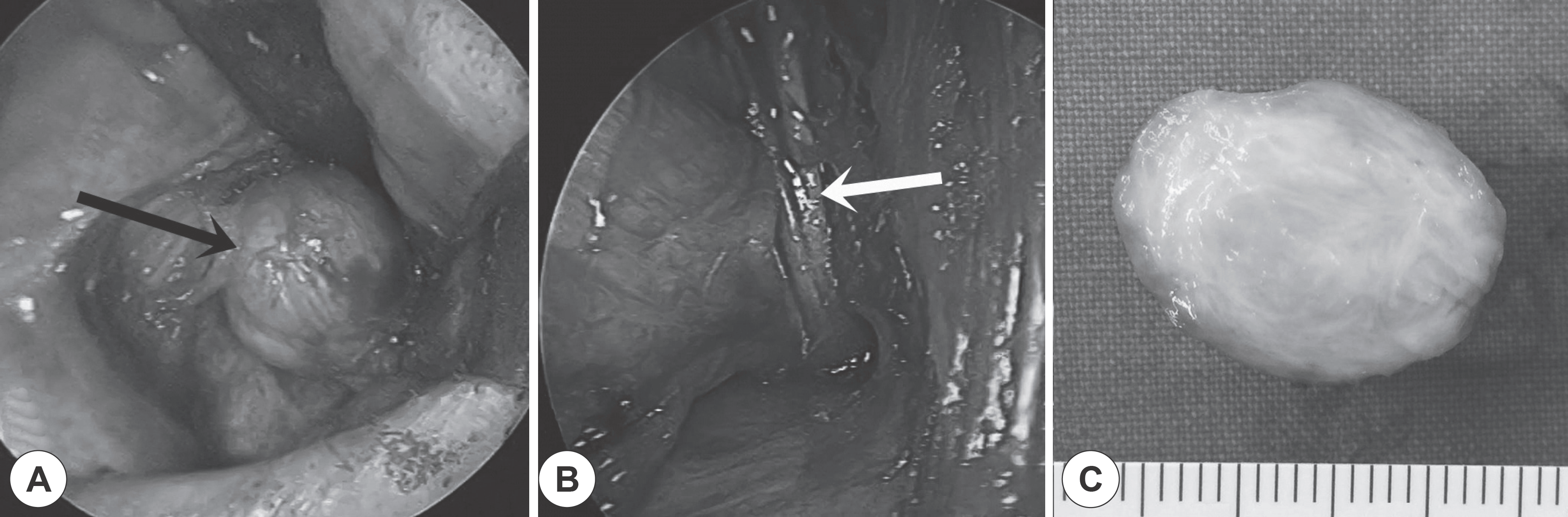
Solitary fibrous tumor is a rare spindle cell neoplasm of mesenchymal origin that occurs most commonly in the pleura. This tumor can be found in various extrathoracic sites that contain soft tissue. There are few reports of solitary fibrous tumors in the head & neck and only 5 cases of solitary fibrous tumors of the cheek have been reported. A 53-year-old man visited our department complaining of a firm mass in the left cheek. We suspected a schwannoma originating from the infraorbital nerve. The mass was removed via a gingivobuccal approach and was diagnosed as a solitary fibrous tumor.
REFERENCES
1). Klemperer P., Rabin CB. Primary neoplasms of the pleura: a report of 5 cases. Arch Pathol. 1931. 11:385–412.
2). Ge W., Yu DC., Chen G., Ding YT. Clinical analysis of 47 cases of solitary fibrous tumor. Oncol Lett. 2016. 12:2475–80.

3). Kim JH., Yim JW., Kim HK., Lee JG. A Case of Solitary Fibrous Tumor of the Nasal Cavity. J Rhinol. 2003. 10:60–3.
4). Kuo WP., Sirois DA., Pemble CW. Locally aggressive solitary fibrous tumor in the infraorbital region: a case report and review of the literature. Oral Surg Oral Med Oral Pathol Oral Radiol Endod. 2001. 92:308–11.
5). Profyris C., Soilleux E., Corkill R., Birch J. Solitary fibrous tumour of the face: a rare case report. J Plast Reconstr Aesthet Surg. 2010. 63:e13–5.
6). Satomi T., Hasegawa O., Abukawa H., Kohno M., Enomoto A., Chika-zu D, et al. Exceptionally large solitary fibrous tumor arising from the cheek: an immunohistochemical and ultrastructural study with a review of the literature. Medical Molecular Morphology. 2014. 47:108–16.
7). Cho KJ., Ro JY., Choi J., Choi SH., Nam SY., Kim SY. Mesenchymal neoplasms of the major salivary glands: clinicopathological features of 18 cases. Eur Arch Otorhinolaryngol. 2008. 265(Suppl 1):S47–56.
8). Gold JS., Antonescu CR., Hajdu C., Ferrone CR., Hussain M., Lewis JJ, et al. Clinicopathologic correlates of solitary fibrous tumors. Cancer. 2002. 94:1057–68.
9). Wilky BA., Montgomery EA., Guzzetta AA., Ahuja N., Meyer CF. Extrathoracic location and “borderline” histology are associated with recurrence of solitary fibrous tumors after surgical resection. Ann Surg Oncol. 2013. 20:4080–9.
10). Kim HJ., Lee HK., Seo JJ., Kim HJ., Shin JH., Jeong AK, et al. MR imaging of solitary fibrous tumors in the head and neck. Korean J Radiol. 2005. 6:136–42.
11). Thway K., Ng W., Noujaim J., Jones RL., Fisher C. The Current Status of Solitary Fibrous Tumor: Diagnostic Features, Variants, and Genetics. Int J Surg Pathol. 2016. 24:281–92.
12). Kwak SG., Kim CD., Kim YJ., Kim SW. A Case of Recurrent and Multiple Schwannomas in the Caudal Septum. J Rhinol. 2015. 22:41–3.
13). Enzinger FM., Smith BH. Hemangiopericytoma. An analysis of 106 cases. Hum Pathol. 1976. 7:61–82.
14). Kayani B., Sharma A., Sewell MD., Platinum J., Olivier A., Briggs TW, et al. A Review of the Surgical Management of Extrathoracic Solitary Fibrous Tumors. Am J Clin Oncol. 2016.
15). Yang XJ., Zheng JW., Ye WM., Wang YA., Zhu HG., Wang LZ, et al. Malignant solitary fibrous tumors of the head and neck: a clinicopathological study of nine consecutive patients. Oral Oncol. 2009. 45:678–82.
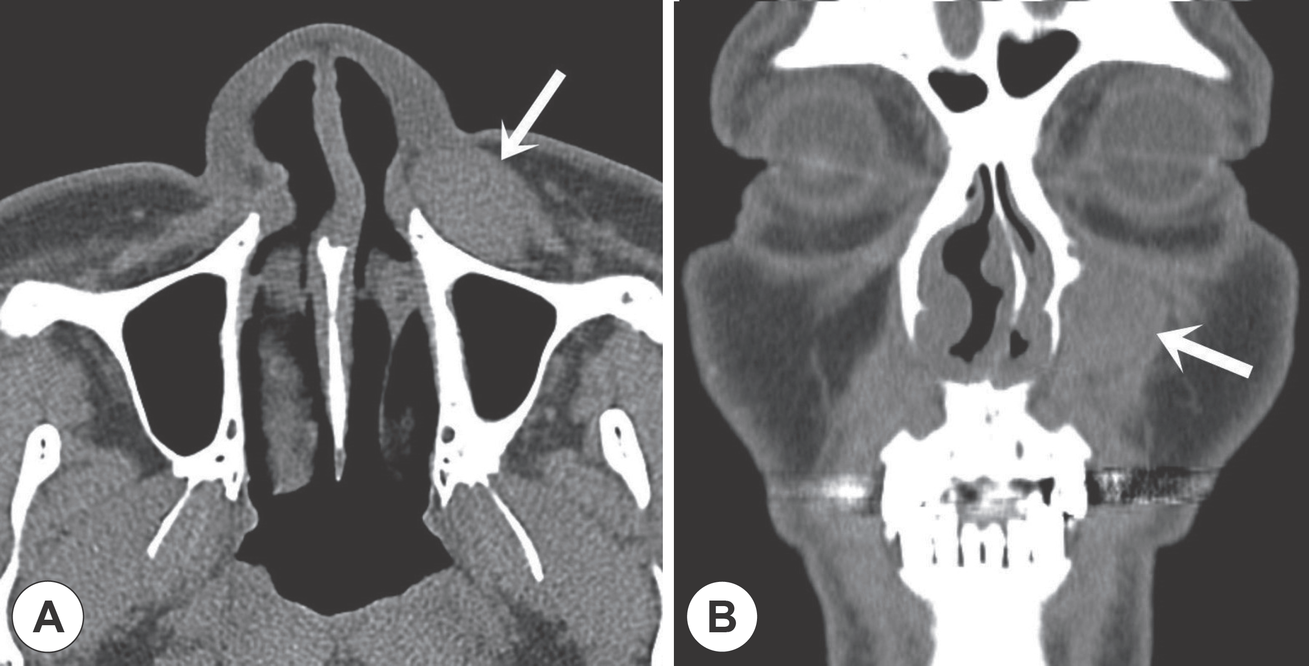
Fig. 1
Preoperative computed tomography scan. The relatively well-circumscribed, isointense mass (white arrow) is shown at the cheek. A: Axial view. B: Coronal view.

 XML Download