

 PDF
PDF ePub
ePub Citation
 Print
Print


Since its initial appearance in 1985, human immunodeficiency virus (HIV) infection and its late-stage manifestation, the acquired immune deficiency syndrome (AIDS) have affected a growing number of persons in Korea and around the world1). As of December 2000, more than 1,280 HIV infections and 286 deaths from AIDS in Korea have been reported to the Korean National Institute of Health (Korean Ministry of Health and Welfare, 2000). Whilst degenerative arthritis may be relatively rare in patients with HIV (who are predominantly of a younger age group), osteonecrosis, inflammatory and post-traumatic problems are seen on a regular basis in areas of high HIV seroprevalence. Whether osteonecrosis is an HIV-related complication, an adverse effect of antiretroviral (ARV) therapy or is caused by another HIV-associated condition remains unclear2). The reported estimated incidence of osteonecrosis of femoral head (ONFH) in patients with HIV ranges from 0.45% to 1.33%3), which is greater than in the general population.
Rapidly destructive coxarthrosis (RDC) is a rare condition with an elusive etiology that involves rapid destruction and collapse of the femoral head within 12 months of symptom onset456). The joint destruction is typically unilateral, more common in elderly women and manifests with severe pain in the hip and claudication. In this case, a patient with HIV infection had sequential arthritis of both sides of her hip, with the left side identified 8 months after the right side. An initial radiographic presentation of either normal anatomy or mild arthritis evolves into rapid destruction of the femoral head within months of the onset of clinical manifestations. In the case presented here, we aim to: i) assess and/or identify additional risk factors of ONFH in an HIV-positive patient and determine if these associations may be multifactorial, ii) explore the alleged association of ONFH and ARV, and iii) assess our patient's early response to hip replacement surgery. In HIV-diagnosed patients, joint pain should not be ignored and should always be observed.
CASE REPORT
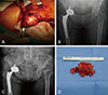
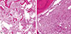
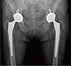
In April 2015, a 71-year-old female patient was referred to our group for osteopathic care after complaining of right hip pain. This patient was diagnosed with HIV 29 months prior (November 2012; 68-years old) and began a continuous ARV treatment regimen (nucleoside reverse transcriptase [lamivudine] and protease inhibitor cocktail [ritonavir+darunavir]) at this time. A plain spine radiograph obtained near the time of referral revealed potential osteonecrosis of the right femoral head. As part of the patient's care for HIV, a medical examination (including a computed tomography scan) was performed 8 months before her referral to our group. We reviewed this assessment and saw no indication of osteonecrosis (Fig. 1). Additionally, the patient had not received steroids since the time of diagnosis with HIV. Following our initial assessment, we recommended a magnetic resonance imaging (MRI) but it was not obtained because of the cost. Clinical (laboratory tests and physical examination) and radiological (pelvic computed tomography with contrast) assessments revealed no signs of infection. Total hip arthroplasty (THA) surgery of the right hip was then performed through a posterolateral approach under general anesthesia and taking all precautions published by AAOS (American Academy of Orthopedic surgeons) for surgery on HIV-positive patients to protect staff against HIV transmission. Specifically, the operating surgeon, two assistants and one scrub nurse wore disposable impervious gowns, helmets with facial shields and a body exhaust system, double hand gloves and impermeable boots. Disposable impervious drapes were used for draping. Instruments contaminated with blood were treated with well-established sterilization methods (e.g., autoclaving) and a 0.5% sodium hypochlorite solution was used to treat any blood spills. Intraoperative finding revealed femoral head collapse consistent with common osteonecrosis (Fig. 2A). The anteroposterior view of an X-ray image confirmed repair of the right hip joint with implant and an intact left hip joint is intact. Bencox ID stem and cup (Corentec, Seoul, Korea) were used (Fig. 2B). The test results for intraoperative bacterial identification was negative, and no signs of infection were observed during outpatient follow-up. No complications were apparent through the 12-month follow up; however, the patient began experiencing left hip pain 17 months after the right-hip THA. Three months later (20 months post right-hip THA), an X-ray revealed loss of the left femoral head (Fig. 2C). A THA of the left hip was then performed using the same technique described above. Intraoperative results revealed more severe femoral head loss and osteolysis of the left hip compared with the right hip, but no signs of infection were observed (Fig. 2D). Furthermore, intraoperative tests for bacteria were negative. Non-specific findings revealing chronic inflammation and fibrotic changes were identified upon histological exam (Fig. 3). No clinical or radiologic manifestations were present 3 months following the second THA (Fig. 4).
DISCUSSION
RDC was first described in 1970 by Postel and Kerboull7). This idiopathic condition usually involves unilateral joint destruction and progressive hip pain that evolves into rapid destruction of the femoral head despite a relatively preserved range of motion, within several months of symptom onset. The exact etiology of RDC is unknown; however, a number of proposed etiological theories have been published. Several authors have alluded to an association between ARV drugs–particularly protease inhibitors–and the onset of RDC2). Additional proposed factors associated with RDC include rheumatoid arthritis, deposition of hydroxyapatite or calcium phosphate crystals, and the use of anti-inflammatory drugs (e.g., indomethacin)8).
Yoo et al.9) suggest that RDC could be a subtype of femoral head osteonecrosis since insignificant histological differences in hip cartilage was observed between osteonecrosis and 29 cases of RDC. In our case report, histological examination revealed fibrosis and chronic inflammation, conditions that may be associated with osteonecrosis of the femoral head.
HIV patients commonly complain of arthralgia and are likely to have localized osteoporosis, fractures and/or fat embolisms due to the long-term use of corticosteroids that may lead to vascular occlusion10). Because multiple arthralgias are common among this patient population, there is a risk of overlooking other potential complications such as avascular necrosis or rapid-destruction arthropathy. Early diagnosis with a highly sensitive tool (e.g., MRI) is recommended. In this case report, the patient was a 73-year old HIV positive woman with no known comorbidities and no history of alcohol consumption or steroid use. Our patient was subjected to prolonged use of antiviral medications–treatments that are associated with an increased risk of femoral head osteonecrosis compared with healthy patients. The patient was later diagnosed with RDC based on X-rays which showed femoral head loss 8 months after symptom onset. A limitation in our case report is that MRI scans were not obtained due to financial restrictions.
The rapid onset of femoral head loss (i.e., within several months of symptom onset) in the present case report distinguishes it from typical femoral head osteonecrosis. The multiple arthralgias that often afflict patients with HIV require pain control and accurate diagnosis with X-ray and MRI. In addition, careful assessment of radiographs taken during follow-up is required. Surgery should be considered as a treatment option for patients with arthralgias, and may help lead to accurate diagnosis and clarify etiology.

 XML Download