

 PDF
PDF ePub
ePub Citation
 Print
Print


Introduction
Thyroid carcinoma accounts for roughly 1% of all new malignant diseases and has increased in incidence due to the use of ultrasonography (US).1) At least 94% of thyroid carcinomas are differentiated thyroid carcinomas, with papillary thyroid carcinoma (PTC) being the most common subtype.1) Fortunately, PTC exhibits indolent, localized features and generally shows good prognosis. Ten-year overall survival rates for PTC are reported to be 93–98%.23) Despite relatively good prognosis, 10-year recurrence rates for PTCs are quite high with values reported to be 14–26%.23) Established unfavorable prognostic factors of PTC are age at diagnosis, tumor size, extrathyroidal extension, lymph node metastasis, histologic subtype (tall cell, columnar cell, diffuse sclerosing, and insular variants), incomplete surgery, and omitted or insufficient radioactive iodine therapy.45) In recent years, there has been wider acceptance about the critical role systemic inflammation plays in the evolution of cancer.678) In the physiologic inflammatory process, wound healing is self-limited by balancing anti- and pro-inflammatory factors. However, cancers are “wounds that do not heal” by persistent and dysregulated inflammatory factors.9) Neutrophils, the first recruited cells in an acute inflammation, are major sources of cytokines/chemokines necessary for inflammatory cell recruitment, activation and reaction.7) Recent studies have suggested that pre-treatment inflammatory parameters based on blood tests (such as the C-reactive protein, albumin, neutrophil and platelet counts) can be predictors of cancer prognosis.10) Among these inflammatory parameters, the pretreatment high neutrophil-to-lymphocyte ratio (NLR) has been especially associated with adverse outcomes in various cancer entities.11) In patients with thyroid cancers, a higher NLR has been associated with larger tumor size,1213) high stage,14) old age,1314) and poorer differentiated cancer.15) Therefore, we investigated whether preoperative NLR is associated with poor prognostic outcomes in conventional PTCs (cPTCs) which comprises the majority of thyroid malignancies.
Materials and Methods
The Institutional Review Board of our institution approved this retrospective study. Neither patient approval nor informed consent for review of patient medical records was required.
Patients
From January 2005 to February 2006, 248 patients who underwent total thyroidectomy for cPTC larger than 10 mm and were followed up for more than 3 years at our institution (a referral center) were initially considered as candidates for our study. After excluding 43 patients without available preoperative blood cell counts (neutrophils and lymphocytes) within a week before surgery, 205 patients were finally included in this study. The NLR was calculated as the absolute neutrophil count divided by the absolute lymphocyte count. The mean follow-up period after surgery was 93.6 months (median, 108.4 months; range, 36–125.4 months). All patients in this study had been included in the study population of a previously published article.16) However, the previous study had different research purposes from our own and did not deal with the same concepts.
Surgery and Pathologic Diagnosis
In our institution, patients with multifocal or bilateral lesions, extrathyroidal extension, or lymph node metastasis underwent total thyroidectomy. Prophylactic central node dissection was routinely performed on all patients, and lateral neck dissection including levels 2, 3, 4 and anterior 5, was selectively performed on patients with preoperatively or intraoperatively suspicious lymph nodes. US-guided fine-needle aspiration or intraoperative frozen biopsy was performed to diagnose metastatic lymph nodes, respectively. Tumor size, extrathyroidal extension, multifocality, and lymph node metastasis were determined from the final pathologic records. Pathologic tumor-node metastasis (TNM) staging was classified according to the eighth edition of the American Joint Committee on Cancer.17)
Follow-up Investigation and Persistence/Recurrence
All patients received thyroid stimulating hormone (TSH) suppressive therapy with levothyroxine after surgery. Radioactive iodine treatment was given to patients with histopathology-proven extrathyroidal extension or lymph node metastasis. Patients who underwent surgery were followed up at 6-month intervals for the first 3 years and annually thereafter. Follow-up investigations included a clinical examination every 6 months; laboratory tests including free T4, TSH, non-stimulated or stimulated thyroglobulin, thyroglobulin antibody (Ab); a chest X-ray; and a neck US examination every 12 months. Iodine-131 whole-body scan or fluorodeoxyglucose positron emission tomography (PET)/computed tomography (CT) were performed in specific cases, such as cases with detectable or persistent serum Tg or anti-Tg antibody without radiological evidence of recurrence. Clinical endpoints were divided into three categories: no clinical evidence of disease (NED), persistence, and recurrence. NED was defined with suppressed serum Tg<1 ng/mL, no detectable TgAb, and no structural evidence of disease. Structural evidence of disease was defined as i) biopsy-proven disease (cytologially or histologically), or ii) highly suspicious finding of lymph nodes or operative bed nodules at neck US examination, or iii) highly suspicious for metastatic disease on cross-sectional imaging modality (US, CT, or magnetic resonance imaging) or functional imaging (radioactive iodine [RAI] scan or PET/CT). Persistence was defined as biochemical (suppressed serum Tg≥1 ng/mL and/or stimulated Tg≥2 ng/mL) or structural evidence of disease that existed after surgery or RAI ablation. Newly detected biochemical, structural, or functional evidence of disease during any period of NED was defined as recurrence.18) The clinical endpoint was persistence/recurrence first detected after initial treatment (including surgical resection and first RAI treatment if performed).
Statistical Analysis
The methods of Contal and O'Quigley were used to determine the optimal cut-off point of NLR with the log-rank test (with log-rank statistics). When the optimal cut-off point which maximized the hazard ratio of disease-free survival was applied, there was no statistical significant difference between the high NLR and low NLR groups (NLR=2.29, p=0.190). Because our data did not show normal distribution by the Kolmogorov-Smirnov test (p<0.01), we used the median NLR value (1.78; mean, 2.237; range, 0.677–14.689) as the reference value. Patients were divided into either the high NLR group or the low NLR group according to the median NLR for statistical analysis. We used the independent two-sample t-test or chi-square test to compare the clinicopathologic differences between the high NLR and low NLR groups. The univariate and multivariate logistic regression analyses were used to evaluate which NLR predicted aggressive PTC features (extrathyroidal extension, central or lateral lymph node metastasis). Odds ratios (ORs) with 95% confidence intervals (CIs) for predicting aggressive PTC features were calculated for all variables. The univariate and multivariate Cox proportional hazard regression models were used to identify variables that influenced persistence/recurrence. Hazard ratios (HRs) estimated from the Cox analysis were reported as relative risks with corresponding 95% CIs. A p value of less than 0.05 was considered statistically significant. Statistical analyses were performed with SAS statistical software (SAS system for Windows, version 9.2; SAS institute, Cary, NC, USA).
Results
Clinicopathologic Characteristics
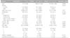
The baseline clinicopathologic characteristics for the included patients are shown in Table 1. The mean age of the 205 patients at diagnosis was 47 years (standard deviation [SD], 13 years; range, 17–80 years); 175 women (mean age, 47 years; range, 17–80 years) and 30 men (mean age, 47 years; range, 25–66 years). The mean tumor size was 18.9 mm (SD, 8.4 mm; range, 11–50 mm). Sixty-six patients (32.2%) had multiple PTCs and 17 patients (8.3%) had pathologic extrathyroidal extension (strap muscle or major organ invasion). Central and lateral LNM were found in 56.1% (115/205) and 22.9% (47/205) of all patients, respectively. Five (2.4%) out of 205 patients had distant metastasis found at initial diagnosis (the lung in 3 patients, lung and bone in one patient, and lung, bone and brain in one patient). Three patients (1.5%) had cancers in advanced stages (stage III-IV). One hundred and seventy-four patients (84.9%) underwent radioactive iodine treatment after surgery; 30 mCi was administered in 159 patients and 50–200 mCi was administered in 190 patients. Persistent disease was observed in 16 patients (7.8%), structural recurrence in 10 patients (4.9%) and both persistent disease and structural recurrence in 4 patients (2.0%).
Predicting Factors for Aggressive Features of cPTC
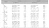
The median value of NLR for all patients was 1.78 and it was defined as the reference value. According to the reference value (1.78), 102 patients had a high NLR (>1.78) and 103 patients had a low NLR (≤1.78). In the high NLR group, there were significantly more males than females (p=0.016). The high NLR group also had a significantly higher presence of lateral LNM (p=0.004) and significantly higher radioactive iodine doses (p=0.006) than the low NLR group. There were no significant differences in mean age, tumor size, extrathyroidal extension, central LNM, multifocality, distant metastasis, higher TNM stage, and persistence/recurrence between the high NLR group and the low NLR group.
High NLR was an independent risk factor for lateral LNM (Table 2). The OR of high NLR was 2.786 (95% CI: 1.358, 5.713; p=0.005) at multivariate analysis. High NLR was not associated with extrathyroidal extension and central LNM at both univariate and multivariate analyses. Tumor size was predictor for central LNM in PTCs. Tumor size and high NLR were predictors for lateral LNM in PTCs.
Predicting Factors for Persistence/Recurrence of cPTC
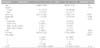
Table 3 compares clinicopathological features of the persistence/recurrence group and the disease-free group. The median NLR value as a continuous variable was not significantly different between the persistence/recurrence group and the disease-free group (2.03 vs. 1.76, p=0.134). Age at operation and gender were similar between the two groups. The persistence/recurrence group had larger tumors (26.4 vs. 18.0 mm, p<0.001) and increased RAI doses (p=0.044). LNM (86.4 vs. 54.6%, p=0.005) and higher TNM stage (13.6 vs. 0%, p<0.001) were more common in the persistence/recurrence group.
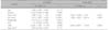
On univariate Cox regression analysis, preoperative high NLR was not significantly associated with persistence/recurrence (Table 4). The mean tumor size and RAI dose were found to be independent predictors of persistence/recurrence on multivariate Cox regression analysis.
Discussion
Neutrophils contribute to tumor growth and metastasis by producing various cytokines/chemokines and reactive oxygen species (ROS) which cause DNA damage and remodel the extracellular matrix leading to tissue destruction.6789) A high neutrophil count has been associated with higher mortality in several advanced or metastatic cancers, such as advanced non-small cell lung cancer,19) metastatic renal cell carcinoma,20) and metastatic melanoma.21) Lymphocytes play a considerable role in cancer immunity by halting the growth of cancer. If a patient has a higher concentration of tumor-infiltrating lymphocytes, he/she shows better prognosis, a phenomenon which was first reported for melanoma22) and breast cancer.23) In different types of cancer, if a cancer patient has a lower peripheral blood lymphocyte count, he or she has a poorer chance of survival.2425) An association between high NLR and worse prognostic outcomes has been well demonstrated in various non-thyroid cancers, including gastric, colorectal, lung, pancreatic, hepatocellular, renal cell carcinoma, cholangiocarcinoma and breast cancer.11)
We found that preoperative high NLR was an independent predictor for lateral LNM in patients with conventional PTCs. Extrathyroidal extension and central LNM, well known as aggressive PTC features, were not associated with a high NLR. Like this study, some studies have reported associations between a high NLR and lymph node metastasis for various malignancies.2627) In 353 non-small cell lung cancer patients, patients with pN1 or pN2 had significantly higher NLRs than patients with pN0 and a high NLR was an independent predictor of regional nodal involvement.26) In 1131 gastric cancer patients, preoperative NLR was significantly positively correlated with the number of metastatic lymph nodes.27) In a meta-analysis studying the prognostic role of NLR in other solid tumors, metastatic diseases were associated with high NLRs with a higher hazard ratio than nonmetastatic diseases (1.80 vs. 1.57, respectively and p=0.12).11) It is unclear why preoperative high NLR in cPTC patients is associated with lateral LNM, while it is not associated with central LNM. Although the mechanism is unclear, we propose more careful preoperative evaluation for lateral neck lymph nodes when a cPTC patient has a high NLR value.
In this study, the overall persistence/recurrence rate was 10.7% and there were no disease-specific deaths. A high NLR was not associated with the persistence/recurrence of conventional PTC. Unlike in patients with non-thyroid cancers, some studies have reported inconsistent results on the prognostic value of high NLR in PTC patients.1328) As noted above, a high NLR has been consistently associated with worse overall survival and secondary survival (such as cancer-specific, progression-free, disease-free survival) in various non-thyroid cancers.11) Lang et al.13) reported that preoperative NLR did not predict disease-free survival in clinically nodal-negative PTCs. Kim et al.28) reported that high NLR was associated with poorer 5-year disease-free survival in advanced PTC patients (stage III and IV). PTC patients showed relatively low NLR levels compared to patients of other non-thyroid cancers.1112) Furthermore, patients with PTC had significantly lower NLR levels than patients with poorly differentiated or anaplastic thyroid cancer.1315) Many studies have demonstrated that PTCs with lymphocytic infiltrates are associated with good prognosis and high disease-free survival.2930) Meanwhile, intratumoral lymphocytic infiltration was intensely reduced or absent in poorly differentiated or undifferentiated thyroid carcinoma.31) Consequently, lymphocytic infiltration in PTC may have protective effects and the loss of protective effects in poorly differentiated or anaplastic thyroid carcinoma may contribute to aggressive behavior and poor prognostic outcomes.293031)
This study has some limitations. First, our study was of retrospective design and 43 patients without available preoperative blood cell counts were excluded. Thus, selection bias is inevitable. Second, our institution selectively performed lateral lymph node dissection when a lateral lymph node was proven to be metastatic on preoperative US-fine needle aspiration or was suspicious for metastasis on preoperative imaging or during surgery. Thus, the incidence of/the effect of lateral LNM might be underestimated. Third, we did not consider reactive inflammatory cervical lymphadenopathy, a common cause of high NLR. The presence of reactive inflammatory cervical lymphadenopathy in patients with cPTC increases the likelihood of tissue sampling, which may result in selection bias. Fourth, the follow-up periods of the included patients varied from 3 to 10 years. Some patients might have not been followed up for a sufficient length of time. Despite these limitations, our study has several potential strengths. Whereas previous studies covered a broad thyroid cancer spectrum from differentiated thyroid cancer to anaplastic thyroid carcinoma,12131415) our study investigated only conventional PTCs. Therefore, our results clarified the clinical significance of preoperative NLR on conventional PTCs which comprise the majority of thyroid malignancies.
In conclusion, preoperative high NLR was significantly associated with lateral LNM in patients with cPTCs, although preoperative high NLR was not significantly associated with persistence/recurrence in patients with cPTCs.
 XML Download