

 PDF
PDF ePub
ePub Citation
 Print
Print


초록
Background and Objectives
The diagnostic performance of shear wave elastography (SWE) combined with ultrasound (US) in the differential diagnosis of thyroid nodules was evaluated.
Materials and Methods
459 articles were collected using KoreaMed, Ovid-MEDLINE, Ovid-EMBASE, and Cochrane Library. The searching words were ‘{(elastography and shear).mp. OR SWE.mp. OR acoustic radiation force impulse.mp. OR ARFI.mp. OR acuson.mp. OR aixplorer.mp.}’. Two authors independently performed article selection and evaluation of the quality of studies with Scottish Intercollegiate Guidelines Network tool.
Results
2582 specimens (thyroid nodules) from 11 studies selected were included in this review. Combined use of US and SWE was reported higher specificity in five literatures, lower specificity in five studies, and no changes in 1 study when compared to US. We performed metaanalysis using data from 10 studies. The pooled sensitivity and specificity of US and SWE group for the differential diagnosis of benign and malignant nodules were 0.91 (I2=83.4%), 0.73 (I2=95.9%). The pooled sensitivity and specificity of US alone group were 0.88 (I2=93.2%), 0.71 (I2=92.7%).
REFERENCES
1). Yi GH. Updated guidelines for the management of thyroid nodule. Korean J Med. 2011; 80(2):158–61.
2). Park YJ, Hyun MK, Kang MJ, Shim JY, Lee JY, Kim KW, et al. Epidemiology of thyroid nodules in screening. Seoul, Korea: National Evidence Collaborating Agency;2015. p. 1–66.
3). Gharib H, Papini E. Thyroid nodules: clinical importance, assessment, and treatment. Endocrinol Metab Clin North Am. 2007; 36(3):707–35. vi.

4). Ross DS. Nonpalpable thyroid nodules–managing an epidemic. J Clin Endocrinol Metab. 2002; 87(5):1938–40.
5). Tan GH, Gharib H. Thyroid incidentalomas: management approaches to nonpalpable nodules discovered incidentally on thyroid imaging. Ann Intern Med. 1997; 126(3):226–31.
6). Yi KH, Park YJ, Koong SS, Kim JH, Na DG, Ryu JS, et al. Revised Korean Thyroid Association Management Guidelines for patients with thyroid nodules and thyroid cancer. Endocrinol Metab. 2010; 25(4):270–97.
7). Tian W, Hao S, Gao B, Jiang Y, Zhang S, Guo L, et al. Comparison of diagnostic accuracy of realtime elastography and shear wave elastography in differentiation malignant from benign thyroid nodules. Medicine (Baltimore). 2015; 94(52):e2312.
8). Xing P, Wu L, Zhang C, Li S, Liu C, Wu C. Differentiation of benign from malignant thyroid lesions: calculation of the strain ratio on thyroid sonoelastography. J Ultrasound Med. 2011; 30(5):663–9.
9). Zhang B, Ma X, Wu N, Liu L, Liu X, Zhang J, et al. Shear wave elastography for differentiation of benign and malignant thyroid nodules: a metaanalysis. J Ultrasound Med. 2013; 32(12):2163–9.
10). Tamsel S, Demirpolat G, Erdogan M, Nart D, Karadeniz M, Uluer H, et al. Power Doppler US patterns of vascularity and spectral Doppler US parameters in predicting malignancy in thyroid nodules. Clin Radiol. 2007; 62(3):245–51.
11). Cappelli C, Castellano M, Pirola I, Gandossi E, De Martino E, Cumetti D, et al. Thyroid nodule shape suggests malignancy. Eur J Endocrinol. 2006; 155(1):27–31.
12). Ophir J, Alam SK, Garra B, Kallel F, Konofagou E, Krouskop T, et al. Elastography: ultrasonic estimation and imaging of the elastic properties of tissues. Proc Inst Mech Eng H. 1999; 213(3):203–33.
13). Lerner RM, Huang SR, Parker KJ. "Sonoelasticity" images derived from ultrasound signals in mechanically vibrated tissues. Ultrasound Med Biol. 1990; 16(3):231–9.
14). Dong FJ, Li M, Jiao Y, Xu JF, Xiong Y, Zhang L, et al. Acoustic radiation force impulse imaging for detecting thyroid nodules: a systematic review and pooled metaanalysis. Med Ultrason. 2015; 17(2):192–9.
15). Zhan J, Diao XH, Chai QL, Chen Y. Comparative study of acoustic radiation force impulse imaging with realtime elastography in differential diagnosis of thyroid nodules. Ultrasound Med Biol. 2013; 39(12):2217–25.
16). Scottish Intercollegiate Guidelines Network. SIGN methodology checklist. [cited February 4, 2018]. Available from. http://www.sign.ac.uk/checklists-and-notes.html.
17). Scottish Intercollegiate Guidelines Network. SIGN 50- a guideline developer's handbook. [cited February 4, 2018]. Available from. http://www.sign.ac.uk/assets/sign50_2015.pdf.
18). Higgins JP, Thompson SG, Deeks JJ, Altman DG. Measuring inconsistency in meta-analyses. BMJ. 2003; 327(7414):557–60.
19). Duan SB, Yu J, Li X, Han ZY, Zhai HY, Liang P. Diagnostic value of two-dimensional shear wave elastography in papillary thyroid microcarcinoma. Onco Targets Ther. 2016; 9:1311–7.
20). Wang F, Chang C, Gao Y, Chen YL, Chen M, Feng LQ. Does shear wave elastography provide additional value in the evaluation of thyroid nodules that are suspicious for malignancy? J Ultrasound Med. 2016; 35(11):2397–404.
21). Liu B, Liang J, Zheng Y, Xie X, Huang G, Zhou L, et al. Two-dimensional shear wave elastography as promising diagnostic tool for predicting malignant thyroid nodules: a prospective single-centre experience. Eur Radiol. 2015; 25(3):624–34.
22). Park AY, Son EJ, Han K, Youk JH, Kim JA, Park CS. Shear wave elastography of thyroid nodules for the prediction of malignancy in a large scale study. Eur J Radiol. 2015; 84(3):407–12.
23). Kim H, Kim JA, Son EJ, Youk JH. Quantitative assessment of shear-wave ultrasound elastography in thyroid nodules: diagnostic performance for predicting malignancy. Eur Radiol. 2013; 23(9):2532–7.
24). Veyrieres JB, Albarel F, Lombard JV, Berbis J, Sebag F, Oliver C, et al. A threshold value in shear wave elastography to rule out malignant thyroid nodules: a reality? Eur J Radiol. 2012; 81(12):3965–72.
25). Kwak JY, Kim EK. Diagnostic performance of quantitative shear wave ultrasound elastography for thyroid cancer. J Korean Thyroid Assoc. 2011; 4(2):109–13.
26). Sebag F, Vaillant-Lombard J, Berbis J, Griset V, Henry JF, Petit P, et al. Shear wave elastography: a new ultrasound imaging mode for the differential diagnosis of benign and malignant thyroid nodules. J Clin Endocrinol Metab. 2010; 95(12):5281–8.
27). Xing P, Chen Q, Yang ZW, Liu CB, Wu CJ. Combination of conventional ultrasound and tissue quantification using acoustic radiation force impulse technology for differential diagnosis of small thyroid nodules. Int J Clin Exp Med. 2016; 9(5):8288–95.
28). Zhang YF, Xu JM, Xu HX, Liu C, Bo XW, Li XL, et al. Acoustic radiation force impulse elastography: a useful tool for differential diagnosis of thyroid nodules and recommending fine-needle aspiration: a diagnostic accuracy study. Medicine (Baltimore). 2015; 94(42):e1834.
29). Zhang YF, Xu HX, Xu JM, Liu C, Guo LH, Liu LN, et al. Acoustic radiation force impulse elastography in the diagnosis of thyroid nodules: useful or not useful? Ultrasound Med Biol. 2015; 41(10):2581–93.
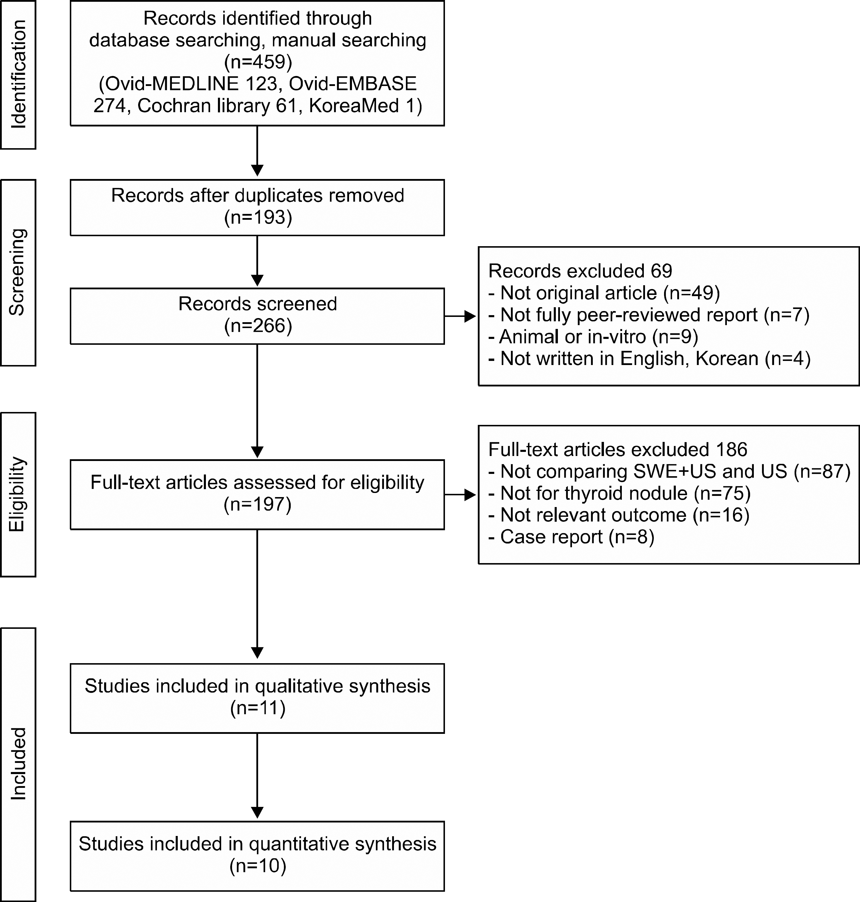
Fig. 2.
Forest plot of sensitivities for SWE+ US and US. CI: confidential interval, NA: not available, SWE: shear wave elastography, US: ultrasonography, VTI: virtual touch tissue imaging, VTQ: virtual touch tissue quantification
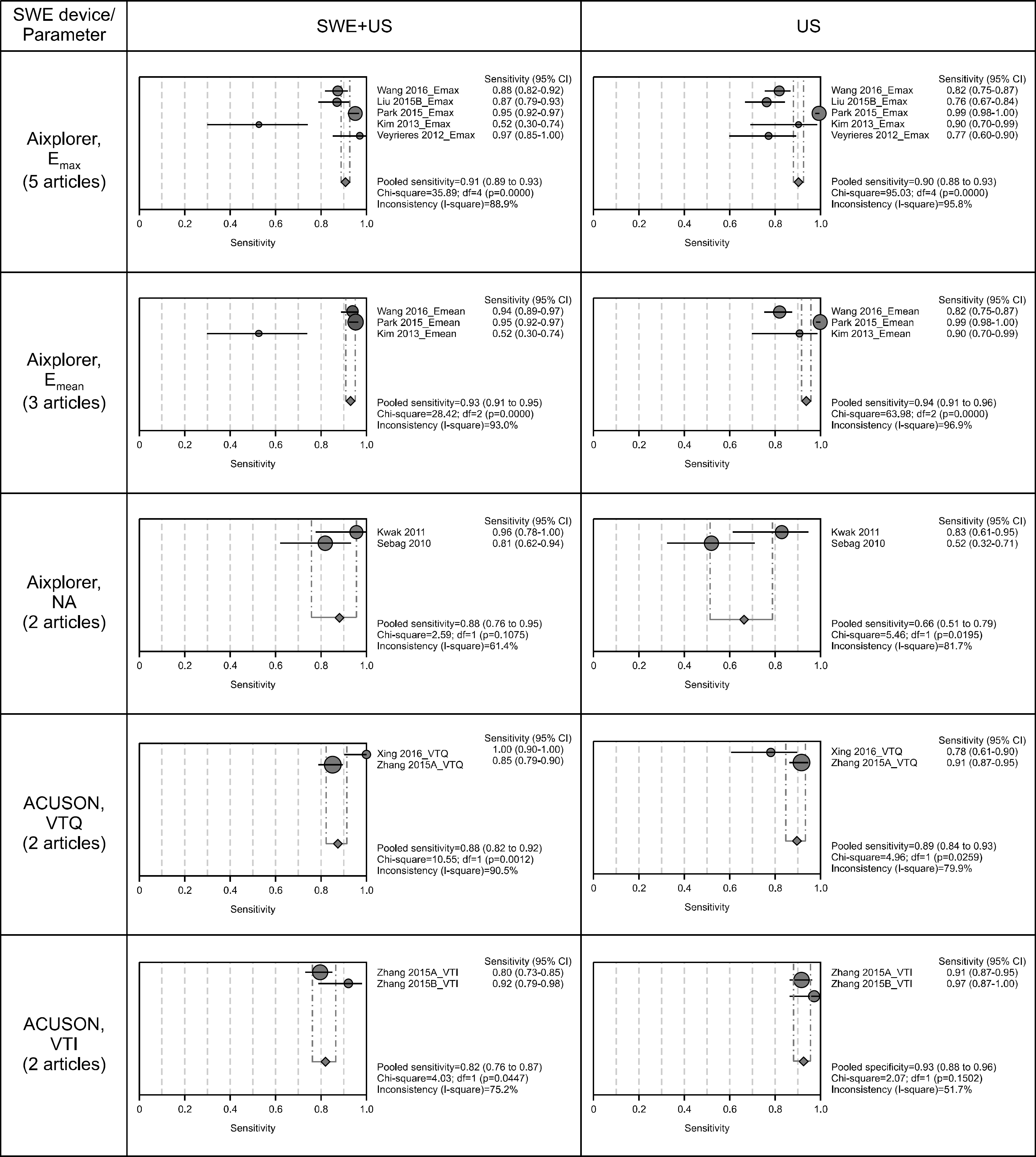
Fig. 3.
Forest plot of specificities for SWE+ US and US. CI: confidential interval, NA: not available, SWE: shear wave elastography, US: ultrasonography, VTI: virtual touch tissue imaging, VTQ: virtual touch tissue quantification
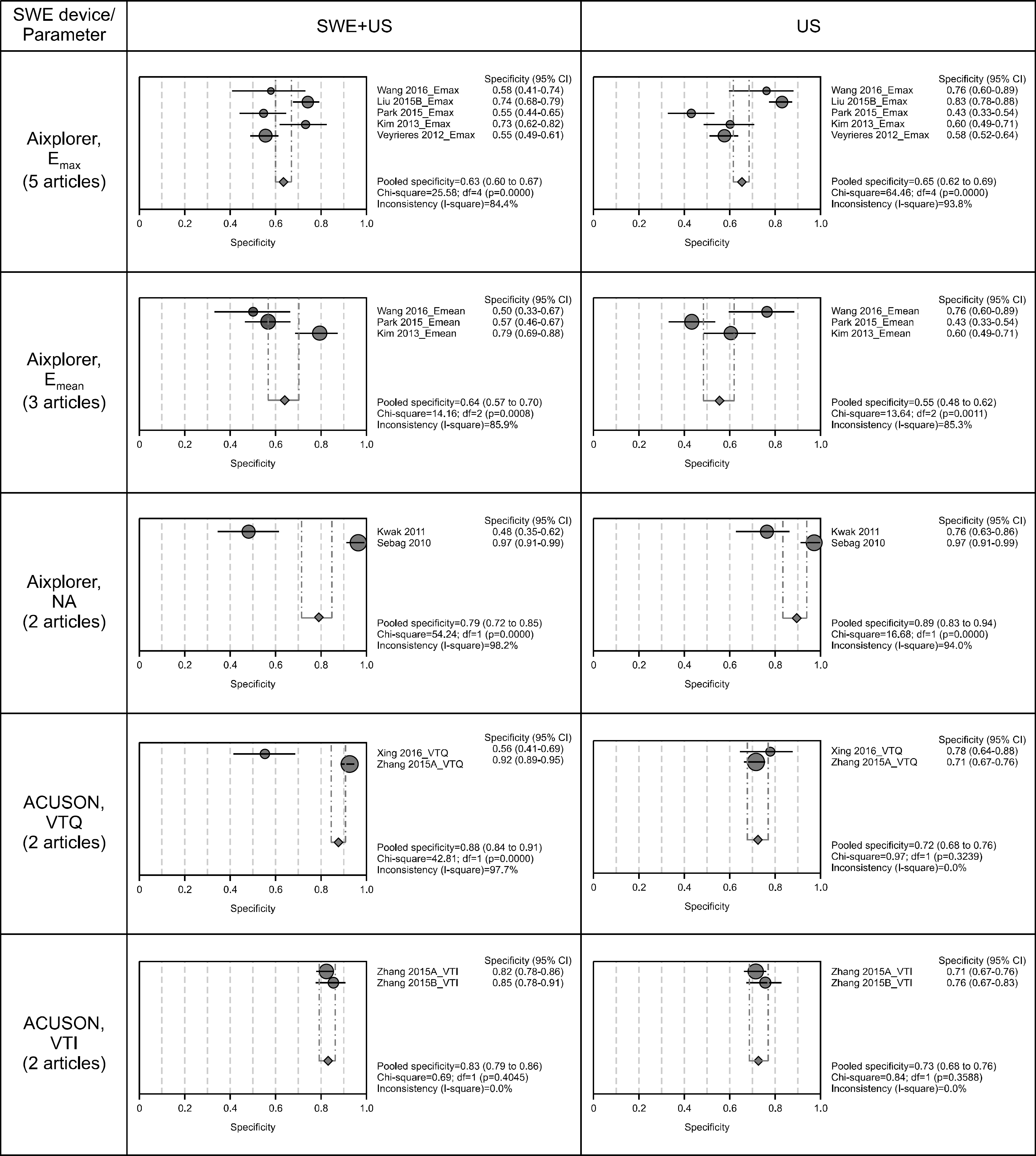
Table 1.
Details of search strategy
Table 2.
Levels of evidence (SIGN criteria)16,17)
Table 3.
Main characteristics of selected studies
Table 4.
Diagnostic accuracy of ultrasonography and combining shear wave elastography with ultrasonography
| Authors | Year | No. of nodules | Ultrasonography | Shear wave elastography and ultrasonography | ||||||||||
|---|---|---|---|---|---|---|---|---|---|---|---|---|---|---|
| Se | Sp | PPV | NPV | Accuracy | Parameter | Diagnosis criteria | Se | Sp | PPV | NPV | Accuracy | |||
| Device: Aixplorer | ||||||||||||||
| Duan et al.19) | 2016 | 137 | 0.89 | 0.90 | 0.82 | 0.93 | 0.90 | E max | 53.2 kPa | 0.96∗ | 0.95∗ | 0.90∗ | 0.98∗ | 0.95 |
| E mean | 34.5 kPa | |||||||||||||
| Wang et al.20) | 2016 | 215 | 0.82 | 0.76 | 0.94 | 0.48 | 0.81 | E max | 65.0 kPa | 0.88 | 0.58 | 0.91 | 0.50 | 0.82 |
| E mean | 26.3 kPa | 0.94 | 0.50 | 0.90 | 0.63 | 0.86 | ||||||||
| Liu et al.21) | 2015 | 331 | 0.76 | 0.83 | 0.66 | 0.89 | 0.81 | E max | 43.8 kPa | 0.87∗ | 0.74∗ | 0.59∗ | 0.93∗ | 0.78∗ |
| E mean | 39.3 kPa | |||||||||||||
| Park et al.22) | 2015 | 476 | 1.00 | 0.43 | 0.87 | 0.96 | 0.88 | E max | 94.0 kPa | 0.95 | 0.55 | 0.89 | 0.74 | 0.87 |
| E mean | 85.2 kPa | 0.95 | 0.57 | 0.90 | 0.74 | 0.87 | ||||||||
| Kim et al.23) | 2013 | 99 | 0.91 | 0.60 | 0.38 | 0.96 | 0.67 | E max | 65.0 kPa | 0.52 | 0.73 | 0.34 | 0.81 | 0.69 |
| E mean | 62.0 kPa | 0.52 | 0.80 | 0.41 | 0.86 | 0.74 | ||||||||
| Veyrieres et al.24) | 2012 | 297 | 0.77 | 0.58 | 0.20 | 0.95 | 0.60 | E max | 66.0 kPa | 0.97 | 0.55 | 0.23 | 0.99 | 0.60 |
| Kwak and Kim25) | 2011 | 81 | 0.83 | 0.76 | 0.58 | 0.92 | 0.78 | NA | 33.3 kPa∗ | 0.96∗ | 0.48∗ | 0.48∗ | 0.97∗ | 0.62∗ |
| Sebag et al.26) | 2010 | 126 | 0.52 | 0.97 | 0.82 | 0.88 | 0.87 | NA | 65.0 kPa∗ | 0.82∗ | 0.97∗ | 0.88∗ | 0.95∗ | 0.94∗ |
| Device: ACUSON | ||||||||||||||
| Xing et al.27) | 2016 | 90 | 0.78 | 0.78 | 0.70 | 0.84 | 0.78 | VTQ | 2.57 m/s | 1.00 | 0.56 | 0.60 | 1.00 | 0.73 |
| Zhang et al.28) | 2015 | 556 | 0.91 | 0.71 | 0.62 | 0.94 | 0.78 | VTQ | 2.87 m/s | 0.85 | 0.92 | 0.85 | 0.92 | 0.90 |
| VTI | 4 (1-6) | 0.80 | 0.82 | 0.69 | 0.89 | 0.81 | ||||||||
| Zhang et al.29) | 2015 | 174 | 0.97 | 0.76 | 0.54 | 0.99 | 0.80 | VTI | 4 (1-6) | 0.92 | 0.85 | 0.64 | 0.97 | 0.87 |
Table 5.
Pooled diagnostic accuracy of US and combining SWE with US
 XML Download