

 PDF
PDF ePub
ePub Citation
 Print
Print


INTRODUCTION
Bifurcation lesions account for 15–20% of all percutaneous coronary interventions (PCIs).1) Coronary bifurcation stenting is still complex and associated with a high risk of stent thrombosis and restenosis even in this era of drug-eluting stent (DES).2)3) Provisional approach (1-stent technique) has been proved to be non-inferior to elective 2-stent technique,4) and even better in terms of peri-procedural myocardial infarction (MI),5) which make it the standard strategy of coronary bifurcation stenting.6) But there is still lack of evidences for multiple steps of the procedure; wiring, predilation, main vessel (MV) stenting, side branch (SB) proximal optimization, SB ballooning, SB stenting, and final kissing ballooning. The treatment of bifurcation lesions is still in some ways an art form, as Dr. Serruys said.7)
This review is not to reiterate all the steps in coronary bifurcation stenting. Comprehensive review of coronary bifurcation lesion was already published in the supplement V of EuroIntervention in 2015, which was organized and edited by European Bifurcation Club (EBC). The EBC released the 12th consensus document on PCI for coronary bifurcation disease recently.8) These 2 papers summarized the contemporary techniques and evidences of coronary bifurcation from computational hemodynamic and bench test to clinical evidences and expert opinions.
For so many years we have been focused on the optimization of SB, but clinical events such as target lesion revascularization (TLR) are mostly on the main vessel. The optimal expansion of MV stent without the compromise of SB is the ultimate goal to achieve in the coronary bifurcation stenting. Understanding the anatomy and physiology of coronary bifurcation lesion should be the most important step to this goal. This specific review will be devoted to review those concepts as well as clinical evidences to support them.
MECHANISMS OF SB COMPROMISE AND HOW TO AVOID IT
Vessel size: the most important concept to understand a bifurcation lesion
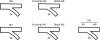
Bifurcation lesion consists of MV and SB. The MV can be divided into proximal MV and distal MV, which is most popular nomenclature for the bifurcation lesion. I prefer, however, to call them as parent vessel (PV) and main branch (MB), just like a tree can be divided into trunk and branches (Figure 1).
Figure 1
Various nomenclature systems of bifurcation lesion.
MB = main branch; MV = main vessel; PV = parent vessel; SB = side branch.
The most important concept to understand a bifurcation lesion is the relationship between the sizes of these vessels. The obvious truth that PV is larger than MB is frequently ignored during the procedure (Figure 2). The first theory that can be applied to explain the relationship was Murray's law.9) It says the cubic of PV diameter (DPV) equals the sum of the cubic of MB diameter (DMB) and the cubic of SB diameter (DSB).
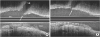
Figure 2
IVUS of coronary bifurcation lesion. (A) Carina (white arrow) is spared of atherosclerotic plaque. (B) Carina shift (white arrow) by the over-expanded stent (IVUs images as the courtesy of Dr. Koo BK).
IVUS = intravascular ultrasonography; MB = main branch; PV = parent vessel; SB = side branch.
Which was calculated mathematically as the physiological principle of minimum work. This theory was proven in normal and diseased coronary bifurcations by intravascular ultrasonography (IVUS) study in our group.10) This study, however, also reported that Murray's law is not correct in the calcified lesion and the culprit lesion of acute coronary syndrome, which is the reason why we need to use IVUS to identify the actual diameters of vessels during the procedure. The first important practical implication of this theory is that the diameter difference of PV and MB is dependent on the size of SB. The larger is the DSB, the larger is the diameter discrepancy between PV and MB. This is why we need to consider the routine proximal optimization technique in the bifurcation lesion with a large SB.11) Secondly, the kissing ballooning with the balloon diameter optimized to MB and SB is always oversized in PV. If the Murray's law is correct, the sum of balloon cross-sectional areas of 2 branches are larger than the cross-sectional area of PV. Kissing ballooning would be better to be conservative with moderate pressure to avoid possible PV injury, according to this theory.
One of the popular methods to calculate the size of PV is Finet's law.12) It says the ratio of DPV to the sum of DMB and DSB is 0.678.
This equation is derived from the quantitative analysis of coronary angiography in normal coronary bifurcations. Unfortunately, the relationship is quite variable according to the vessel size. For example, if DSB is small enough, the calculated value of DPV is smaller than DMB, which cannot be true.
As a summary, understanding the relationship of different vessel sizes in PV, MB, and SB is the key to optimal final kissing ballooning and proximal optimization, which will be reviewed in more detail below. Considering the common variations of vessel size, IVUS examination is required for the optimal result. IVUS guidance was reportedly associated with a better cardiovascular outcome after coronary bifurcation stenting.13)
Plaque shift and carina shift
The occlusion of SB after MV stenting is one of the most common complications during bifurcation stenting. It seemed to be reasonable to assume that the major mechanism of SB compromise is plaque shift from MV to SB, for the plaque burden in MV as well as in SB is the major risk factor of SB compromise.14) That is why most of the classifications of coronary bifurcation lesion were based on the plaque distribution of MV and SB.15)16) A pathological study, however, revealed that the flow divider region (carina) was spared of atherosclerotic plaque burden, whereas plaques were mostly observed in the lateral wall.17) This distribution was also confirmed in IVUS study.18) Scanty amount of plaque in the carina cannot be a cause of major plaque shift, which suggests that the contribution of plaque shift may have been overestimated. Instead, the carina structure itself can be shifted to SB, which can be the major cause SB compromise (Figure 2).
The first paper suggesting the critical role of carina shift was based on the complex angiographic analysis of coronary bifurcation lesion.19) The predicted SB minimal lumen diameter (MLD) was calculated by the geometric assumption that the carina shift was a major mechanism of SB compromise. Of note, the predicted percent diameter stenosis of SB ostium with full carina shifting is calculated as a cosine of bifurcation angle, which means more carina shift with narrower bifurcation angle. Predicted SB MLD was well correlated with the observed MLD (r=0.91, p<0.001). This result suggested the initial assumption that the carina shift is the major mechanism, but this is indirect morphological evidence. More definite evidence came from IVUS and pressure wire measurement, but SB was not imaged in the study.20) Our group measured carina shift and plaque shift directly in the IVUS images of MV and SB before and after MV stent implantation in 44 patients.21) SB compromise was well correlated with carina shift (r=0.94, p<0.001), but not with plaque shift (r=−0.02, p=0.90). Moreover, carina shift accounted for 85% of SB compromise examined by IVUS. So it seems evident that the carina shift is a major contributor of anatomical SB ostial compromise.
Functional study, however, showed an opposite result. A study examined the MV and SB by pressure wire as well as IVUS in 40 patients.22) This study found that abnormal fractional flow reserve (FFR) in the SB after MV stenting was always accompanied by the plaque shift, whereas the carina shift was mostly not associated with a significant drop of FFR in SB. It has been well-known that the anatomical significance was not well correlated with the functional significance measure by FFR in SB after MV stenting.23) The reason why the carina shift is functionally not significant, I think, is because the carina shift is mostly short and eccentric. Angiographically the carina shift looks exaggerated by the negative shadow of MV stent across SB ostium.
A large bifurcation stenting registry data also confirmed the importance of plaque shift, again.24) A subgroup analysis of The Second Korean Coronary Bifurcation Stenting (COBIS II) analyzed the predictor of SB compromise in 2,227 patients. SB compromise (thrombolysis in myocardial infarction [TIMI] flow <3) was noted 187 patients (8.4%) just after MV stenting. Notably, this study found that significant stenosis in ostial SB, significant proximal MV disease, and acute coronary syndrome were independent predictors of SB compromise, which suggests that the plaque shift is the major mechanism. Similar finding was noted from computed tomography angiography study.25) The previous IVUS study in our group showed plaque shift is coming from proximal MV, which comes in line with the result of these 2 studies.21) The Bifurcation angle was not the significant predictor, which suggested carina shift is not an important cause of SB compromise.
As a summary, the anatomical compromise of SB after MV stenting is not functionally so significant than it looks, because it is mostly explained by carina shift, which is not the major cause of functional compromise. The plaque shift superimposed on carina shift appeared to be necessary to cause a hemodynamically significant SB stenosis. The plaque is shifted mostly from the proximal MV, which explains that the plaque burden of proximal MV is the significant risk factor of SB functional compromise or occlusion. This concept is practically important to avoid SB compromise after MV stenting, which will be discussed below.
MV stenting and optimization technique
As described above, the large the size of SB, the larger the discrepancy of PV and MB vessel size. The first step of MV stenting is the selection of stent with optimal size to distal vessel diameter (I call it distal optimization). The diameter of the vessel is better to be assessed by IVUS, for angiography is frequently misleading. When the distal reference vessel is disease free, the stent size should be the same size of vessel size. When the distal reference vessel is abundant in atherosclerotic plaque, common knowledge is to select the average of lumen and vessel diameters. In the reference segment is calcified, stent size should be smaller to avoid distal stent edge dissection.
Then the stent should be more expanded in the PV. The proximal optimization technique (POT) is post-dilating the MV stent just proximal to the carina, with a short non-compliant balloon sized for the reference diameter of PV. Originally it was invented to facilitate the passage of a wire and a balloon into the distal struts on MV stent.26) It also improves a proximal MV stent apposition and eccentricity.27) According to the Murray's law, POT is mandatory when SB size large than 2.3 mm, for the discrepancy between PV and MV size is mostly larger than 1.0 mm. The subgroup analysis of COBIS II registry showed that the POT significantly reduced the restenosis rate in MV in the bifurcation lesions with SB size ≥2.5 mm in core-lab quantitative coronary angiography (unpublished data). Interestingly, when final kissing ballooning was performed, there was no benefit of POT. Maybe it is because the PV is already fully expanded by 2 balloons used for kissing technique.
How to prevent SB occlusion
The risk of SB occlusion during the procedure is the major cause of the complexity of coronary bifurcation stenting. The maneuvers to avoid SB occlusion may be the cause of suboptimal MV stent expansion, which may be the major cause of stent thrombosis and restenosis. The SB compromise was reported to be associated with the increased risk of peri-procedural cardiac mortality and myocardial infarction (MI).24) The peri-procedural MI, however, was not associated with long-term adverse outcomes.28)
There have been several studies for the predictors of SB occlusion,29)30) but most of them were small studies. Recent analysis of COBIS II registry included 2,227 patients who were treated with provisional approach.24) The significant independent predictors were SB ostial disease and lesion length, PV stenosis, acute coronary syndrome, and non-left main disease. Jailed wire technique, SB predilatation, and IVUS guidance were not predictors. Jailed wire technique, however, is the significantly predictor of reopening the occluded SB. Unfortunately, this study could not catch any significant procedural predictor we can apply in the real practice.
Our IVUS study in the coronary bifurcation lesion showed the stent expansion in MB is significantly associated with carina shift, and the stent expansion in PV was associated with plaque shift.21) The operators should consider the risk of carina and plaque shift based on this observation when they select optimal size of stent and POT balloon. The proximal and distal optimization technique can be a good solution for the optimal stent expansion avoiding SB compromise.
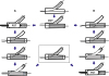
My personal recommendations based those studies are as follows (Figure 3); 1) start with the wiring the MV and a large SB. 2) Predilate the MV. Predilate the SB with severe ostial stenosis. 3) MV stenting with a size just optimal to distal MV, avoiding stent overexpansion (distal optimization). 4) Rewiring the SB using the wire inserted in SB using. Wire prolapse technique is useful to avoid wire undermining of the stent. 5) Proximal optimization technique may help wiring SB, and is also important for the stent apposition in the PV. You can do POT before SB rewiring according to the EBC consensus, and 6) SB ballooning with or without final kissing ballooning and SB stenting.
Figure 3
POT and DOT. (A) POT is performed before SB rewiring by the recommendation of EBC. (B) My personal recommendation. POT is performed after SB rewiring with wire prolapse technique.
DOT = distal optimization technique; EBC = European Bifurcation Club; POT = proximal optimization technique; SB = side branch.
MULTIPLE STEPS OF CORONARY BIFURCATION STENTING
Predilation of SB
Predilation lowers the risk of SB compromise after MV stenting, and also relieves the ischemia in the myocardial territory of SB. But it may complicate the procedure with the higher risk of peri-procedural MI, and increase the risk of SB dissection. In the provisional approach group in COBIS II registry, predilation was not predictor of SB occlusion.24) Recent randomized trial, however, showed that the predilation reduced the risk of SB compromise after the MV stenting.31) Long-term clinical outcome was not improved by predilation in both studies.
So predilation is reasonable way to prevent SB compromise in the high-risk lesion. But the operators should be careful not to make dissection in SB, which will complicate the SB rewiring after MV stenting, if needed.
SB ballooning and final kissing ballooning
After MV stenting, the ostium of SB is jailed by the stent struts across the MV, frequently along with SB ostial stenosis. The purpose of SB ballooning is to free the SB from jailed strut, dilating the SB ostium. Consensus is that final kissing ballooning (FKB) is mandatory after SB ballooning. But, there have a lot of debates on the indication of SB ballooning after MV stenting. SB ballooning deforms MV stent struts, often not fully corrected by FKB.32) The effect of FKB is quite variable in many studies including 2 randomized controlled trials.32)34) Of note, in the COBIS I registry, 2-year major adverse cardiac event (MACE) was worse in FKB group (FKB group 9.5%, non-FKB group 4.5%, p=0.02), mostly because of higher rate of TLR in MV. On the contrary in COBIS II registry, 3-year MACE rate was lower in FKB group (FKB group 6.8%, non-FKB group 9.7%, p=0.02), again mostly because of lower rate of TLR in MV. The major discrepancy of 2 studies is the average SB size. The enrollment criteria of COBIS I includes SB ≥2.0 mm, whereas that of COBIS II was SB ≥2.3 mm. The larger the SB, the larger the PV compared to MB, so further proximal stent expansion by kissing balloon may have played an important role. The most important goal in coronary bifurcation stenting is the optimal stent expansion both in PV and MB, which explains the variable results of FKB studies. So many years, the protection and the treatment of SB is the key issue of coronary bifurcation stenting, but the clinical outcome is highly dependent on the MV stent expansion, particularly in the patients treated with 1-stent technique. The TLR is very infrequent in SB in many papers in COBIS II registry.
Next question is what the indication of SB ballooning is. According to SMart Angioplasty Research Team-Optimal STRATEGY for Provisional Side Branch Intervention in Coronary Bifurcation Lesions (SMART-STRATEGY) trial, TIMI flow less than 3 may be the optimal indication in non-left main bifurcation, and residual stenosis >70% in left main bifurcation.35) More aggressive treatment of SB did not improve the clinical result, whereas the peri-procedural MI risk is higher. When in doubt, the measurement of FFR is sometimes helpful in a very large SB. The clinical outcome, however, was proved not to be improved by the FFR-guided treatment of SB compared to conventional strategy.36)
POT-side-POT (re-POT)
As mentioned above, the major benefit of FKB is not the SB treatment, but the optimal stent expansion in MV. So FKB can be replaced by the final POT. POT is also beneficial to facilitate the cross of wire and balloon after MV stenting. So, first POT is to be done just after MV stenting for this original purpose. If SB treatment is needed, SB is rewired and treated with SB ballooning. SB ballooning will result in the MV stent deformation and stent carina shift into MV, which can be corrected by second POT, instead of FKB. Compared to conventional FKB, POT-side-POT (also known as re-POT) is simpler and can be done through a smaller guiding catheter. Bench test showed that re-POT was associated with better stent apposition and circularity of MV stent compared with FKB.37) The most challenging part of re-POT is correct location of POT balloon. It should cover the proximal edge of stent carina, which can be done by aligning the proximal edge of distal balloon marker with the tip of stent carina (Figure 4). The clinical impact of this new technique should be tested in the clinical trial.
Figure 4
POT-side-POT (re-POT) (A) MV stent is under-expanded after SB ballooning. (B) Correct positioning of a post-dilating balloon, aligning the proximal edge of distal balloon marker with the tip of stent carina. (C) Post-dilation. (D) MV stent is expanded after post-dilation.
MV = main vessel; POT = proximal optimization technique; SB = side branch.

Indication of SB stenting in the provisional approach
Current consensus is that the provisional approach is the standard strategy for the most of coronary bifurcation stenting. The indication of SB treatment, however, is not clear in the provisional approach. The indication of SB stenting was most conservative (TIMI 0) in NORDIC trial,38) and most aggressive in SIRIUS Bifurcation study (residual stenosis ≥50% only).39) More recent Compression versus Anticoagulant treatment and compression in symptomatic Calf Thrombosis diagnosed by UltraSound (CACTUS) trial adopted residual stenosis ≥50% or dissection type B or more as an indication.40) SMART-STRATEGY trial was designed to answer this question.33) In conservative group, SB stenting is indicated if TIMI flow <3 in non-left main bifurcation, and diameter stenosis >50% or dissection in left main bifurcation. In aggressive group, SB stenting is indicated if diameter stenosis >50% or dissection in non-left main and diameter stenosis >30% or dissection in left main bifurcation. SB was stented in 7% of conservative group and 3% in aggressive group. Target vessel failure (TVF), the primary endpoint was similar between 2 groups (9.4% vs. 9.2%, p=0.97). Interestingly, TLR was numerically higher (7.8% vs. 5.4%, p=0.43), and mortality was numerically lower (0.8% vs. 2.3%, p=0.62) in conservative group, although they were not significant. Peri-procedural MI was not included in TVF, but was significantly lower in conservative group (5.5% vs. 17.7%, p=0.002). Current EBC consensus recommended SB stenting only in very complex lesions with large calcified SBs with ostial disease extending >5 mm from the carina and in bifurcations with SBs whose access is particularly challenging and where the SB should be secured by stenting once accessed.8) However, this recommendation is not based on evidences but on expert consensus. European Bifurcation Coronary TWO (EBC TWO) trial compared provisional 1-stent technique with elective 2-stengting in the large caliber true bifurcation lesions (SB diameter ≥2.5 mm) and significant ostial disease length (≥5 mm), but MACE was not different between 2 groups. Currently EBC-MAIN trial is ongoing to see if 2-stent technique is better in this important bifurcation with a large SB. As a conclusion, the indication of SB stenting is better to be conservative.
What is the best 2-stent technique?
Most of bifurcation lesion can be treated with the provisional approach, but still we have some cases we have to consider 2-stent technique. There have several trials to find the best elective 2-stent techniques, but the results are quite variable. Bifurcations Bad Krozingen (BBK) II trial found that culotte technique is better than T-stenting in terms of restenosis rate.41) But culotte technique showed a similar result compared with crush technique in NORDIC Stent Technique study42) and was even inferior to double kissing (DK)-crush technique in DK-CRUSH III trial.43) I think the best 2-stent technique is the technique you are most familiar with. Maybe the optimal result especially in term of stent expansion is much more important than the selection of a specific 2-stent technique. Currently most popular techniques are T-stent and small protrusion, mini-crush technique, mini-culotte technique, and DK-crush technique. I prefer T-stenting and small protrusion technique, because it is simple, provisional in nature, and above all the most familiar to me.
FUTURE PERSPECTIVES
Even after so many studies, still we have more questions than answers. We do not know whether the elective 2-stenting is better with next generation DES. We do not know the future roles of dedicated bifurcation stent and fully bioresorbable scaffold in the bifurcation lesion. The best clinical come is the most important goal of coronary bifurcation stenting. Good question and persistent study will make it happen.
 XML Download