

 PDF
PDF ePub
ePub Citation
 Print
Print


INTRODUCTION
A bronchobiliary fistula (BBF), also called biliobronchial fistula, is a rare condition caused by an abnormal connection between the biliary and bronchial trees. It is characterized by pathognomonic bilious sputum exhibiting pneumonic infiltration. BBF has rarely been reported since Peacock first described the condition in 1850.1 In the past, infectious diseases, such as a hydatid infection, have been known to cause BBF, but recent reports have suggested its association with tumors.2 Bilious sputum is pathognomonic and aids in the diagnosis of patients with BBF, but a definitive treatment is unclear and controversial. Therefore, case-specific procedures and treatments, such as invasive surgical procedures and simple conservative treatment, differ between centers.2 We report two cases of BBF treated by interventional radiology.
CASE
Case 1
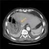
A 66-year-old man visited the emergency room complaining of greenish sputum and dyspnea. He underwent a pylorus-preserving pancreaticoduodenectomy (PPPD) in 2012 to treat distal common bile duct cancer. In May 2016, local recurrence of cancer was found in the right hepatic duct; hence a right hemihepatectomy was performed. During the operation, severe adhesion between the liver dome and right diaphragm was observed, and adhesiolysis was carefully performed to avoid injury to the diaphragm. Because he had undergone PPPD before, the previous hepaticojejunostomy was revised, using the left hepatic duct and the jejunum. Because a follow-up computed tomography (CT) scan taken on post-operative day (POD) 7 showed very little fluid collection at the operation site (Fig. 1), the patient was discharged on POD 10.
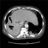
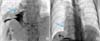
He visited the emergency room 3 days after his discharge, complaining of greenish sputum and mild dyspnea. A chest X-ray revealed suspicious pneumonic infiltrates and pleural effusion in the right lower lung zone. Additionally, an abdominal CT scan showed the same previous fluid collection at the operation site (Fig. 2). Because the patient complained of mild dyspnea and showed signs of decreased oxygen saturation, he was admitted to the intensive care unit. A chest tube was placed to treat a small right pleural effusion, and serous fluid was drained. The amount of bilious sputum was 200–300 ml/day. We assumed that the cause of this symptom was BBF and performed percutaneous transhepatic biliary drainage (PTBD) (Fig. 3A). After PTBD tube insertion, the greenish sputum improved greatly within 24 hours. Initially, to shrink the abscess cavity, the tip of the PTBD tube was placed in the fluid cavity outside the liver through a leaking bile duct. A few days later, it was repositioned inside the bile duct and then clamped. After confirming that the bilious sputum had not recurred, we removed the PTBD tube, and he was discharged.
Since his discharge, he has been visiting an outpatient clinic periodically without any other abnormalities. In addition, there was no recurrence of cancer or abnormal findings in his recent CT scans.
Case 2
A 38-year-old female with a cirrhotic liver underwent a right hemihepatectomy in 2008 to treat hepatocellular carcinoma. A partial resection with a primary repair of the diaphragm was performed, because cancer invasion was observed. During the follow-up period, her cancer recurred and transarterial chemoembolization (TACE) was performed several times until 2015. She visited the emergency room complaining of bilious sputum in June 2015. After insertion of a PTBD tube through the left bile duct, her symptoms improved, and cholangiography showed contrast leakage from the biliary to the bronchial tree (Fig. 3B). But after 9 months of PTBD insertion, we observed recurrence of the bilious sputum, and the PTBD tube was repositioned to treat BBF. Unfortunately, she still has the PTBD tube, often has recurring symptoms, and visits the emergency room. She is in poor condition because of tumor progression.
DISCUSSION
BBF is an abnormal connection between the biliary and bronchial trees, and its etiology remains unclear. The BBF apparently can have several causes, such as hydatid infection, trauma, bile duct obstruction, tumor invasion, and iatrogenic injuries, such as those caused by TACE and radiofrequency ablation.34 Recent advancements in surgical techniques have led to the development of major hepatectomy, which can cause BBF, as described in this report. The following three conditions might be needed to form a BBF: postoperative collection of bile or pus, any small injury to the diaphragm during liver mobilization, and adhesion between the pleura and the lungs. The first two conditions usually cause only abdominal or pleural fluid collection, but the most important and usually undetectable condition is adhesion between the pleura and the lungs.
Bilious sputum is a pathognomonic symptom of BBF.2 For a definite diagnosis, imaging studies that can show the fistula tract are mandatory. PTBD cholangiography, magnetic resonance cholangiopancreatography, and hydroxyl iminodiacetic acid scans are useful for identifying the connection between the biliary and bronchial trees, which is essential for diagnosis.56
Although clear and definitive treatment for BBF has not yet been established, surgical or non-surgical interventional procedures, such as endoscopic retrograde cholangiopancreatography (ERCP) or PTBD, are frequently used. A systematic review reported differences in cure rates between procedures; interventional procedures were slightly more effective than surgical procedure (97% vs. 85%).2 This report considered surgical procedures as the last option for BBF treatment, that is, only when interventional procedures had failed.2 In performing surgical assessment for BBF treatment, a two-stage approach is recommended.1 The first step is external biliary drainage or direct percutaneous drainage of an abscess, and the second is treatment of the underlying disease. In some cases, n-butyl cyanoacrylate or histoacryl embolization through the fistula tract under bronchoscopic guidance were used for BBF management.78 Yamaguchi et al.9 reported a case of congenital BBF in which the patient was diagnosed by bronchoscopy and bronchography, and treated by excision of the fistula.
We believe that there should be two key procedures for the management of BBF. One is drainage of the subphrenic abscess to shrink the abscess cavity that has caused the development of the fistula tract. The other is biliary drainage to minimize the amount of bile leakage into the abdominal cavity. Shrinking the abscess cavity closes the fistula tract, and biliary drainage prevents the recurrence of the fistula.
In conclusion, BBF can occur in patients who have fluid collection near the diaphragm after liver resection. Because the pathogenesis of BBF is still unclear, BBF should be suspected in patients who have undergone liver resection and have fluid collection near the diaphragm, with bilious sputum exhibiting pneumonic infiltration. Either ERCP or PTBD can be used for the primary management of BBF.
 XML Download