

 PDF
PDF ePub
ePub Citation
 Print
Print


INTRODUCTION
With a Western lifestyle, an increasingly aging society, and improvements in diagnostic modalities such as abdominopelvic CT, the incidence of colonic diverticular disease is on the rise and seems likely to increase in the future [1]. This disorder may be complicated by the presence of inflammation, bleeding, abscess formation, feculent peritonitis, and fistula. Patients diagnosed with colonic diverticulitis present with a varied disease spectrum in terms of severity. Uncomplicated diverticulitis has a relatively self-limiting clinical course that can even be treated without antibiotics [2]. Severe complicated diverticulitis is a catastrophic illness that presents with septic shock, and usually requires emergency surgery and fecal diversion because of generalized peritonitis.
Several grading systems have been developed to classify colonic diverticulitis depending on the severity of the disease [3], and clinical decision making and management tends to differ according to the various classification systems. If there is a need for an operation, the surgical approach, technique, and necessity for emergency action may differ in accordance with the classification system being used. Today most classification systems, such as the modified Hinchey Classification [4] and the Ambrosetti classification [5], are based on radiologic features, particularly via CT scanning. CT shows many characteristics of colonic diverticulitis, such as colonic wall thickening, pericolic fat stranding, pericolic air in the inflamed diverticula, and the presence of free fluid or distant intraperitoneal air.
Although treatment strategies for colonic diverticulitis are constantly under discussion, there is an ongoing debate about the optimal treatment guidelines for surgical intervention. In this study, we aimed to define the impact of the various CT findings of colonic diverticulitis on the need for surgery. We conducted comprehensive analyses of patients' clinical status, laboratory indices, and the classification systems to help clinicians manage colonic diverticulitis and demonstrate which factors can affect the need for operative treatment.
METHODS
This study was approved by the Institutional Review Board and Ethics Committee of Daegu Catholic University Medical Center (CR-18-072). The need for informed consent was waived because of the retrospective nature of the study.
Patients diagnosed with colonic diverticulitis by employing abdominopelvic CT at the Daegu Catholic University Medical Center from January 2010 to July 2016 were retrospectively evaluated. First, patients were identified by a search for the International Classification of Diseases, 10th edition (ICD-10) code “K57” related to the terms “diverticulitis,” “diverticulum,” and “diverticulosis.” A total of 357 patients diagnosed with colonic diverticular diseases were enrolled for analysis. Patients without CT evidence of diverticulitis were excluded from the study. After further exclusion of patients who had pure diverticulosis or diverticular bleeding, a colon cancer that mimicked acute diverticulitis, or relevant data deficiencies, the remaining 178 patients were reviewed.
Patient data were extracted from electronic medical records. For each patient, demographics and clinical features including age, sex, body mass index (BMI), comorbidities, previous episodes of diverticulitis, and initial vital signs were obtained from patients' records. In addition we collected data regarding leukocyte count, CRP, length of hospital stay, CT findings, types of surgery, clinical outcomes, and recurrence.
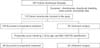
Patients were categorized into 2 groups: a successful nonoperation group and an operation group. However, the 2 groups showed significant differences in the basic demographics. To eliminate these confounding variables between, we conducted a propensity score matching (PSM) analysis using the matching variables age, sex, American Society of Anesthesiologists (ASA) physical status classification, and BMI. The operation group was then matched 1:2 with the nonoperative treatment group (Fig. 1).
A radiologist blinded to the patient's clinical demographics, operation, and final outcomes retrospectively reviewed the CT scans. The CT images were reviewed and assessed for signs of diverticulitis (colonic wall thickening >5 mm, pericolic fat stranding), segment of the colon involved, presence of a fecalith or pericolic air in the inflamed diverticula, and presence of free fluid or distant intraperitoneal air. Pericolic air was defined as air bubbles or air collection within 5 cm of the affected bowel segment without an abscess. Distant intraperitoneal air was defined as air bubbles or air collection within the abdominal cavity with distance of greater than 5 cm of the affected bowel segment. Free fluid in the abdominal cavity was defined as fluid over 30 mm in anteroposterior direction in the pelvis. The CT findings were categorized according to the modified Hinchey classification [4]. Complicated diverticulitis was defined as the presence of abscess, distant intraperitoneal air, fistula, or obstruction at the time of diagnosis. Uncomplicated diverticulitis was defined by CT findings of thickened colonic wall, pericolic fat inflammation, and/or pericolic air.
Diverticulitis was also considered moderate or severe according to the Ambrosetti classification [5]. Moderate diverticulitis was defined as a localized thickening of the colonic wall of 5 mm or more and signs of inflammation of the pericolic fat. It was considered severe when in addition to these signs one or more of the following findings were present: abscess and/or extraluminal air and/or extraluminal contrast.
Perforation of diverticulitis tends to originate from either side of the taenia mesenterica and near the mesenteric borders of the antimesenteric taeniae such as taenia omentalis and taenia libera [6]. This anatomic location of perforation was defined as mesenteric perforation. Conversely, peritoneal perforation was characterized by free or antimesenteric perforation confined to the area between the antimesenteric taeniae.
Patients' preoperative status was categorized according to the ASA physical status classification. The territory of the colon was divided, with diverticulitis occurring from the cecum to the transverse colon considered right-sided and that occurring from the splenic flexure to the sigmoid colon as left-sided. Patients who presented with a diffuse abdominal guarding and/or signs of systemic sepsis were operated on without any further investigations. The decision regarding conservative or surgical treatment was at the discretion of the surgical team.
Postoperative complications were stratified according to the Clavien-Dindo classification of surgical complications [7]. The total number of postoperative complications was recorded for all events related to morbidity.
Categorical variables were compared by using the chi-square and Fisher exact tests, whereas continuous variables were compared by using Student t-tests. A logistic regression analysis was used for the multivariate analysis. Results with P-values of <0.05 were considered statistically significant. To minimize the influence of potential confounders on the selection bias, PSM was performed using the MatchIt package in the R program. One-to-two matching between the groups was accomplished using the nearest-neighbor matching method. Factors with a P-value of <0.2 were included in the stepwise multivariate logistic regression to evaluate potential predictor variables. We performed statistical analyses using IBM SPSS Statistics ver. 21.0 (IBM Co., Armonk, NY, USA). Differences were considered statistically significant at P < 0.05.
RESULTS
Patients' demographics and clinical characteristics
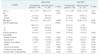
Before PSM, the clinical characteristics of overall patients in the 2 groups were compared (Table 1). Patients in the operation group were significantly more likely to be older (68.9 ± 15.7 vs. 47.1 ± 15.5, P < 0.001) and have a higher ASA physical status classification (III: 4 [14.3%] vs. 6 [4.0%]; II: 18 [64.3%] vs. 35 [23.3%]; I: 6 [21.4%] vs. 109 [72.7%]; P < 0.001). Patients in the operation group were more likely to be female (18 [64.3%] vs. 73 [48.7%], P = 0.129) and have a lower BMI (22.3 ± 3.5 kg/m2 vs. 23.6 ± 3.4 kg/m2, P = 0.080), but without significant differences.
After 1:2 fixed-ratio PSM, 48 patients from the successful nonoperation group and 24 from the operation group were retained for comparison. There were no significant differences regarding patient demographic characteristics between the 2 groups.
Clinical parameters and CT findings
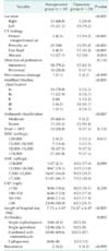
Table 2 summarizes the clinical parameters and CT findings after PSM analysis. The need for surgery was associated more with the left side than the right side, and was statistically significant (P < 0.001). However, both groups were similar with regard to fever, white blood cell count, absolute neutrophil count, and CRP.
In terms of the findings from CT scans, distant intraperitoneal air, pericolic air, and free fluid were significantly more frequent in the operation group. However, the presence of abscess showed marginal significance. The rate of percutaneous drainage was similar in both groups. The operation group had higher stages to a significant extent in terms of modified Hinchey classification and Ambrosetti classification. The total length of hospital stay was longer for the operation group than the nonoperation group (15.57 ± 6.47 days vs. 7.73 ± 4.87 days, P < 0.001) and the rate of combination with metronidazole and the use of carbapenem were higher in the operation group.
Operative characteristics
Among the 24 patients managed by surgery, 8 patients underwent Hartmann's operation, 14 were treated by primary resection and anastomosis, and 2 underwent wedge resection (Table 3), one of whom eventually underwent sigmoidectomy after recurrence in the same location. The types and rates of morbidities were similar between the Hartmann procedure and the other surgeries. There were no significant differences in allcomplication severity by Clavien-Dindo classification between the types of operation. Of 8 Hartmann's operation patients, 2 had postoperative septic complications that led to death. Because of old age and poor performance status, 3 patients were unable to undergo colostomy reversal.
Analysis of risk factors for operations
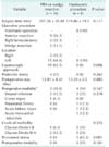
Table 4 shows the results of the univariate analysis of factors associated with the operations. Age > 65 years, left location, peritoneal perforation, distant intraperitoneal air, pericolic air, free fluid, abscess, and CRP had a P-value of less than 0.2 and were included in the multivariate analysis.
Upon multivariate analysis of these factors, distant intraperitoneal air showed statistical significance (P = 0.046) and pericolic air and left location showed a significant trend (P = 0.071 and P = 0.067, respectively).
DISCUSSION
The role of CT in diagnosing colonic diverticulitis, evaluating its severity, and determining the need for surgery has now been mostly proved [15]. It is understandably important that the clinical classification based on the detailed findings of CT scans has some impact on the management of colonic diverticulitis. In our analysis, we found that the presence of distant intraperitoneal air on CT is the only significant parameter that correlated with operative management of colonic diverticulitis. Patients with a left-sided location or pericolic air had a relatively higher operation rate but with marginal significance. The modified Hinchey classification and Ambrosetti classification based on the CT findings were valuable in terms of grading the severity and deciding on the management. However, against expectations, this analysis identified that abscess, pericolic air, direction of perforation, and laboratory indices have no significance with regard to the operation rate.
The present study found that before PSM, patients in the operation group were significantly older than those in the successful nonoperation group. The advanced age in the operation group was associated with higher ASA physical status classification, left-sided location, and more comorbidities. Young patients with right-sided diverticulitis were predominant, similar to results from other Asian studies [89]. In Western countries where left-sided diverticulitis is more prevalent, younger age and right-sided location was usually considered to be severe colonic diverticulitis, contrary to our results [10]. To control the potential confounding factors, PSM was used to adjust for significant differences between the basic characteristics of both groups.
Colonic diverticulosis refers to the presence of a diverticulum or several diverticula [11]. Colonic diverticula usually develop at weak points where the vasa recta penetrate from either side of the taenia mesenterica and near the mesenteric borders of the antimesenteric taeniae, such as taenia omentalis and taenia libera. Perforation of diverticulitis occurs between the mesenteric and antimesenteric taeniae, usually leading to intramesenteric or retroperitoneal abscess/air. Conversely, peritoneal perforation is characterized by free or antimesenteric perforation confined to the area between the antimesenteric taeniae that can develop into generalized peritonitis. We expected that the direction of perforation may be associated with failure of medical treatment. One previous study showed that retroperitoneal abscesses are at higher risk for recurrent diverticulitis [12]. However, our results revealed that, unexpectedly, the need for surgery was no different with regard to the direction of perforation.
Nowadays most patients with suspected colonic diverticulitis presenting at the emergency department are examined by a CT scan. CT has been shown to be both highly sensitive and specific regarding the presence of diverticulitis. It is also considered the method of choice to evaluate the severity and decide on treatment strategy. In the absence of standardized treatment of colonic diverticulitis, physicians must decide on appropriate management depending on the clinical status of patients, the hemodynamic status, laboratory indices, and CT findings. Especially in hemodynamically stable patients, CT has an important role in identifying patients who may best respond to nonoperative treatment and those who need surgery.
Ambrosetti et al. [13] found that the risk of recurrence in patients who were treated conservatively for the first episode of acute diverticulitis was statistically higher in patients with severe diverticulitis such as abscess formation and extracolonic contrast or air. Poletti et al. [14] found that the presence of an abscess and pockets of extraintestinal gas 5 mm or larger in diameter were predictive of failure of nonoperative treatment. Hall et al. [12] found that a long segment of involved colon (>5 cm) and retroperitoneal abscess were at higher risk for recurrence of diverticulitis. This study revealed that patients who present with right-sided diverticulitis are at low risk for recurrent disease. Buchs et al. [15] found that free air on initial CT was of borderline significance for the risk of recurrence, and reported that a raised serum level of CRP (>240 mg/L) during the first attack was associated with early recurrence. However, there are no absolute parameters to predict the clinical course or recurrence of colonic diverticulitis, although several studies have attempted to identify the risk factors for recurrence and complications, and have proposed a method of risk scoring or a staging system based on the clinical factors and CT findings [1617].
The modified Hinchey Classification is the most widely used method to determine disease severity. This classification is currently insufficient to decide upon treatment options for acute colonic diverticulitis because colonic perforation without generalized peritonitis could eventually be treated nonoperatively. Another classification published by Ambrosetti et al. [5] categorizes diverticulitis into severe or moderate disease. Shaikh and Krukowski [18], using the Ambrosetti classification, found that patients who had severe diverticulitis on CT were in greater need of surgery during both index admission and follow-up in comparison with mild diverticulitis. Trenti et al. [19] prospectively studied 560 patients admitted for a first episode of colonic diverticulitis and evaluated their long-term recurrence according to the Ambrosetti classification guided by CT scans. After conservative treatment, the rate of recurrence showed no significant difference between mild and severe diverticulitis, but the risk of severe recurrence was statistically greater in the group with an initial severe diverticulitis. However, recent studies have reported that complicated colonic diverticulitis with extraluminal air or abscess can be managed conservatively [202122]. Improved detection using recent CT technology is able to manage more patients with complicated diverticulitis nonoperatively. For this reason, the Dharmarajan classification has emerged as a system focusing on complicated diverticulitis only, especially on the quantity of free air or abscess [23].
This study has several limitations, including its retrospective design and the fact that it was conducted at a single hospital where all patients were Asian. Moreover, only inpatients were included; no outpatients had mild diverticulitis. In addition, the number of patients with left-sided diverticulitis was much smaller than the number with right-sided diverticulitis. Because of the small number of cases of left colonic diverticulitis in our study, we were unable to conduct a subgroup analysis between right and left colonic diverticulitis by each CT stage. Moreover, 3 patients (6.3%) in nonoperation group was classified into III and IV according to modified Hinchey classification. Whereas, 14 patients (58.3%) in operation group was classified into III and IV. Therefore, patients in the operation group had more severe peritonitis than those in the nonoperation group. And the indication for surgical intervention should be inconstant by surgeon's preference. A prospective, randomized controlled study would help to resolve such limitations, which unfortunately are common in retrospective studies.
In conclusion, CT plays a significant role in the diagnosis and treatment of colonic diverticulitis. In the present study we found that distant intraperitoneal air is the most important factor in the need for surgical treatment of colonic diverticulitis. Other CT findings and clinical characteristics, such as the left-sided location and pericolic air, can be used to make the appropriate treatment decision. It is hoped that further study will identify more detailed CT findings and verify their significance, and aid in the design of practical scoring and classification systems.
 XML Download