Abstract
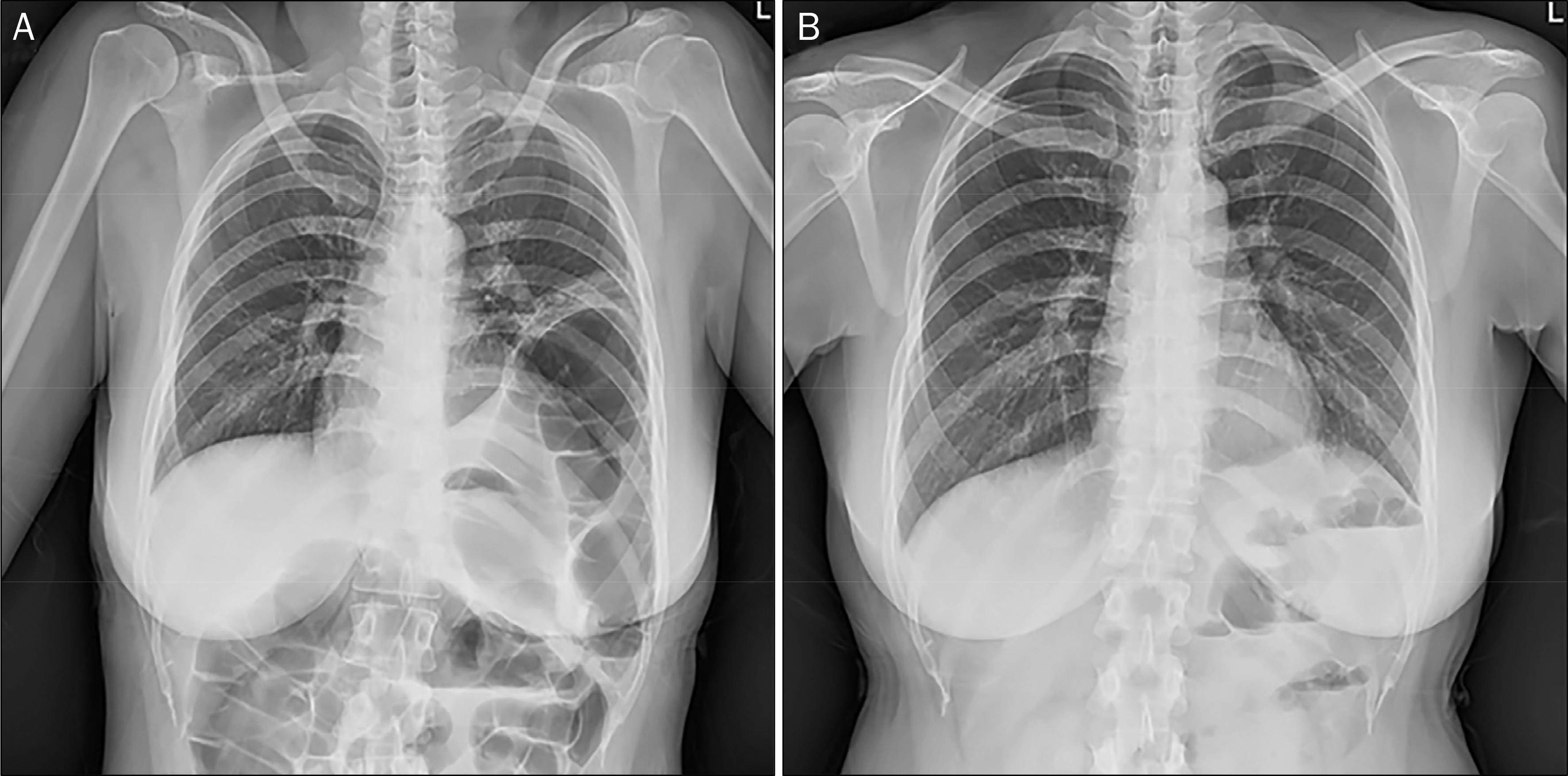
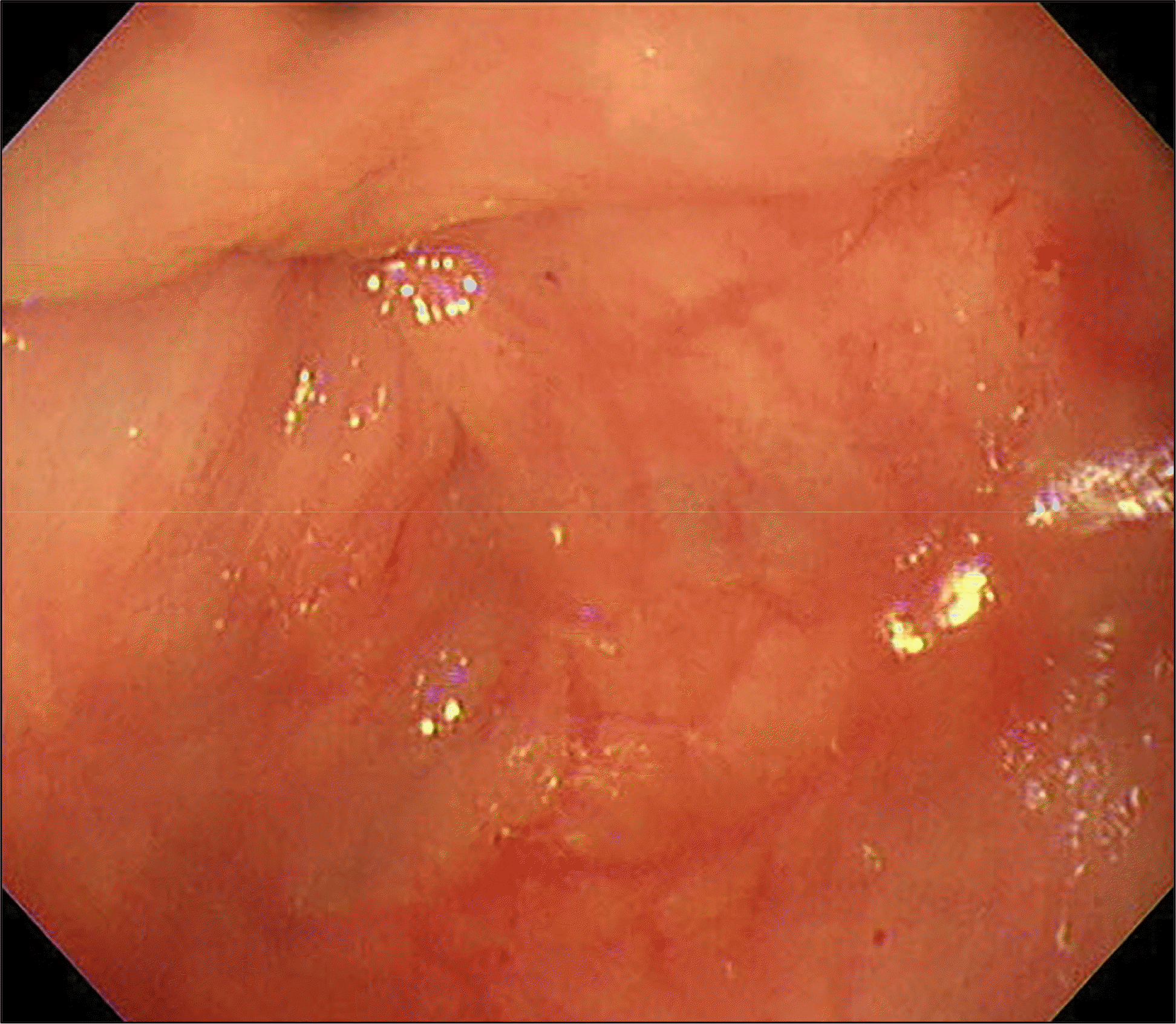
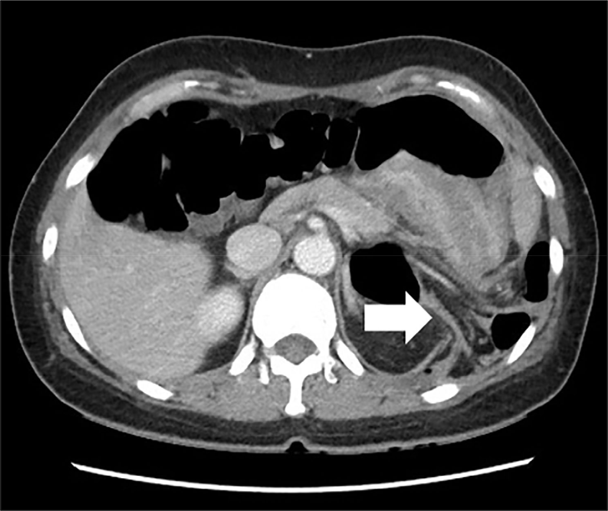
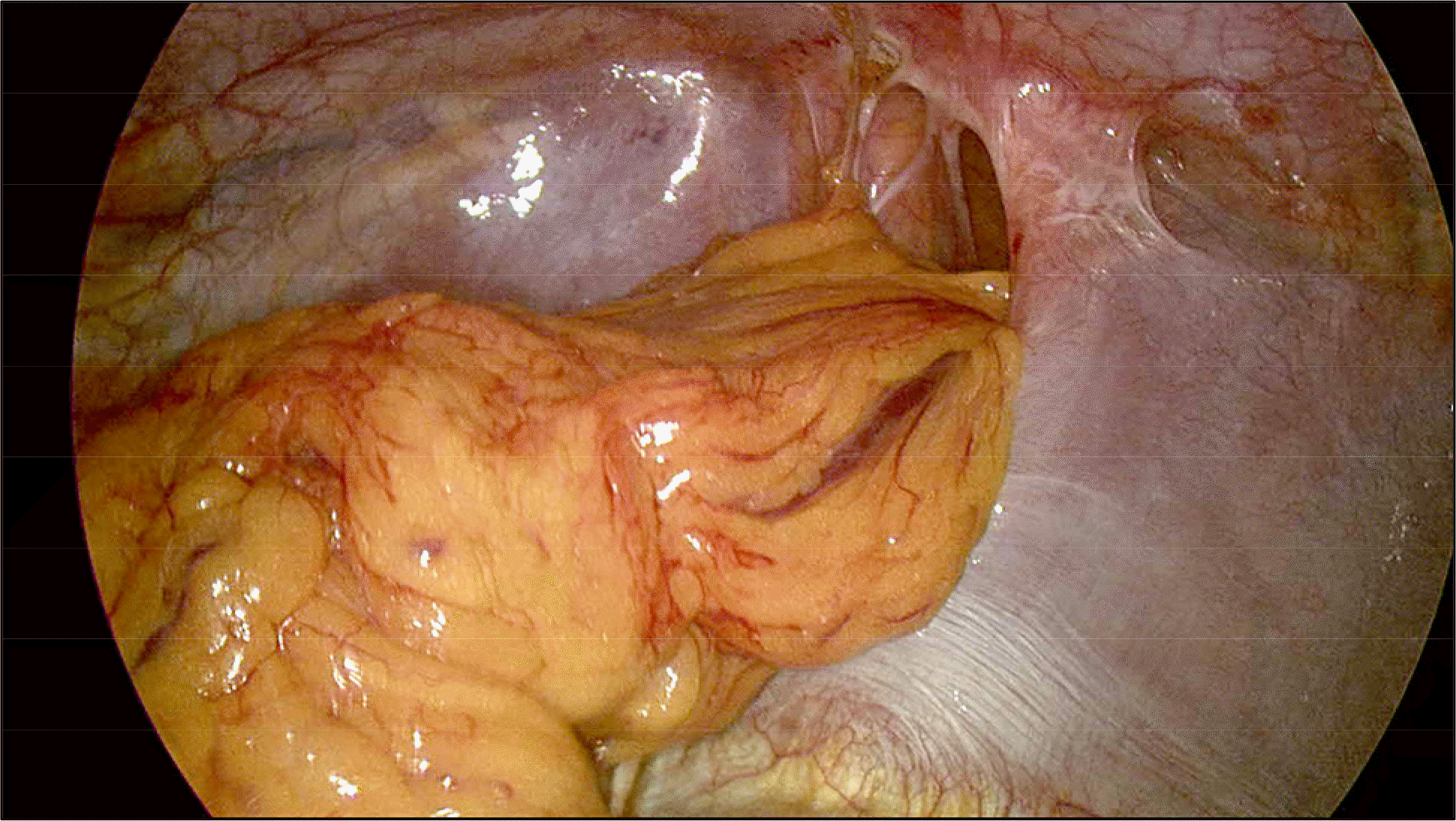
Bochdalek hernia (BH) is defined as herniated abdominal contents appearing throughout the posterolateral segment of the diaphragm. It is usually observed during the prenatal or newborn period. Here, we report a case of an adult patient with herniated omentum and colon due to BH that was discovered during a colonoscopy. A 41-year-old woman was referred to our hospital with severe left chest and abdominal pain that began during a colonoscopy. Her chest radiography showed colonic shadow filling in the lower half of the left thoracic cavity. A computed tomography scan revealed an approximately 6-cm-sized left posterolateral diaphragmatic defect and a herniated omentum in the colon. The patient underwent thoracoscopic surgery, during which, the diaphragmatic defect was closed and herniated omentum was repaired. The patient was discharged without further complications. To the best of our knowledge, this case is the first report of BH in an adult found during a routine colonoscopy screening.
References
1. Mullins ME, Stein J, Saini SS, Mueller PR. Prevalence of incidental Bochdalek's hernia in a large adult population. AJR Am J Roentgenol. 2001; 177:363–366.

2. Senkyrik M, Lata J, Husová L, et al. Unusual Bochdalek hernia in puerperium. Hepatogastroenterology. 2003; 50:1449–1451.
3. Losanoff JE, Sauter ER. Congenital posterolateral diaphragmatic hernia in an adult. Hernia. 2004; 8:83–85.

4. Brown SR, Horton JD, Trivette E, Hofmann LJ, Johnson JM. Bochdalek hernia in the adult: demographics, presentation, and surgical management. Hernia. 2011; 15:23–30.

5. Shin MS, Mulligan SA, Baxley WA, Ho KJ. Bochdalek hernia of diaphragm in the adult. diagnosis by computed tomography. Chest. 1987; 92:1098–1101.
6. Sugimura A, Kikuchi J, Satoh M, Ogata M, Inoue H, Takishima T. Bilateral bochdalek hernias in an elderly patient diagnosed by magnetic resonance imaging. Intern Med. 1992; 31:281–283.

7. Megremis SD, Segkos NI, Gavridakis GP, et al. Sonographic appearance of a late-diagnosed left bochdalek hernia in a middle-aged woman: case report and review of the literature. J Clin Ultrasound. 2005; 33:412–417.

8. Swain JM, Klaus A, Achem SR, Hinder RA. Congenital diaphragmatic hernia in adults. Semin Laparosc Surg. 2001; 8:246–255.

9. Weissberg D, Refaely Y. Symptomatic diaphragmatic hernia: surgical treatment. Scand J Thorac Cardiovasc Surg. 1995; 29:201–206.

10. Ninos A, Felekouras E, Douridas G, et al. Congenital diaphragmatic hernia complicated by tension gastrothorax during gastroscopy: report of a case. Surg Today. 2005; 35:149–152.

11. Harinath G, Senapati PS, Pollitt MJ, Ammori BJ. Laparoscopic reduction of an acute gastric volvulus and repair of a hernia of bochdalek. Surg Laparosc Endosc Percutan Tech. 2002; 12:180–183.

12. Brusciano L, Izzo G, Maffettone V, et al. Laparoscopic treatment of bochdalek hernia without the use of a mesh. Surg Endosc. 2003; 17:1497–1498.

Fig. 2.
(A) Chest radiography after colonoscopy showing colonic air shadow protruding into the left thoracic cavity. (B) Chest radiography before colonoscopy with normal results.


 PDF
PDF ePub
ePub Citation
 Print
Print


 XML Download