

 PDF
PDF ePub
ePub Citation
 Print
Print


Introduction
Middle East Respiratory Syndrome coronavirus (MERS-CoV) is an emerging novel coronavirus known to cause acute severe respiratory infections in humans. Despite limited transmission in community settings, large outbreaks have been reported in healthcare settings [12], raising concerns regarding the potential for global virus transmission.
A MERS outbreak in South Korea that occurred in 2015 was started by a single infected traveler returning from the Arabian Peninsula. He was hospitalized in a hospital in Pyeongtaek, where he transmitted MERS-CoV to twenty eight individuals [3]. These secondary cases visited multiple healthcare facilities in different regions of Korea, including Seoul, Daejeon and Gyeonggi-do, which initiated new hospital outbreaks of MERS and facilitated the nationwide spread of MERS [45]. Furthermore, the MERS outbreak was amplified in healthcare facilities due to superspreading events on an unprecedented scale. As a result, a total of 186 cases were confirmed to have MERS-CoV infection including 38 deaths [67].
In contrast to the hospital outbreak of MERS-CoV in Saudi Arabia where the continuous influx of MERS cases from the community might complicate the identification of the timing and source of exposure [28], in Korea, there was no additional importation of MERS cases during the outbreak period. In addition, all MERS case were thoroughly traced and their contacts were closely monitored for symptom onset as early as possible whereas severe cases were more likely to be identified in Saudi Arabia. For these reasons, the MERS outbreak in Korea gave us the unique opportunity to further understand the epidemiologic characteristics and observe the wide range of clinical severity of MERS-CoV infection.
In Daejeon, a single secondary case of the first Korean case of MERS (the index case in the Daejeon cluster) caused outbreaks of MERS-CoV infection in two hospitals, leading to 23 secondary cases and 3 tertiary cases. Because this index case was transferred from one hospital to another, individuals in each hospital were infected by this index case at different disease stages of disease.
Therefore, we describe two hospital outbreaks of MERS-CoV infections in Daejeon and compare the epidemiologic parameters and clinical outcomes between the two hospital clusters in order to explore the patterns of transmission and disease severity of MERS-CoV infection among those infected according to the clinical course of the index patient,
Materials and Methods
1. Settings
Daejeon, a geographic center of South Korea, has a population of >1.5 million. Hospital A is a 300-bed general hospital that provides acute and long-term care specialized for geriatric patients. Hospital B is a 730-bed, secondary university-affiliated hospital.
2. Assessment of close contacts and data collection
Potential contacts and possible exposure dates were traced by follow-up patient interviews, reviewing hospital records, reviewing staff rotations and patient assignments, and monitoring security video footage of each MERS case. Publicly available data were also retrieved from daily reports of the Ministry of Health and Welfare if needed [9]. Symptoms, laboratory and radiological findings, and clinical courses were retrospectively collected from the medical records. This study was approved by Institutional Review Board at each hospital (CNUH 2015-07-021-002, KYUH 2015-07-018) with a waiver of informed consents.
3. Definitions
MERS-CoV infection was confirmed by positive real-time reverse transcriptase polymerase chain reaction assays targeting two genes (upE and ORF1a) [10] in two consecutive respiratory samples collected 48 hours apart.
Close contacts were defined as individuals who 1) had any contact within 2 meters or were in the same care area as confirmed cases without wearing personal protective equipment (PPE; gowns, gloves, respirators, and eye protection); 2) had direct contact with respiratory secretions from MERS cases; or 3) shared hospital equipment with confirmed cases or received care from a healthcare worker (HCW) who was caring for confirmed cases [11]. The risk of transmission from asymptomatic cases was assumed to be negligible. The time of symptom onset was defined as the first identified time of persistent fever (≥37.5℃) [11] >24 hours among febrile patients and as the first day of new relevant symptoms among afebrile patients. Based on the duration and proximity of contact, symptoms of cases, and adequacy of PPE, the level of infection risk was assessed, and the most likely source of exposure was determined for those who had been possibly exposed to more than one symptomatic case (Supplementary Table 1) [12] The duration of exposure days was defined as the total number of hospital days of those who stayed in the same ward as the index case.
4. Sequencing and phylogenetic analysis
Total RNAs were extracted from respiratory tract samples obtained from the index case as well as four secondary cases and one tertiary case from each hospital. The partial S2 domain of the MERS-CoV spike (corresponding to nucleotides 23781-24395 of the MERS-CoV EMC genome JX869059) was amplified from the extracted RNAs using primers VS804 (5'-TCAGGTTGATCAACTTAATAGT-3') and VS805 (5'-TTGAGTAATGCCAACACCGTT-3') [13]. The 11 new partial S2 sequences from this cluster were aligned with those of 20 published MERS-CoV genomes including two MERS-CoV strains isolated from Korean patients [12131415] (Supplementary Table 2) using the MUSCLE method [13] implemented in Molecular Evolution Genetics Analysis, software version 6 [16]. A PhyML tree was generated using Seaview4 software [17] and the approximate likelihood ratio test based on a Shimodaira-Hasegawa-like procedure was applied using the general time-reversible substitution model, as previously described [13].
5. Statistical analyses
The incubation period was estimated by identifying the earliest and latest possible dates of exposure and the time of symptom onset. The serial interval was estimated by identifying the time between symptom onset in the index case and symptom onset in secondary cases. Because the data were interval-censored, we fitted them to a gamma distribution and maximum-likelihood estimates were identified using the coarseDataTools package for R [18]. The parametric distribution was compared with empirical cumulative density functions of the observed incubation period (the time between the midpoint of the exposure duration and symptom onset) and serial intervals. For sensitivity analysis, we estimated the parameters excluding those with possible alternative sources of exposure (Supplementary Data).
Clinical characteristics were compared between the two hospital clusters using Fisher's exact test and the Wilcoxon rank-sum test. The non-parametric incubation periods were compared between two hospital clusters using Kaplan-Meier method. R statistical package (version 3.1.2, R Foundation for Statistical Computing, Vienna, Austria) was used for all analyses.
Results
1. Description of the outbreak and infection control measures
The index case of the Daejeon cluster was hospitalized in the same ward as the first Korean case of MERS from May 15 to May 17. After being discharged from the original hospital, the index case moved to Daejeon and became ill on May 20. Unaware of his exposure status, he was hospitalized in a 4-bed room at Hospital A on May 22. At the time of admission, he had pneumonia with mild respiratory symptoms and diarrhea. However, as symptoms worsened and pneumonia progressed bilaterally, he was subsequently transferred to Hospital B on May 28. In Hospital B, he was Hospitalized in a 6-bed room in the pulmonary ward. He received nebulizer therapy and underwent bronchoscopy. On May 30, hospital B was notified of the index case's exposure status by the Korea Centers for Disease Control and Prevention. Hospital A was also notified the next day. The following infection control measures were immediately implemented. 1) The index case and patients who had shared the same room were isolated in single rooms with negative pressure ventilation. 2) The hospital ward and all possible contaminated areas were disinfected. 3) The affected ward was closed to new admissions. Patients who remained on the same ward were quarantined in the hospital for 14 days and their movement was restricted. 4) Airborne and contact precautions were implemented while caring for all quarantined patients. Quarantined individuals wore surgical masks and were asked to appropriately perform hand hygiene. 5) Individuals quarantined in the hospital were monitored every 4-6 hours for the development of fever and new symptoms, and were isolated as soon as they developed symptoms. Once patients were confirmed to have MERS-CoV infection, they were transferred to the regional medical centers designated for MERS treatment. 6) Close contacts who left the hospital were quarantined at home in the same manner.
As of June 2, the infection control measures were intensified. The affected wards were completely closed to both admissions and discharges and this strategy was expanded to the neighboring wards. Caregivers on the same ward were also quarantined in the hospital.
A total of 27 confirmed cases of MERS-CoV infection were identified in the Daejeon cluster, including 23 secondary cases and 3 tertiary cases (Fig. 1). No secondary transmission occurred among family members of the index case and individuals on different wards.
1) Hospital A
There were 13 secondary cases of MERS-CoV infection in Hospital A (Fig. 2, Supplementary Fig. 1A). Two cases (A1 and A2) who shared the same room as the index case developed fever on May 29 and 30, respectively, and were isolated on May 31. Among quarantined individuals on the same ward, six additional patients (A4, A6, A7, A10, A11, and A12) and three caregivers (A8, A9, and A13) developed symptoms. One caregiver (A3) developed symptoms on May 31 and was quarantined at home from June 1 until the diagnosis was confirmed on June 3. Before being quarantined, Case A3 infected another caregiver (A14) during a brief conversation (<30 minutes). A temporary computer technologist (A5), who left Hospital A and moved to Busan on May 30, developed fever on June 2. Case A5 visited four different hospitals for 10 days before diagnosis and infected Case A15 in Hospital C.
2) Hospital B
Between May 31 and June 8, there were 10 secondary cases of MERS-CoV infection in Hospital B (Fig. 2, Supplementary Fig. 1B). Eight of these cases (five patients, one paid caregiver and two family caregivers) had shared the same room as the index case. Of these eight, five (B1, B2, B3, B6, and B7) developed symptoms during isolation, and three (B8, B9, and B10) developed symptoms during quarantine. Case B4, a family caregiver, was presumed to have close contact with the index case in the emergency department and in the ward. Case B5 was in a different room on the same ward and was cared for by the same staff as the index case.
There was only one tertiary case in Hospital B (B11). Transmission occurred while Case B11 was doffing the contaminated PPE after cardiopulmonary resuscitation of Case B3.
2. Comparisons between the secondary cases in the two hospitals
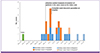
A total of 123 close contacts (70 patients, 19 caregivers, and 34 HCWs) and 117 close contacts (70 patients, 17 caregivers, and 30 HCWs) were identified at Hospitals A and B, respectively. Among patient- and caregiver close contacts on the same ward, the secondary attack rate was similar between the two hospitals: 15.8% (12/76) in Hospital A and 14.3% (10/70) in Hospital B (P = 0.51). However, considering the duration of exposure, the incidence rate of MERS-CoV infections was higher in Hospital B (7.7/100 exposure-days) than Hospital A (3.4/100 exposure-days) (incidence rate ratio 2.3; P < 0.001).
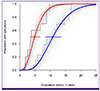
Among the 23 secondary cases, the median incubation period was estimated to be 7.8 days (95% confidence interval [CI], 6.0-10.0) (Supplementary Fig. S2). The parametric estimate of median incubation period was significantly shorter in the Hospital B cluster (4.8 days [95% CI, 3.4-6.7]) than that in the Hospital A cluster (10.8 days [95% CI, 8.4-13.5]; Fig. 3) where the incubation periods varied widely. The non-parametric estimates of incubation periods between two hospitals clusters also showed a significant difference: median 3.6 days (95% CI, 1.8-8.7) vs. 11.0 days (95% CI, 5.8-14.7) (P = 0.003). The median serial interval for secondary cases was estimated as 14.6 days (95% CI, 12.9-16.5; Supplementary Fig. S2).
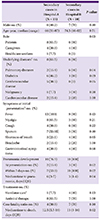
Demographic and patient characteristics were similar between the two hospital clusters although a greater proportion of cases in Hospital B had pulmonary diseases (Table 1). Secondary cases in Hospital B were more likely to develop rapidly progressing pneumonia with a median time to pneumonia development from symptom onset of 3 days (interquartile range [IQR], 1-4 days) compared with 6 days (IQR, 3-7 days) in Hospital A. Mortality was also higher in the Hospital B cluster (70%, 7/10) than in the Hospital A cluster (30.8%, 4/13; odds ratio [OR], 5.3; 95% CI, 0.9-31.6; P = 0.07).
3. Phylogenetic analysis
A phylogenetic analysis revealed that all S2 fragment sequences of MERS-CoV from the Daejeon cluster formed a monophyletic clade, B1 (Fig. 4). This clade showed the closest similarity to two recently reported genomes, KOR/KNIH/002_05_2015 (KT029139) [19] and ChinaGD01 (KT006149) [15] obtained from Korean patients, and to the Saudi Arabian isolates (KT026453 and KT026454) collected from Riyadh in February and March 2015.
Discussion
We have described MERS outbreaks in two hospitals in Daejeon initiated by a single patient. We found that exposure to the MERS case in the late stage (2nd week) of disease appeared to increase the risk of transmission and was associated with shorter incubation periods and rapid disease progression among those infected.
Given that transmission occurred quickly and explosively in Hospital B as compared with Hospital A in the Daejeon cluster, it seems that MERS-CoV was be more efficiently transmitted during the later stage of disease in the index case when pneumonia progressed. These findings was consistent with the report of another MERS case in Korea, which caused a superspreading event where the majority of transmissions occurred when the infector had advanced pneumonia [20].
Multiple factors may influence this pattern of transmission. Firstly, an increase in the viral loads as disease progresses could be an important factor. Despite limited evidence about the viral shedding kinetics of MERS-CoV over the disease course, it is expected that viral load increases with disease progression up to a point [21]. Therefore, it seems plausible that close contacts in Hospital B were exposed to a higher infective dose than those in Hospital A. In addition, worsening respiratory symptoms and the consequent need for respiratory procedures during the later stage of disease could facilitate viral transmission [2223]. Since the index case received nebulizer therapy in a six-bed room, aerosols generated by the nebulizer might amplify viral transmission to those who shared the room in Hospital B. The shorter incubation period and rapid disease progression among secondary cases in Hospital B are consistent with these explanations. In contrast, individuals in Hospital A might have been exposed to different levels of viral loads depending on when and for how long they were exposed during the disease progression in the first week of illness of the index case. These differences in exposure can partly explain the wide range of incubation periods and diverse clinical features among secondary cases in Hospital A. Secondly, underlying comorbidities in secondary cases can affect the explosive nature of transmission and the clinical consequences. Given that the index patient was hospitalized in the pulmonary ward in Hospital B, a large proportion of affected patients had underlying pulmonary diseases. This comorbidity combined with a high infective dose could exacerbate the disease progression, resulting in the high mortality among secondary cases in Hospital B.
The study results highlight the critical role of early case detection and isolation in preventing a MERS outbreak in healthcare facilities and averting resultant mortality due to MERS-CoV infection. To that end, healthcare facilities should be highly vigilant about the possibility of MERS among travelers returning from the Middle East, and infection control practices be properly implemented for patients with respiratory infections. More importantly, an effective risk communication between health authorities and healthcare facilities is mandatory in the initial stage of MERS outbreak.
We observed heterogeneity in transmission in this Daejeon cluster as described in the previous epidemiologic studies [2425]. One secondary case infected only one patient among numerous contacts, whereas one index case generated 23 secondary cases. The disease stage of an infector was not the sole determinant for disease severity in infected individuals as one tertiary case who was infected by a secondary case on the day of symptom onset suffered severe pneumonia requiring extracorporeal membrane oxygenation. Also, the disease severity was often unpredictable in that there were severe cases among those without comorbidities or those with the long incubation period. Studies on genetic predisposition and susceptibility of hosts may help provide a clue to such heterogeneities in MERS-CoV infection.
The phylogenic analysis revealed that all viruses from this cluster belong to the same clade as strains from Riyadh. This indicates that the MERS outbreak in South Korea was caused by introduction of the MERS-CoV strain prevailing in Riyadh, where there have been repeated and ongoing MERS outbreaks since 2014 [2627]. To date, there is no evidence of genetic mutations affecting viral transmissibility [28]. However, a recent surge of MERS in Riyadh [29] highlights the need for intensified research on strain-specific viral characteristics and continuous monitoring of genetic variations of the virus.
There are some limitations in this study. First, the viral shedding kinetics was not assessed and whole-genome sequence analysis was not performed. Although the MERS-CoV variant typing method based on the S2 fragment sequence provides reliable and timely information regarding the source of infection [13], whole-genome sequences should be analyzed in detail to determine the mechanisms that might underlie the potential genomic adaptation of MERS-CoV in the MERS outbreak in South Korea.
Despite these limitations, this study provides valuable information regarding the natural history of MERS transmission before implementing infection control measures, and provides insights into the factors that might facilitate the spread of MERS-CoV in healthcare settings. Our results also emphasize the critical role of early case detection and isolation in preventing a MERS outbreak. Such strategies might help to reduce disease severity and mortality.


 XML Download