

 PDF
PDF Citation
 Print
Print


INTRODUCTION
Endodontic surgery is a resource used in cases when conventional endodontic treatment fails. The surgical method involves the production of a cavity in the apical region with subsequent sealing using a retrograde filling material. For successful surgical therapy, good adaptation of this retrograde filling material to the dentin walls is extremely important, in order to prevent fluids from percolating into the canal space [1].
Mineral trioxide aggregate (MTA) is one of the most widely used materials for retrograde fillings [2]. This cement is widely known for its biological properties, which include stimulation of tissue repair and induction of mineralization [3]. Despite its adequate properties, some characteristics of MTA have been questioned [45]. The consistency of this material is sandy and dry, which makes it difficult to insert into cavities [4]. To improve the handling characteristics of MTA, some authors have evaluated different mixing vehicles, including propylene glycol [4]. Bismuth oxide, present in MTA, has been reported to cause negative interference with MTA [678] and for this reason, other options for radiopacifiers have also been tested [910].
A novel calcium silicate-based cement (CSC) containing zirconium oxide as a radiopacifier and propylene glycol/water in its liquid form was recently developed [1011]. Zirconium oxide has been tested as a substitute for bismuth oxide due to its adequate radiopacity [1012] and because it does not interfere with the hydration of CSC [13]. Moreover, propylene glycol associated with distilled water has positive effects on the handling characteristics, flowability, and film thickness of CSCs [411].
Sealer 26 (S26; Dentsply, Petrόpolis, RJ, Brazil) is an epoxy resin-based endodontic sealer that contains calcium hydroxide [14]. Calcium hydroxide promotes calcium ion release and alkalinity, which favor tissue repair [15]. S26 has a satisfactory sealing ability for preventing bacterial leakage [1617]. A thicker consistency of this sealer was suggested for use as a retrograde filling material [17]. After testing, an adequate biological response was found, and S26 showed similar results to those of MTA [17].
Ultrasonic energy has been used to improve the flow of various materials within root canals [18192021]. Ultrasonic activation (US) of an endodontic sealer promotes greater sealer penetration into the dentinal tubules and improves the sealer/dentin interface [21]. The acoustic microstreaming energy transmitted improves the cleaning ability of irrigating solutions, diffusion of medicaments, and the interfacial adaptation of root canal sealers [182021]. US of retrograde filling materials has previously been tested in MTA [22]. A method of inserting the material into root canals using US was evaluated in comparison with hand condensation [2324]. Yeung et al. [23] found denser material when MTA was inserted using hand condensation followed by indirect US. The US may have increased penetration into the dentinal tubules and influenced the push-out bond strength.
The aim of the study was to evaluate the effect of US of retrograde filling materials (MTA, S26, and CSC) on adaptation to the dentinal walls and push-out bond strength. The null hypothesis tested was that the US would not improve the adaptation and push-out bond strength of the retrograde filling cements.
MATERIALS AND METHODS
The study was approved by the Ethics Committee of Bauru Dental School, University of São Paulo (080/2011). Sixty extracted maxillary canine teeth with a curvature of less than 5° were selected, as described by Schneider [25]. The coronal portions were sectioned using a 0.3-mm Isomet saw (Buehler, Lake Bluff, IL, USA), and the root canal length was standardized at 15 mm. The working length was established by measuring the penetration of a size 10 K-file (Dentsply Maillefer, Ballaigues, Switzerland) until it reached the apical foramen and then subtracting 1 mm. The root canals were shaped using the Reciproc system (VDW, Munich, Germany) to size R50/0.05 and then irrigated with 2 mL of 2.5% sodium hypochlorite. A final flush of 2 mL of 17% ethylenediaminetetraacetic acid (Biodinâmica, Ibiporã, PR, Brazil) for 3 minutes was used to eliminate the smear layer, and the canals were finally washed with 2 mL of distilled water and dried with paper points (Dentsply Maillefer). The canals were filled with size R50/0.05 (VDW) gutta-percha single cones coated with AH Plus sealer (Dentsply Maillefer). The excess of gutta-percha was removed with a heated instrument and vertically compacted. Filled roots were stored in 100% humidity at 37°C for 48 hours to allow the sealer to set. Then, the specimens were embedded in polyester resin, fixed in an apparatus using the Godiva impression compound (Nova DFL, Rio de Janeiro, RJ, Brazil), and horizontally sectioned at 3 mm from the apex, at an angle of approximately 90° to the long axis of roots, using No. 699 steel burs (KG Sorensen, Cotia, SP, Brazil) under continuous water-cooling.
The specimens were fixed in a device to create the retrograde cavities and to favor parallelism of the dentin walls, according to Vivan et al. [26]. The retrograde cavities were prepared with a 12/90D diamond insert coupled to a piezoelectric ultrasonic device (Piezon master 200, EMS, Nyon, Switzerland) measuring 3 mm in depth and 1.5 mm in diameter, with constant irrigation.
The teeth were divided into 6 groups (n = 10) according to the cement used and whether US was performed. In the S26 group, S26 alone was used, while in the S26 US group, S26 was used with US. Likewise, in the MTA group, white MTA (Angelus, Londrina, PR, Brazil) alone was used, and in the MTA US group, white MTA (Angelus) was ultrasonically activated. In the CSC group, only CSC was used, and in the CSC US group, CSC was ultrasonically activated. Before the push-out test, all specimens were analyzed by low-vacuum scanning electron microscopy to verify marginal adaptation.
The CSC was prepared using a proportion of 80% calcium silicate (Portland Cement, Irajazinho, Cimento Rio Branco, Rio de Janeiro, Brazil) to 20% zirconium oxide (Sigma-Aldrich, St. Louis, MO, USA) by weight. The powder-to-liquid ratio used for manipulating CSC was 4:1. The liquid was composed of 80% water and 20% propylene glycol (C3H8O2; Pharmácia Specífica, Bauru, SP, Brazil) by volume. MTA was prepared according to the manufacturer's instructions. S26 was mixed to a denser consistency, with a powder-to-resin ratio of 5:1 [4].
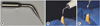
After mixing, the cements were inserted and vertically compacted into retrograde cavities with a condenser (S. S. White, Rio de Janeiro, RJ, Brazil). In the S26 US, MTA US, and CSC US groups, the cement was activated using an ultrasonic unit (Jet-Sonic Four Plus, Gnatus, Ribeirão Preto, SP, Brazil) set to the Endo mode and 30% power for 1 minute (30 seconds in the mesio-distal direction and 30 seconds in the buccal-lingual direction), with a mini Irrisonic tip (Helse Ultrasonic, Santa Rosa do Viterbo, SP, Brazil, Figure 1), which was a prototype of an ultrasonic tip that will be commercially produced. The samples were stored at 37°C and 100% humidity for 72 hours.
Figure 1
Mini Irrisonic tip used for ultrasonic activation (US) of the cements (A). US of the cement on retrograde cavity in the lateral (B) and top views (C). Images depicting agitation of the filling materials.
Sample preparation
From the apical portion of the specimens, 3 mm was horizontally sectioned using a 0.3 mm Isomet saw (Buehler) at 200 rpm with continuous water cooling to prevent frictional heat. Then, the surfaces were polished using 200- and 600-grit SiC abrasive paper (Buehler) under running water (Politriz, Arotec, Cotia, SP, Brazil) until 1.5-mm thick sections were obtained (Figure 2A). The thickness of the discs was controlled using a digital micrometer (Starrett Indústria & Comércio Ltda., Itu, SP, Brazil).

Interfacial analysis
The specimens were viewed under a low-vacuum scanning electron microscope (SEM; PSEM eXpress, Aspex Corp., Delmont, PA, USA), which did not require metallization of the sample, so that the samples could be reused. Scanning electron micrographs of the material-tooth interface were recorded at ×50 and ×250. The higher magnification was applied to confirm the presence or the absence of gaps. Image analysis was performed using Image J (National Institutes of Health, Bethesda, MD, USA). The images were analyzed for marginal adaptation of the retrograde cement to the dentinal walls. The total perimeter of the canal and the perimeter of gaps at the cement/dentin interface were used to calculate the adaptation of each cement to the dentin. After evaluating the interface, all the specimens were subjected to the push-out test, and the type of failure (adhesive, cohesive, or mixed) was then verified and quantified using a stereomicroscope at ×20 magnification (Stemi 2000C, Carl Zeiss, Jena, Germany) and the AxioVision software (Carls Zeiss).
Push-out bond strength
The samples were subjected to the push-out bond strength test to determine bond strength at the cement/dentin interface. For this purpose, each resin/dentin/root-end filling material disc was placed in a mechanical test machine (EMIC DL 2000, Instron, Barueri, SP, Brazil). A vertical force was applied in the middle of the canals, from the cervical to the apical direction, using the metal point of the test machine (EMIC DL 2000) at a speed of 1 mm/min (Figure 2B). The area (mm2) under load was calculated by the cylinder lateral surface area formula: bonding area = 2πrh, where r is the radius of the preparation circumference, and h is the thickness of the root slice (1.5 mm). The push-out strength value in megapascals (MPa) was calculated by dividing the load (N) by the bonding area (mm2). The maximum force to dislocate the cement from canal was measured in newtons (N). The bond strength was calculated in MPa by dividing the force in N by the area of the canal in mm2 [26].
Statistical analysis
Statistical analysis was performed using the non-parametric Kruskal-Wallis and Dunn tests for the interface test (p < 0.05), because the Shapiro-Wilk test did not confirm a normal distribution. Analysis of variance and the Tukey's test were used to analyze push-out bond strength, because a normal distribution was confirmed by the D'Agostino-Pearson test (p < 0.05).
RESULTS
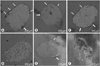
The median, minimum, and maximum values of interfacial adaptation are shown in Table 1. There was a statistically significant difference in the comparison of S26 US with MTA (p < 0.05). The cement in the S26 US group presented no gaps at the dentin/cement interface. The other comparisons showed no statistically significant differences (Figure 3). The type of failure observed was predominantly adhesive for all the tested materials.
Table 1
Median, minimum, and maximum values of interfacial adaptation (gaps in dentin/cement interface) and number of samples according to the type of failure

Figure 3
Representative images of interfacial adaptation at ×50 magnification by scanning electron microscope in the (A) mineral trioxide aggregate (MTA), (B) MTA/ultrasonic activation (US), (C) calcium silicate-based cement (CSC), (D) CSC/US, (E) Sealer 26 (S26), and (F) S26/US groups. The white arrows indicate the presence of interfacial gaps.
The mean and standard deviation of the push-out bond strength of the tested cements are presented in Table 2. US significantly improved the bond strength of all the tested cements (p < 0.05). Comparisons of the cements without US showed similar push-out bond strengths among them.

DISCUSSION
US improved the push-out bond strength of cements at the dentin/cement interface. Interfacial adaptation was significantly altered only in the comparison between the S26 US and MTA groups (p < 0.05). Thus, the null hypothesis was partially rejected. The push-out bond strength analysis, when only the cements without US were compared, showed no statistically significant difference; this was also the case when only the cements with US were compared. This suggests that the greatest influence on the push-out bond strength was the use of US, not the type of cement used.
The use of ultrasound has been proposed for various endodontic treatment procedures, from the agitation of irrigants to the activation of root canal sealers [181920212728]. Promising results were found for improvements in the intratubular penetration of sealers [21]. We tested whether agitation of MTA during its insertion increased its adaptation to the root canal walls. High levels of cement adaptation at the dentin/cement interface favors sealing, thereby preventing bacterial leakage [2930]. Previous studies have tested the US of MTA and obtained good results in terms of solubility, density, and sealing capacity [232428]. Aminoshariae et al. [22] used a different methodology that applied US as a cement placement method, and not as a complement to the hand condensation method. In the present investigation, the cements were activated after being previously inserted by means of hand condensation, as previously described [23]. This method resulted in a denser material than was achieved with simple hand condensation [23]. Those results are in accordance with the results of the present study, in which push-out bond strength was enhanced with the use of US. This suggests that the density might have increased the push-out bond strength of the materials tested.
A CSC containing zirconium oxide as a radiopacifier was recently developed [1011]. This material was previously suggested as a sealer with a powder-to-liquid ratio of 0.3 [11]. In the present study, for manipulating the CSC, a powder-to-liquid ratio of 4:1 was used to obtain a denser consistency for retrograde fillings. The formulation of this cement contained liquid propylene glycol, as previously described [4]. The addition of 20% propylene glycol to distilled water improved the handling characteristics of MTA, enhanced the bond to dentinal walls, and did not interfere with its biological properties [48]. In the present study, no differences in interfacial adaptation or push-out bond strength were found among CSC, MTA, and S26.
The interfacial adaptation values of the cements to the root canal walls were higher in the groups in which US was applied; however, no statistically significant differences were found when the values were compared with those of the same cement, either ultrasonically activated or not. The only statistically significant difference was found between the S26 US and MTA groups (p < 0.05). In this case, in addition to ultrasonic agitation, the composition of the materials could have influenced the results. S26 is a resinous material that has been reported to show high flowability [14], while MTA is based on tricalcium silicate, which is known for its low flow rate [4]. Thus, in addition to S26 having a higher flow rate than MTA, when treated with ultrasonic agitation, this flow rate was even higher, thereby improving the penetration of the cement and its interfacial adaptation.
The type of failure observed was predominantly adhesive. This suggests that the bond of these materials to dentin was not effective. For MTA and CSC, this was expected, as these materials were hydraulic and not resinous. For S26, the remnant smear layer present on the dentin surface before cement application might have obstructed the tubules, preventing adequate penetration.
In this study, the push-out bond strength increased with the US of the cements (p < 0.05). High interface bond strength to the canal walls was found for all the ultrasonically activated groups. This property is crucial for preventing cement displacement from the cavity and subsequent gaps at the interface. A previous report showed that US improved the penetration of root canal sealers into the dentinal tubules, resulting in superior interfacial adaptation [21].
Taking into account the limitations of this study, in which the samples were subjected to humidity, whereas clinically the retrograde cavity would be exposed to blood, further studies are needed to demonstrate whether blood interferes with interfacial adaptation and push-out bond strength. Additional studies are needed to determine whether the effect of US on improving the push-out bond strength of cements is related to changes in their structures or limited only to their increased adaptation to the canal walls.
 XML Download