

 PDF
PDF Citation
 Print
Print


INTRODUCTION
Interest in aesthetic dentistry in achieving a beautiful smile has prompted the invention of tooth bleaching treatments [12]. In today's dentistry, 3 approaches are used for vital tooth bleaching: in-office, at-home, and over-the-counter (OTC) bleaching [3]. During in-office treatments, clinicians lighten teeth rapidly in a single visit, and the results appear to be as good as those with home bleaching treatments [456]. Home bleaching treatments are applied via custom-fabricated trays loaded with low levels of bleaching gels for at least 2 weeks (for 2–8 hr/day). In-office treatments are for professional use in clinical applications; they contain high concentrations of hydrogen peroxide (HP; 35%–38%) or carbamide peroxide (CP; 30%–35%), whereas at-home bleaching gels contain low concentrations (3%–16%) of carbamide or HP [78].
Interest in whitening the teeth for a better aesthetic appearance had led to the use of these bleaching materials. Patients' demands for bleaching procedures and the introduction of new bleaching products have prompted much research concerning the side effects of these materials. Free radicals released during bleaching procedures affect the properties of restorations such as microhardness, surface roughness, color, and microleakage [9].
Tooth mineral loss after bleaching may reduce the strength of the bond between restorative materials and enamel [1011] because of changes in the mechanical properties of the enamel and dentin [112]. The decrease in microhardness or residual oxygen interferes with resin tag formation and inhibits resin polymerization through a free radical mechanism, thus affecting the bond strength [1314]. To solve these clinical problems related to bleaching procedures, delaying bonding and waiting up to 10 days have been suggested [815].
The selection of adhesive type might influence bonding to bleached enamel [1216]. Recent investigations in adhesive technology have led to the production of multi-mode universal adhesives. Clinicians can use universal adhesives in etch and rinse, self-etch, or selective etching mode depending on the case. Some in vitro studies [171819] have evaluated the bond strength of universal adhesives, but the effects of bleaching procedures on their bonding efficiency in different modes have not been examined. As bleaching procedures have become popular, it is crucial to evaluate the bonding ability of different universal adhesives to bleached enamel. The aim of this in vitro study was to assess the shear bond strength (SBS) of 2 different universal adhesives in etch and rinse and self-etch modes after in-office and at-home bleaching treatments. The null hypotheses tested were as follows: 1) bleaching treatments would not affect the bond strength values of the tested universal adhesives, and 2) there would be no difference in bond strength values between the etch and rinse and self-etch modes.
MATERIALS AND METHODS
This in vitro study was approved by the Institutional Ethical Committee of Hacettepe University (ethical approval No. GO 16/711-13). The materials used in this study and the study design are presented in Table 1 and Figure 1.
Table 1
Materials used in the study
Figure 1
Study design.
SU, Single Bond Universal; GU, Gluma Universal; ER, etch and rinse; SE, self-etch; HP, hydrogen peroxide; CP, carbamide peroxide.
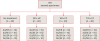
Extracted sound, human anterior teeth (n = 160) were gathered, and the calculus, plaque, and remaining tissue were removed with scaling instruments and pumice using a rubber cup. The teeth were stored in 0.1% thymol for 1 week at room temperature and transferred to distilled water at 4°C until specimen preparation. A water-cooled diamond disc (Isomet, Buehler, Lake Bluff, IL, USA) was used to separate crowns from roots. After examination under a stereomicroscope (Leica, Meyer Instruments, Houston, TX, USA) for surface structural damage or defects, the teeth were embedded in a block of acrylic resin (Meliodent, Heraeus/Kulzer, Hanau, Germany) with the buccal surface positioned up for surface treatment and composite bonding. Enamel surfaces were polished with 200, 400, and 600 grit silicon carbide papers. The specimens were then randomly divided into four groups (n = 40). The negative control group received no treatment, while the three experimental groups were treated with either 35% HP (Total Blanc Office H35, Nova DFL, Rio de Janeiro, Brazil), 16% CP (Total Blanc Home C16, Nova DFL), or 7.5% CP (Total Blanc Home C7.5, Nova DFL) following the manufacturer's instructions as follows:
Total Blanc Office H35 (n = 40): A 1-mm layer of Total Blanc H35 was applied to the enamel surface for 20 minutes. All specimens in this group received 3 applications at 3-day intervals.
Total Blanc Home C16 (n = 40): A 1-mm layer of Total Blanc C16 was applied to the enamel surface for 6 hours daily for 14 days.
Total Blanc Home C7.5 (n = 40): A 1-mm layer of Total Blanc C7.5 was applied to the enamel surface for 6 hours daily for 14 days.
The enamel specimens were dried using cotton pellets before the bleaching procedure. A soft toothbrush was used to remove the bleaching gel under running tap water, and the samples were stored in distilled water at 37°C. The distilled water was changed daily for 14 days. After 14 days, the specimens were divided into four groups according to the adhesive material and application mode (n = 10) as presented in Figure 1. The adhesives Single Bond Universal (SU) and Gluma Universal (GU) were applied according to the manufacturers' instructions (Table 1). The adhesives were cured using a light-emitting diode (LED) light-curing unit (Radii Plus, SDI, Victoria, Australia) in standard mode. The output of the light-curing unit was regularly checked to ensure that it was 1,200 mW/cm2. After adhesive application, a nanohybrid universal composite resin (Herculite XRV Ultra Universal, Kerr Corp., Orange, CA, USA) was inserted into a Teflon tube (3 mm wide and 2 mm high) seated perpendicularly against the prepared enamel surfaces and light-cured for 40 s using the LED light-curing unit. After curing, the Teflon tube was removed carefully. The specimens were stored in distilled water at 37°C for 24 hours and thermocycled for 5,000 cycles between 5°C and 55°C with a dwell time of 25 seconds each. The cylinders were then loaded with a chisel-shaped loading metal rod parallel with and close to the bonding interface at 1 mm/min in the shear mode until fracture occurred (LR50K, Lloyd Instruments, Fareham, UK). SBS (MPa) values were calculated by dividing the failure load (N) by the cross-sectional area (mm2) of the cylindrical composite. The SBS data exhibited a normal distribution according to the Kolmogorov-Smirnov test; therefore, a three-way analysis of variance and Tukey post hoc test at a significance level of 0.05 were used to analyze differences. The failure modes were observed at 40× magnification using a stereomicroscope (SZX7, Olympus, Hamburg, Germany) and classified as either adhesive (between the enamel and bond or composite and bond), cohesive in composite, cohesive within the enamel, or mixed (a combination of adhesive and cohesive failure).
RESULTS
The mean SBS values and standard deviations of each group are presented in Table 2. The specimens in the no treatment group in which the composite was bonded with SU using the etch and rinse mode exhibited the highest mean SBS (34.8 ± 6.98 MPa). No statistically significant differences in SBS values were observed among the bleaching treatment groups (no treatment, 35% HP, 16% CP, and 7.5% CP; p = 0.346). There were no significant differences in the SBS values between adhesives in the same application modes. However, the self-etch groups showed significantly lower SBS values compared to the etch and rinse groups (p = 0.001). Specimens bleached with 16% CP with SU applied in self-etch mode (18.5 ± 7.35 MPa) and specimens bleached with 35% HP with SU applied in self-etch mode (18.9 ± 6.75 MPa) showed the lowest mean SBS values (p < 0.05).
Table 2
Shear bond strength (SBS) values of universal adhesives to enamel according to etch and rinse or self-etch modes

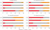
A failure mode examination showed that 39.3% of the specimens had adhesive failures, whereas 37.5% had cohesive failures and 23.1% had mixed failures (Figure 2).
DISCUSSION
The present study compared the SBS of a nanohybrid resin composite to human enamel that was bleached with one in-office (35% HP) and two home bleaching materials (16% CP and 7.5% CP) and bonded with 2 universal adhesives in etch and rinse and self-etch modes. Adhesive procedures were conducted at 14 days following the bleaching treatments.
Many types of bleaching systems are currently available with different concentrations of HP or CP as active agents. The vital bleaching agents have direct contact with the enamel surface and might cause chemical and morphological changes in the enamel structure [202122]. Declines in bond strength may be caused by bleaching treatment or the immediate placement of adhesive restorative materials after bleaching treatment. Furthermore, a number of studies revealed that residual oxygen from the bleaching agent present on the enamel surface might be the cause of failure in adhesive procedures after bleaching [142021]. The adverse effects of bleaching materials on adhesive systems have been investigated widely, and these studies showed that waiting time is crucial for better bond strength values [101415]. In vitro studies emphasized that the immediate SBS values of bleached enamel were low, whereas the values obtained at 14–21 days after bleaching were nearly the same as those of untreated controls [101415]. Similarly, in the present study, after 14 days no significant differences were detected between the bleached and no-treatment groups. In addition, an in vitro study [23] compared waiting times after bleaching treatments prior to adhesive application and reported similar SBS values after 7–14 days of waiting.
There was no significant difference in the SBS values of bleached enamel with 16% CP, 7.5% CP, and 35% HP. The first hypothesis is thus supported. Since the different concentrations of home bleaching agents did not affect the bond strength values, we concluded that an increased concentration did not prolong the time needed prior to bonding. The specimens that were not treated with any bleaching gel showed similar results to the bleached specimens. Conversely, an in vitro study reported that office bleaching treatment with 40% HP reduced SBS values but home bleaching treatment with 10% CP had no effect [24]. However, the adhesives were applied after 1 day in that study. Similar to these findings, Akın et al. [25] showed that 10% CP bleaching did not significantly reduce SBS values but 38% HP bleaching did. On the other hand, Gungor et al. [26] suggested that both home and office treatments adversely affected SBS values, but the home bleaching group had lower SBS values than the office bleaching group, unlike in other studies.
Universal adhesives represent the most recent generation of adhesives and can be used in different modes [2728]. The adhesive systems compared in the present study had different solvents; the ethanol-based adhesive (SU) and acetone-based adhesive showed similar SBS values in the etch and rinse mode. Montalvan et al. [16] also reported in their study that after bleaching with 35% HP, the SBS values of an ethanol-based adhesive and an acetone-based adhesive were similar. Additionally, groups that did not receive bleaching treatments and were bonded in etch and rinse mode demonstrated no significant differences. Previous studies [2930] showed findings similar to ours (i.e., that adhesives with different solvents [ethanol or acetone] showed no difference in SBS tests without bleaching treatments).
Laboratory studies [183132] showed that universal adhesives showed significantly higher SBS values on enamel in the etch and rinse mode than in self-etch mode, as in the present study. Similar to these findings, Vermelho et al. [33] suggested that SU adhesive produced superior SBS values in etch and rinse mode compared to self-etch mode. Additionally, another study comparing bonding efficiency in self-etch and etch and rinse modes of a different universal adhesive (G-Bond Plus, GC Corporation, Tokyo, Japan) clearly indicated that phosphoric acid etching significantly increased the strength of bonding to enamel [34]. Also, Suzuki et al. [19] compared the enamel bond durability of several universal adhesives in different etching modes and found results similar to ours. McLean et al. [27] also reported that the etching procedure increases SBS values, whereas a self-etch adhesive (Clearfil SE, Kuraray Dental, New York, NY, USA) showed significantly higher SBS values than SU. The results of the current research show the superiority of etch and rinse mode compared to self-etch mode for the 2 tested universal adhesive systems; therefore, the second hypothesis is rejected. Therefore, the treatment of dental tissues prior to adhesive application is an important step for the clinical success of restorations. Two recent clinical studies [3536] on the performance of universal adhesives mentioned that no significant differences were seen between the etch and rinse and self-etch modes in any criteria evaluated, but long-term observations are needed.
The universal adhesives used in the present study contain 10-methacryloyloxydecyl dihydrogen phosphate (MDP monomer), which provides chemical bonding to hydroxyapatite. At the etched enamel substrate, chemical bonding of MDP monomers could significantly increase long-term enamel bonding [37]. The pH value of SU (pH 2.7) is considered ultramild, whereas the pH value of GU (pH 1.8) is mild. Therefore, the additional etching step clearly improved micromechanical bonds between the composite resin and the highly mineralized enamel substrate. Nonetheless, neither the acidity of the adhesive agent nor the thickness of the hybrid layer nor the length of the resin tags is solely responsible for bonding effectiveness and stability for all adhesives. Although the universal adhesives in this study had different pH values, the SBS values were not affected by acidity.
The failure modes were mostly cohesive for SU in etch and rinse mode, whereas they were adhesive in self-etch mode. Mixed mode failures were observed mostly with GU in the etch and rinse mode after 7.5% CP application. These adhesive failures demonstrate the weak link between the composite resin and enamel surface due to the absence of phosphoric acid etching treatment prior to adhesive applications. Cohesive and mixed failures were found at a high frequency on specimens treated using the etch and rinse mode.
 XML Download