

 PDF
PDF Citation
 Print
Print


INTRODUCTION
Root canal treatments have been a successful treatment modality in terms of maintaining the function and esthetics of natural teeth [12]. Recent studies reported a 95% success rate after conventional root canal treatment in cases of irreversible pulpitis [34]. Eighty-five percent of teeth with necrotic pulp were treated successfully using non-surgical endodontic treatment [56].
However, the success rate of non-surgical retreatment is markedly lower, at 74% [7]. Iterative endodontic treatments may not be able to resolve the problem because of the complex anatomy of the root canal system, such as the reticular structures of the lateral and accessory canals as well as oval-shaped root canals, which make complete cleaning and shaping difficult [891011]. Untreated root canal areas allow bacterial proliferation, and the remaining pulp tissue, necrotic tissue, and microorganisms are considered to be the main cause of failure of non-surgical retreatment [11121314]. Necrotic pulp tissue and bacterial byproducts may infiltrate into the dentin adjacent to the root canal through the dentinal tubules, and this contamination cannot be identified by cone-beam computed tomography. Despite this, few studies have investigated contaminated dentin around the root canal.
This study examined the cross-sectional anatomy and discolored root dentin of mandibular first molars from Korean patients in which non-surgical retreatment failed, at the level of 3 mm apically. The influence of the complex root canal structure on the failure of conventional endodontic treatment was investigated. The short-term clinical outcomes of intentional replantation (IR) was also investigated.
MATERIALS AND METHODS
This study was approved by the Seoul National University Bundang Hospital Institutional Review Board (IRB No. B-1109/136-104). A total of 115 mandibular first molars treated with IR due to persistent symptoms, such as pain on chewing or percussion, and a persistent sinus tract after repeated endodontic treatment at the Department of Conservative Dentistry of Seoul National University Bundang Hospital from September 2009 to March 2014 were examined. The teeth included in this study were obtained from patients who agreed to receive IR as a last resort before simple extraction and placement of a dental implant, as they had teeth with favorable root morphology for surgery. The inclusion criteria were 1) immobile teeth with normal periodontal pocket depth, 2) teeth in which peri-radicular surgery was not indicated due to the apex being closed to the inferior alveolar nerve and mental foramen, and 3) teeth with an apical lesion on the disto-lingual root when the root morphology was feasible for the tooth to be extracted without root fracture and with sufficient mobility gained after pre-surgical orthodontic treatment. Teeth were excluded if intra-canal instrument separation took place or if they contained a calcified canal that was not negotiated, untreated or noticeably underfilled canals, horizontal and vertical cracks, or radiographic evidence of perforation. All the patients were in adequate health to agree to undergo IR after receiving an explanation of its advantages and disadvantages as the final option.
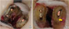
After pre-surgical orthodontic extrusion for 3 weeks and receiving 400 mg of ibuprofen 30 minutes before surgery for pain control, the patients were anesthetized with 2% lidocaine (with 1:80,000 epinephrine, Huons, Hwasung, Korea) using conventional inferior alveolar nerve block techniques. Patient preparation was done following routine protocols for minor dental surgery. An atraumatic extraction of the target tooth was performed using Physics forceps (Golden Misch, Detroit, MI, USA), taking care not to damage the root structure, surface periodontal ligament, or surrounding alveolar bone [15]. After extraction, the tooth was wrapped with saline-soaked gauze to prevent the periodontal ligament from drying. Using a diamond bur (FG 6856014, Mani, Tokyo, Japan), the root was resected at 3 mm from the apex; the resected root surface was observed using an OPMI pico dental microscope (Carl Zeiss, Oberkochen, Germany) at ×25 magnification. To record the number of roots and root canals, the presence of accessory canals and isthmuses, and discolored dentinal area, clinical images were obtained using an 85-mm macro lens and a digital camera (Nikon D90, Nikon, Tokyo, Japan) from a distance of 14 mm (magnification ratio, 2.6:1; Figure 1). Retrograde preparation of 3 mm using a high-speed handpiece and #330 carbide bur and a thin tapered diamond bur (Mani), retrofilling with mineral trioxide aggregate (ProRoot MTA, Dentsply Tulsa, Tulsa, OK, USA), and meticulous curettage of the socket were performed before the tooth was replanted into its original location to complete surgery. A resin wire splint was used to fix the replanted tooth for 1–2 weeks.
Figure 1
Examples of the inclusion and exclusion criteria. (A) An example of an included tooth: a cross-sectional image at 3 mm from the apex, showing the apical portions of a mandibular first molar with 2 roots: a type V mesial root and a type I distal root. (B) An example of an excluded tooth: the distal root shows a vertical crack (arrow).
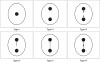
The digital images were used to examine the configuration type of the mandibular molar, the number of roots and root canals, and the presence of accessory canals and isthmuses. The root canal configurations were classified into 6 types, following the classification used by Tam and Yu [16] (Table 1 and Figure 2). The number (1 or 2) of main canals in the root canal was recorded. If there were 2 canals, a note was made of whether the isthmus, defined as a narrow extension from the root canal, was incomplete or complete; the presence of accessory canals away from the main canal was also recorded.
Table 1
Classification of the root canal configurations at 3–5 mm from the apex, based on Tam and Yu [16]

Figure 2
Classification of the root canal configurations at 3–5 mm from the apex, based on Tam and Yu [16].
The discolored area that spread into the root dentin around the endodontically treated area was also examined. Discoloration was observed in the mesial and/or distal roots. The total and obturated areas of the root surfaces and discolored dentinal areas were measured using image analysis software (AxioVision Software 4.11, Carl Zeiss, Munich, Germany). Two examiners agreed on the results of the discoloration measurements in all 115 specimens. The entire surface area of each root was determined by the software. Then, each original root canal treatment area and the discolored dentin area were determined. Each ratio was calculated and analyzed.
Using these calculations, the extent of dentin discoloration on the root surface was quantified. The difference between the originally-treated areas and the discolored dentinal areas was compared statistically using the Wilcoxon signed-rank test. Furthermore, the prevalence of dark-discolored dentin at 3 mm from the apex in either the mesial or distal roots was statistically evaluated by the χ2 test using SPSS 18 (IBM SPSS, Inc., Chicago, IL, USA).
RESULTS
Of the 115 teeth treated by IR, the average time required for extraction was 5 minutes 35 seconds ± 2 minutes 52 seconds. A root tip fracture of less than 3 mm during extraction occurred in 3 cases with a disto-lingual root, but this did not adversely affect the IR procedure because it was similar to the length of the root resection. After extraction, an average time of 12 minutes 24 seconds ± 2 minutes 43 seconds was required for the extra-oral procedure. Conventional prosthetic treatment was performed 2–3 months after surgery according to the symptoms and mobility. All teeth survived throughout the follow-up period (12.9 ± 17.9 months), with periodic follow-up examinations.
Tables 2 and 3 list the frequencies of the configuration types of the mandibular first molar roots. Of the 115 teeth, 92 had 2 roots (80%) and 23 teeth (20%) showed 3 roots (mesial, disto-buccal, and disto-lingual). In the 92 teeth with 2 roots, most of the mesial roots showed 2 root canals (95.65%), with 4 teeth (4.35%) showing a single root canal. In the distal roots, most showed 1 root canal (65.22%), and 34.78% showed 2 root canals. In the 23 teeth with 3 roots, all the mesial roots had 2 root canals, and all the distal roots showed 1 root canal. In the teeth with 2 roots, the distribution of configuration types of the mesial roots was as follows: type I, 4.35%; type II, 9.78%; type III, 6.52%; type IV, 28.26%; type V, 33.70%; type VI, 17.39%. These results indicate that the majority of mesial roots were types IV, V, and VI, which accounted for 79.35% of the mesial roots. The distal roots showed the following configuration types: type I, 65.22%; type II, 3.26%; type III, 1.09%; type IV, 4.35%; type V, 18.48%; type VI, 7.61%. This indicates that the majority of distal roots were type I (65.22%). In the teeth with 3 roots, the distribution of the configuration type of the mesial roots was as follows: type I, 0%; type II, 13.04%; type III, 4.35%; type IV, 26.09%; type V, 30.43%; type VI, 26.09%. Therefore, the majority of mesial roots of the teeth with 3 roots were types IV, V, and VI; these types accounted for 82.61% of the roots, which is similar to the results in the teeth with 2 roots. The canal configuration type of all the disto-buccal roots in the teeth with 3 roots was type I. In addition, the type of the disto-lingual root was type I in 100% of the teeth.
Table 2
The number and configuration type of canals in 2-rooted teeth at 3 mm from the apex

Table 3
The number and configuration type of canals in 3-rooted teeth at 3 mm from the apex

Thirty-nine mesial roots of the 92 two-rooted mandibular molars showed black discoloration around the root canal (42.39%). Ten mesial roots of the 23 three-rooted molars showed black discoloration (43.48%). Thirty-two distal roots of the 92 two-rooted molars showed discoloration (34.78%). Two disto-buccal roots showed discoloration (8.7%) and 3 disto-lingual roots showed discoloration (13.04%). The prevalence of a black discoloration was significantly higher in the mesial root than in the distal root of the 2-rooted molars and the disto-buccal and disto-lingual roots (χ2 test, p = 0.014).
Table 4 presents the ratio of the original root canal treatment area to the total root surface and the ratio of the area of the spread of discoloration of the root dentin to the total root surface. In the mesial root, the original endodontically treated area comprised 2.08% ± 0.36% of the total root surface, whereas the discolored root dentin occupied 6.84% ± 0.28% of the total root surface. The maximum proportion of the discolored dentin area was 26.90% of the total root surface of the mesial root. In the distal root, the original endodontically-treated area comprised 2.53% ± 0.30% of the total root surface, and the discolored dentin occupied 5.01% ± 1.68% of the total root surface. The maximum proportion of the discolored dentin area was 16.67% of the total root surface of the distal root. The ratio of discoloration of the mesial root was significantly higher than that of the distal root at the apical 3 mm level (Wilcoxon signed-rank test, z = −1.636).
Table 4
Discoloration around the root canals of mesial and distal roots at 3 mm from the apex in mandibular first molars

DISCUSSION
The mandibular first molar is frequently subjected to endodontic treatment. This molar generally has 2 roots: a mesio-distally flattened mesial root and an oval-shaped distal root [17]. Occasionally, a third disto-lingual root is found, and its presence is associated with specific ethnic communities [18]. Usually, the mesial root has 2 root canals characterized by complex inter-canal communications, such as isthmuses and accessory canals [19]. Previous studies of the frequency of mandibular first molars with 3 roots reported a rate of 22.7% in Japan, indicating that East Asians have a higher probability of mandibular first molars with 3 roots [20]. In this study, 20% of mandibular first molars had 3 roots. Therefore, the number of roots was not related to the success rate of non-surgical retreatment in the mandibular first molar, in accordance with previous studies. However, clinicians should consider the possible presence of a third root. Nonetheless, the cross-sectional aspect of the disto-buccal canal and disto-lingual canal is almost a circular shape (type I), which is relatively easy to shape using a rotary instrument.
The presence of an accessory canal and isthmus at 3 mm from the apex was observed in 85.87% of the mesial roots of mandibular first molars with 2 roots and in 86.6% of those with 3 roots. The mesial root anatomy of the 2 types of mandibular first molars is similar. These results are higher than those of a previous study, which reported that 60.2% of mesial roots had anatomical complexities [21]. This may have been a reason for the failure of non-surgical retreatment. An isthmus is a narrow, ribbon-shaped communication between 2 canals with pulp tissue, and the untreated isthmus is a reservoir of bacteria that can cause persistent discomfort, even after root canal treatment, making it very difficult to clean and shape using a rotary instrument [22]. Any remnants can contaminate the nearby dentin through the dentinal tubules [232425]. Therefore, clinicians should use effective techniques to clean and disinfect this complex form of root canal anatomy. For example, lasers may be an effective treatment option, enabling better root canal disinfection than when only mechanical instrumentation is used [2627].
To our knowledge, 1 previous study investigated the cross-sectional anatomy of mesio-buccal roots of the maxillary first molar [16]. Few studies have investigated the cross-sectional anatomy of the mandibular molars, even though the morphology of the root canal system has been extensively investigated, with at least 23 types being classified [19]. However, some overlap might exist among these types at certain cross-sectional levels, making it hard to standardize. These factors led us to adopt the classification proposed in the previous study of this topic. A new classification of the cross-sectional anatomy of the mandibular first molar should be developed as a future study.
The amorphous smear layer, with organic and inorganic phases found on the instrumented dentinal walls, can be contaminated by bacterial remnants that penetrate the dentinal tubules for distances of up to 10–150 μm [2528]. The acid produced by microorganisms can dissolve the smear layer and allow bacteria to pass into the dentinal tubules [29]. Therefore, increased porosity may allow bacterial byproducts to settle, which causes dentin discoloration [30]. Furthermore, blood from the apical foramen may also be a reason for dentinal discoloration, as 1 tooth showed a discolored area of 26.9% of the total surface. This can only be explained by blood infiltration through the dentinal tubule [31]. More than 40% of the mesial roots and distal roots of the 2-rooted mandibular molars showed discoloration in this study. Discolored dentin should be examined by culturing the bacteria and inspecting them with an electron microscope to investigate their nature and toxicity. Such an examination could not be performed on the teeth included in this study that were extracted for IR. Further studies should be conducted of discolored dentin to evaluate its nature, toxicity, and effects on the clinical outcomes of root canal treatment. All discolored areas were removed during the IR procedures, but it is unclear whether the removal of discolored dentin affected the clinical outcomes. The nature and toxicity of the discolored dentin made an unclear contribution to the failure of the previous root canal treatment. In the cases examined in this retrospective study, the operator removed any possible factor that might have contributed to the failure of the previous root canal treatment. The apical 3 mm of the most complex portion of the root is removed during peri-radicular surgery and IR, but little is known regarding the width of the retrograde preparation. Further studies need to be done to clarify the nature of discolored dentin, its effects on the clinical outcomes of non-surgical root canal treatment, and the appropriate width and depth of retrograde preparation for clinicians to use as a standard when performing retrograde endodontic treatment.
CONCLUSIONS
The root anatomy of mandibular first molars that failed non-surgical retreatment was found to be more complex than average. The mesial root of these molars had a more complex inter-canal anatomy than the distal root, making complete cleaning, shaping, and disinfection more difficult. The prevalence of disto-lingual roots in the mandibular first molars that failed endodontic retreatment was 20%, but the number of roots did not affect the clinical outcome. The discoloration of dentin around the root canal was quite common in the mandibular first molars that failed non-surgical retreatment, and was significantly more extensive in the mesial root, regardless of the number of roots. However, the cause of the discoloration and its clinical implications are unclear. Further investigation of the effects of discoloration will be needed to improve the clinical outcomes of non-surgical retreatment.
 XML Download