

 PDF
PDF Citation
 Print
Print


INTRODUCTION
One of the ultimate goals of modern dentistry is to link basic research findings with their clinical significance. This can be achieved by synthesizing state-of-the-art scientific knowledge from conflicting results, which may limit the translation of research findings into daily clinical practice. The philosophy of evidence-based dentistry was developed to support both clinicians and academicians in making ‘well-justified’ decisions and judgments. This medical philosophy incorporates standardized scientific skills and tools (e.g., systematic reviews and meta-analyses) to strengthen the current scientific evidence on controversial research topics [1]. Systematic reviews and meta-analyses depend mainly on analyzing the current available scientific knowledge to reach the highest level of evidence [2].
In the past 3 decades, the field of adhesive dentistry has been comprehensively investigated. This has led to significant developments in the chemistry of dental adhesives, allowing greater preservation of the tooth substrate. The dental substrate is a complex structure that consists of enamel, dentin, and cementum. Enamel is a homogenous hard tissue consisting of hydroxyapatite (HAp) (96 Wt%) crystals [3]. Conversely, dentin is a heterogeneous tissue consisting of 20 Wt% inorganic crystals (HAp) that envelop the dentinal collagen fibers (mainly type I fibers) [4]. Previous laboratory studies [5678910] reported that enamel exhibited higher bond strength values than dentin. The most difficult challenge in bonding to dentin is its relatively high-water content, which may interfere with the bonding of hydrophobic dental adhesives to the collagen scaffolds of dentin [5]. This problem seems to be more obvious in bonding to caries-affected dentin, which has a porous nature and contains high water percentages [11].
A thin layer, referred to as the ‘smear layer,’ is generated when cutting into dentin [121314]. This layer covers the superficial dentin surface and may extend into dentinal tubules, forming smear ‘plugs.’ This layer consists of depleted hydroxyapatite crystals, denatured collagen fibrils, saliva, and blood and food debris. The smear layer plays a significant role in the bonding of resin-based adhesives to dentin. Current dental adhesives can be classified into 4 categories according to how they deal with the smear layer: etch-and-rinse (E&R), self-etch (SE), multi-mode ‘universal,’ and resin-modified glass ionomer adhesives. In E&R adhesive systems, dentin surface treatment is performed using phosphoric acid etching gels to totally remove the smear layer and open the dentinal tubules. Theoretically, this technique can enhance resin infiltration into the partially demineralized collagen network.
However, the surface treatment of dentin with phosphoric acid solutions faces 2 major challenges. The first challenge is the excessive dehydration of the dentin collagen caused by over-air-drying, which is referred to as dentin desiccation. Prolonged air drying of dentin collapses the micro-spaces (created after the demineralization of dentin) of its collagenous fibril network and subsequently reduces the infiltration of resin adhesives. The second challenge associated with dentin etching is deep demineralization beyond the resin-infiltration level, which leads to poor hybridization with dentin.
SE adhesives were introduced to overcome the problems of E&R adhesives. SE adhesives depend on a smear layer-modifying (dissolving) bonding strategy. Nevertheless, the demineralization depth of SE adhesives is less than that of E&R adhesives, and many studies have shown that the quality of the hybrid layer produced by SE adhesives is much better than that generated using E&R adhesives [151617]. The presence of water is essential for the ionization of the acidic moieties of SE adhesives to form oxonium ions (H3O+) [18], which demineralize the dentin surface [18]. Currently, the SE approach is widely accepted by practicing dentists, and most manufacturers claim that these categories of adhesives are more user-friendly, have fewer application steps and a shorter application time, and do not require complicated technique-sensitive procedures [1920]. Due to the incomplete removal of the smear layer, SE adhesives exhibit a marked reduction in postoperative sensitivity [2122]. SE adhesives can be classified as either one-step (1-S) or two-step (2-S) adhesives. The acidulated primer can be either provided in a separate bottle (2-S) or combined with the hydrophobic resin adhesive in the same bottle as ‘all-in-one’ (1-S) SE adhesives [23]. Furthermore, SE adhesives can be classified according to their acidity: ultra-mild (pH > 2.5), mild (pH ≈ 2), intermediately strong (pH 1 to 2), and strong (pH ≤ 1) [24].
An important technique aiming to enhance resin/dentin hybridization involves pretreatment of the dentin surface with a deproteinizing agent, such as sodium hypochlorite (NaOCl) or hypochlorous acid (HOCl) solution [25]. This dentin surface pretreatment method is referred to as the smear layer deproteinizing process. Deproteinizing agents can dissolve the organic content of the smear layer, and they exhibit marked antibacterial activity [26]. Several studies have reported that the pretreatment of dentin with either NaOCl or HOCl deproteinizing agents could dissolve the organic components of the smear layer, leaving the inorganic crystals to act as filler with the hybrid layer [272829]. Nevertheless, NaOCl exhibited a strong non-specific proteolytic response, and it has been reported that it may adversely affect the intact ‘sound’ collagen [3031].
The aim of this systematic review was to critically analyze previously published studies of the effects of dentin surface pretreatment with deproteinizing agents on the bonding of SE adhesives to dentin. Additionally, a meta-analysis was performed to quantify the effects of the above-mentioned surface pretreatment methods on the bonding of SE adhesives to dentin. The key questions of this systematic review were “Do deproteinizing agents promote bonding of SE adhesives to dentin?” and “What are the ideal smear layer deproteinizing protocols (concentration and application time) to obtain adequate bond strength?”
MATERIALS AND METHODS
Protocol development and eligibility criteria
The protocol of this systematic review was designed following the Preferred Reporting Items Systematic Review and Meta-Analysis (PRISMA) guidelines [32]. The methodologies of the previous laboratory studies were comprehensively assessed. The reviewed studies were subjected to a meta-analysis to quantify the effects of the application time and concentration of NaOCl and HOCl deproteinizing agents on bonding to dentin. The meta-analysis was conducted using Comprehensive Meta-Analysis software (version 5, Biostat, Englewood, NJ, USA) with 95% confidence intervals.
Search strategies/inclusion and exclusion criteria
The initial online search was performed by 1 of the authors (K.A.) using the following databases: Scopus, PubMed, and ScienceDirect. The online search was performed using the following keywords: ‘dentin’ or ‘hypochlorous acid’ or ‘sodium hypochlorite’ and ‘self-etch adhesive.’ An additional hand search was performed to check for non-online resources. The initial screening of the articles depended on the title, abstract, and full text (when needed). All articles found by both electronic and hand searching were collected onto a single sheet, of which 3 copies were printed and distributed among the 3 authors. Each author individually checked the eligibility criteria for each study, and the agreement of at least 2 authors was essential for exclusion/inclusion of the study for the systematic review. The selected manuscripts were discussed and the selections were made in face-to-face meetings.
This review included studies that stated clear objectives and detailed testing methodologies. The selected studies had at least a 2-arm design; in the test group, the dentin surface pretreatment was performed using a deproteinizing agent, while in the control group, no dentin surface pretreatment was used. Studies that utilized carious or bovine teeth were excluded. The bond strength testing of included studies was performed by a standard microtensile bond strength (μTBS) method. Accordingly, studies that utilized other bond strength testing methodologies (e.g., macro-tensile or shear bond strength) were excluded.
The following categories were excluded during the assessment process: non-English articles, randomized controlled trials (RCTs), case reports, animal studies, and review articles. The studies that were included investigated the bonding of SE adhesive to dentin; therefore, studies that evaluated the bonding of E&R adhesives to dentin were excluded. Any study that failed to present an appropriate and logical statistical analysis was excluded. The goal was to include studies that evaluated the bonding of resin composite to dentin; therefore, studies that were conducted to evaluate the bonding of glass ionomer cements, resin-modified glass ionomer cements, or compomer to dentin were excluded.
RESULTS
Search results
The initial search of the ScienceDirect, PubMed, and Scopus databases resulted in 124 articles being identified. Three review articles were excluded. Another 8 studies were also excluded because they were conducted to evaluate the bonding of resin luting cement to dentin. Of the remaining 113 studies, 1 was an animal study, 6 utilized bovine teeth, and 3 were RCTs; these 10 studies were excluded. In addition, 22 studies were excluded because they utilized laser-treated (2 studies), bleached (5 studies), carious (12 studies), or deciduous (3 studies) teeth. Moreover, 1 oral bioscience study that evaluated stem cells and 2 studies that used ethylenediaminetetraacetic acid (EDTA)-treated teeth were excluded. All studies that evaluated the bonding of E&R adhesives to deproteinized dentin surfaces were excluded (12 studies).
From the remaining 63 articles, 11 were excluded because they used NaOCl as a storage medium, not for dentin surface treatment. Another 23 endodontic studies that utilized root canal-treated teeth were also excluded. Of the remaining 29 studies, 3 that did not use deproteinizing agents to optimize the hybrid layer and 1 that evaluated hardness properties were excluded. In addition, 16 studies were excluded for the following reasons: 8 studies evaluated nanoleakage patterns, while the remaining 8 used a shear bond strength testing method. Finally, 9 studies fit the inclusion criteria of this systematic review (Table 1). The detailed study selection procedure is illustrated in Figure 1.
Table 1
Summary of methodologies and results of the included studies
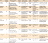
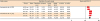
| Study | Sample size (molars) | Method, test machine, and speed | Adhesive system | Number, diameter, and shape of beam | Storage time | NaOCl concentration and time | Result |
|---|---|---|---|---|---|---|---|
| Taniguchi et al. [28] | 40 | - µTBS | 1-S SE and 2-S SE | Three hourglass-shaped specimens with a cross-sectional area of approximately 1 mm2 | 24 hr water storage | a. 6% NaOCl for 30 and 15 sec | Pretreatment of dentin with NaOCl for 30 sec adversely affected the bonding of SE adhesives to dentin |
| - Testing machine: EZ-test, Shimadzu Co., Kyoto, Japan | b. Control group: rinse with water | ||||||
| - Cross-head speed: 1.0 mm/min | |||||||
| Kunawarote et al. [33] | 39 | - µTBS | 2-S SE | Five hourglass-shaped specimens with a cross-sectional area of approximately 1 mm2 | 24 hr water storage | a. 6% NaOCl | The longer the dentin pretreatment time with NaOCl, the lower µTBS values were obtained |
| - Testing machine: EZ-test, Shimadzu Co., Kyoto, Japan | b. 50 ppm HOCl for 30, 15, and 5 sec | ||||||
| - Cross-head speed: 1 mm/min | c. Control group: rinse with water | ||||||
| Cecchin et al. [38] | 30 | - µTBS | 1-S SE | Four hourglass-shaped specimens with a cross-sectional area of approximately 1 mm2 | 24 hr water storage | a. 1% NaOCl applied to the dentin for 1 hr | The deproteinizing did not deteriorate the bonding of SE adhesive (XENO III, DENTSPLY, Tulsa, OK, USA) to dentin |
| - Universal testing machine (Emic DL 2000) at a cross-head speed of 0.5 mm/min | b. Control group: DI water | ||||||
| Farina et al. [37] | 60 | - µTBS | 2-S SE | Four hourglass-shaped specimens with a cross-sectional area of approximately 1 mm2 | 24 hr water storage | a. 1% NaOCl was applied to the dentin surface for 40 min | Dentin surface pretreatment with 1 % NaOCl reduced the bonding of SE to dentin |
| - Universal testing machine (Emic DL 2000) at a cross-head speed of 0.5 mm/min | b. Control group: DI water | ||||||
| Ozturk and Ozer [35] | 40 | - µTBS | 2-S SE | Three rectangular sticks (1.0 ± 0.03 mm2) | 24 hr water storage | a. 5% NaOCl for 1 min | Dentin surface pretreatment with NaOCl reduced the bonding of SE to dentin |
| - Testing apparatus (Bencor-Multi T, Danville Engineering Co., Danville, CA, USA) at a cross-head speed of 1 mm/min | b. Control group: DI water | ||||||
| Prasansuttiporn et al. [39] | 24 | - µTBS | 2-S SE | Four hourglass-shaped specimens with a cross-sectional area of approximately 1 mm2 | 24 hr water storage | a. 6% NaOCl for 30 sec | The NaOCl-treated group exhibited lower bond strength than the control group |
| - Universal testing machine (EZ-test, Shimadzu Crop., Kyoto, Japan) at a cross-head speed of 1 mm/min | b. Control group: DI water | ||||||
| Kunawarote et al. [34] | 40 | - µTBS | 2-S SE | Five hourglass-shaped specimens with a cross-sectional area of approximately 1 mm2 | 24 hr water storage | a. 806 mM NaOCl, | None of the pretreatments demonstrated a negative influence on the bonding of SE adhesives to normal dentin |
| - Testing machine (EZ-test, Shimadzu, Kyoto, Japan) at a cross-head speed of 1 mm/min | b. 0.95 or 1.91 mM HOCl for 5 sec | ||||||
| c. Control group: DI water | |||||||
| Prasansuttiporn et al. [40] | 36 | - µTBS | 1-S SE and 2-S SE | Five hourglass-shaped specimens with a cross-sectional area of approximately 1 mm2 | 24 hr water storage | a. 6% NaOCl for 30 sec | The recorded bond strength values of the deproteinized dentin group were significantly lower than those of the control group |
| - Universal testing machine (EZ-test, Shimadzu Crop., Kyoto, Japan) at a cross-head speed of 1 mm/min | b. Control group: DI water | ||||||
| Sacramento et al. [36] | 90 | - µTBS | 1-S SE and 2-S SE | Fourteen sticks with a surface area of about 1.0 mm2 | 24 hr water storage | a. 0.5% NaOCl for 30 min | The NaOCl-treated group exhibited lower bond strength than the control group |
| - Universal testing machine (Instron model 4411, Canton, MA, USA) at a cross-head speed of 0.5 mm/min. | b. Control group: DI water |
NaOCl, sodium hypochlorite; μTBS, microtensile bond strength; 1-S, one-step; 2-S, two-step; SE, self-etch; HOCl, hypochlorous acid; DI, distilled water.
![]()
Assessment of the deproteinizing agent concentrations/application time periods
Only laboratory studies were included in this systematic review, regardless of the concentration of the deproteinizing agent and exposure time. Two studies used HOCl solution with different concentrations, while the remaining 7 studies used NaOCl solution with different concentrations. All included studies used SE adhesive; 8 of them used 2-S SE adhesives and 4 used 1-S SE adhesives. In the reviewed studies, NaOCl solution was used at the following concentrations: 6% (4 studies), 1% (2 studies), 5% (1 study), and 0.5% (1 study). Only 1 study used a molar concentration formula (806.02 mM) to describe the concentration of the NaOCl solution. The application time varied among the reviewed studies. Four studies applied 6% NaOCl for 30 seconds, while 2 studies applied the same concentration for 15 seconds. Only 2 studies applied NaOCl and HOCl for 5 seconds [3334]. Moreover, the following application times were used for smear layer deproteinization by NaOCl solution: 60 seconds [35], 30 minutes [36], 40 minutes [37], and 1 hour [38].
Assessment of μTBS: testing setup
Six of the included studies used a crosshead speed of 1 mm/min [283334353940], and 3 used a crosshead speed of 0.5 mm/min [363738]. Seven of the included studies used hourglass-shape specimens for microtensile testing [28333437383940], while the remaining 2 used rectangular beams [3536]. Seven of the selected studies used bonded specimens with a surface area of 1 mm2, while the remaining 2 utilized bonded specimens with a surface area of 0.7 mm2.
In all the included studies, bonded specimens were tested after 24-hour water storage; then, the fractured dentin surfaces were gold sputter-coated and observed under a scanning electron microscope to assess the fracture modes. The failure modes were classified into; 1) adhesive if 100% of the bonded interface failed between the dentin and bonding agent, 2) cohesive in dentin if 100% of the failure occurred within the dentin, 3) cohesive in resin composite if 100% of the failure occurred within a resin composite restoration, or 4) mixed failure if a combination of adhesive and cohesive failures in the dentin and/or resin composite was observed. A significant increase in the number of mixed failures was observed after dentin surface treatment with 6% NaOCl for prolonged times [28333940]. Two studies [3334] reported that the surface treatment of dentin with 50 ppm HOCl showed more mixed failures than were observed in the NaOCl groups. The use of 1% NaOCl for 40 minutes showed more adhesive failures, and similar findings were reported when using 5% NaOCl for 60 seconds [3537].
The results of this review revealed that μTBS values significantly decreased following dentin surface treatment with high concentrations of HOCl. Additionally, the adhesive failure mode was the predominant fracture pattern in this group. Moreover, the concentration of deproteinizing solution had a significant effect on the failure mode. Studies that utilized NaOCl showed a significant increase in the mixed failure percentage associated with increased NaOCl concentration. It was also shown that 2-S SE adhesives exhibited significantly higher μTBS values than 1-S SE adhesives [283334353637383940]. The μTBS results of the reviewed studies are shown in Table 2.
Table 2
Overall analysis of μTBS and fracture modes reported in the reviewed studies
| Study | SE adhesive system | Deproteinizing agent | Time | Mean µTBS (MPa) | Mode of failure (%) | |||
|---|---|---|---|---|---|---|---|---|
| Cohesive in resin | Cohesive in dentin | Mixed | Adhesive | |||||
| Taniguchi et al. [28] | Bond Force (1-S) | 6% NaOCl | 30 sec | 30.4 | 4 | 4 | 83 | 4 |
| 15 sec | 43.7 | |||||||
| Clearfil SE Protect (2-S) | 6% NaOCl | 30 sec | 34.4 | 0 | 4 | 91.5 | 4 | |
| 15 sec | 42.0 | |||||||
| Kunawarote et al. [33] | Clearfil SE Bond (2-S) | 6% NaOCl | 30 sec | 27.19 | 0 | 0 | 90 | 10 |
| 15 sec | 38.43 | 0 | 20 | 65 | 15 | |||
| 5 sec | 40.34 | 0 | 35 | 58 | 7 | |||
| 50 ppm HOCl | 30 sec | 36.87 | 0 | 17 | 55 | 28 | ||
| 15 sec | 37.64 | 0 | 60 | 23 | 17 | |||
| 5 sec | 41.97 | 38 | 10 | 25 | 27 | |||
| Cecchin et al. [38] | XENO III (1-S) | 1% NaOCl | 1 hr | 19.41 | NA | NA | NA | NA |
| Farina et al. [37] | Clearfil SE Bond (2-S) | 1% NaOCl | 40 min | 19.08 | 0 | 0 | 27 | 73 |
| Ozturk and Ozer [35] | Clearfil SE Bond (2-S) | 5% NaOCl | 60 sec | 15.58 | 13.5 | 6.5 | 80 | |
| Prasansuttiporn et al. [39] | Clearfil Protect Bond (2-S) | 6% NaOCl | 30 sec | 43.6 | 7.5 | 7.5 | 85 | 0 |
| Kunawarote et al. [34] | Clearfil SE Bond (2-S) | 806.02 mM NaOCl | 5 sec | 40.87 | 0 | 40 | 50 | 10 |
| 0.95 mM HOCl | 5 sec | 41.93 | 35 | 15 | 35 | 15 | ||
| 1.91 mM HOCl | 5 sec | 41.24 | 27 | 7 | 38 | 28 | ||
| Prasansuttiporn et al. [40] | Clearfil s3 bond (1-S) | 6% NaOCl | 30 sec | 33.6 | 7 | 14.5 | 78.5 | 0 |
| Bond force (1-S) | 6% NaOCl | 30 sec | 26.9 | 22 | 0 | 64 | 14 | |
| Clearfil protect bond (2-S) | 6% NaOCl | 30 sec | 43.6 | 7.5 | 7.5 | 85 | 0 | |
| Sacramento et al. [36] | Clearfil protect bond (2-S) | 0.5% NaOCl | 30 min | 30.60 | 70 | 0 | 30 | 0 |
| Adper Prompt L-Pop (1-S) | 0.5% NaOCl | 30 min | 20.67 | 25 | 0 | 75 | 0 | |
μTBS, microtensile bond strength; SE, self-etch; 1-S, one-step; 2-S, two-step; NaOCl, sodium hypochlorite; HOCl, hypochlorous acid; NA, not available.
![]()
Meta-analysis results
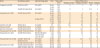
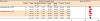
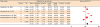
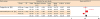
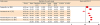
The results of this meta-analysis revealed that the pooled average μTBS values of dentin pre-treated with deproteinizing agents (15.71 MPa) were significantly lower than those of the non-treated control group (20.94 MPa) (Figures 2 and 3). However, since the majority of the reviewed studies were performed using NaOCl solution, the overall average seems to be an inappropriate basis for making judgments. Therefore, a specific meta-analysis for each deproteinizing solution was conducted. This analysis revealed that the mean μTBS values of the HOCl group (40.17 MPa) were significantly higher than those of the NaOCl group (15.87 MPa) (Table 3, Figures 4 and 5). Additionally, long exposure to the deproteinizing agent adversely affected bonding to dentin (Table 3, Figures 6 and 7). For the deproteinizing groups, the results of the meta-analysis showed that the 2-S SE adhesives exhibited higher mean bond strength values than the 1-S SE adhesives (Table 3, Figures 8 and 9).
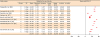
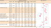 | Figure 2Overall meta-analysis results of the mean μTBS of SE adhesives bonded to NaOCl/HOCl-treated dentin.
μTBS, microtensile bond strength; SE, self-etch; NaOCl, sodium hypochlorite; HOCl, hypochlorous acid; CI, confidence interval; A, 6% NaOCl; B, 50 ppm HOCl; C, 1% NaOCl; D, 5% NaOCl; E, 806.02 mM NaOCl; F, 0.95 mM HOCl; G, 1.91 mM HOCl; H, 0.5% NaOCl.
|
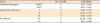
 | Figure 3Meta-analysis results of μTBS for control groups.
μTBS, microtensile bond strength; SE, self-etch; C, no dentin surface treatment was performed; CI, confidence interval.
|
Table 3
Results of applying the medical statistical model of Borenstein et al. [70] to the meta-analysis outcomes
Data are shown as means ± standard deviations. Groups identified by different superscript letters within the rows for each factor were significantly different at p < 0.05.
μTBS, microtensile bond strength; NaOCl, sodium hypochlorite; HOCl, hypochlorous acid; SE, self-etch; 1-S, one-step; 2-S, two-step.
![]()
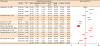 | Figure 4Meta-analysis results of the mean μTBS for SE adhesive bonded to NaOCl-treated dentin.
μTBS, microtensile bond strength; SE, self-etch; NaOCl, sodium hypochlorite; CI, confidence interval; A, 6% NaOCl; B, 1% NaOCl; C, 1% NaOCl; D, 806.02 mM NaOCl; E, 0.5% NaOCl.
|
 | Figure 5Meta-analysis results of the mean μTBS for SE adhesive bonded to HOCl-treated dentin.
μTBS, microtensile bond strength; SE, self-etch; HOCl, hypochlorous acid; CI, confidence interval; A, 50 ppm HOCl; B, 0.95 mM HOCl; C, 1.91 mM HOCl.
|
 | Figure 6Meta-analysis results of the mean μTBS for 5 second dentin surface treatment with a deproteinizing agent.
μTBS, microtensile bond strength; CI, confidence interval; A, 6% NaOCl; B, 50 ppm HOCl; C, 806.02 mM NaOCl; D, 0.95 mM HOCl; E, 1.91mM HOCl.
|
 | Figure 7Meta-analysis results of the mean μTBS for 30 second dentin surface treatment with a deproteinizing agent.
μTBS, microtensile bond strength; SE, self-etch; CI, confidence interval; A, 6% NaOCl; B, 50 ppm HOCl.
|
Discussion
Currently, evidence-based dentistry is an essential approach for detecting research gaps and synthesizing conclusions from the current literature despite conflicting opinions. The ultimate goal of this scientific approach is to summarize, disseminate, and critique the currently available scientific knowledge, while aiming to translate this knowledge into clinical recommendations. A systematic review is a powerful tool in this scientific approach that helps achieve its objectives [1]. The majority of selected studies in this review did not follow methodologically ideal testing techniques, and consequently, considerable variation in the results was observed among the studies. Thus, the rationale behind conducting this review was to obtain well-justified conclusions, which may help both researchers and clinicians to judge the efficacy of using deproteinizing agents as a dentin surface pretreatment method for modifying the smear layer.
Dentin is a natural composite structure and is considered a challenging substrate for dental adhesion. Dentin has a heterogeneous nature and consists of a complex inorganic/organic structure [4]. The presence of the smear layer represents another major challenge for successful bonding to dentin [4142]. It is well known that a micromechanical adhesion mechanism plays an essential role in the adhesion of resin-based bonding agents to dentin, in which adhesive primers infiltrate into the superficial demineralized collagen fibers of ‘hybridized’ dentin [43]. However, previous studies showed that resin primers could not totally infiltrate the demineralized dentin, leaving behind some gaps and denuded collagen. These negative spaces can act as pathways for microorganisms and may influence bond stability, particularly when water seeps in [4445464748].
The results of this systematic review showed that the surface pretreatment of dentin with either NaOCl or HOCl solutions led to low μTBS values compared with non-treated surfaces. Additionally, it showed that the μTBS values of dentin treated with HOCl solution were significantly higher than those of NaOCl-pretreated dentin. This may be attributed to the chemistry of the NaOCl solution, which has a low surface tension and a high potential to disrupt both sound and denatured collagen. It has been reported that applying NaOCl to the smear layer removed only the superficial ‘loosely attached organic component, without opening the dentinal tubules’ [28495051]. However, it may deteriorate the mechanical properties of dentin via the degradation of the sound collagen fibers [31]. NaOCl solutions may degrade the collagen scaffolds of dentin, consequently reducing the number of bonding sites for adhesive primers. This impairs resin hybridization with dentin, leading to a marked reduction in the μTBS [43525354].
Furthermore, the low bond strength of NaOCl-treated dentin may be attributed to the strong oxidizing action of NaOCl, which leads to the formation of chloramine-derived radicals. These reactive radicals could interfere with the free radical polymerization of resin-based adhesives [2655565758]. Additionally, bonding to dentin might be influenced by the residual NaOCl entrapped in the porous structure of mineralized dentin [59]. The residual chemical substances in the fluid may interact with the adhesive system and affect the light polymerization of the monomer in the demineralized dentin, causing a marked reduction in bond strength [3760].
Moreover, Taniguchi et al. [28] investigated the surface pH of NaOCl-treated dentin and reported that these surfaces exhibited significantly higher pH values than non-treated dentin surfaces, even after copious rinsing with water for sufficient time periods. The high alkalinity of NaOCl-treated surfaces could be explained by the high concentration of hydroxyl (OH) groups on the dentin surface [516162]. The alkalinity of NaOCl might buffer the acidity of SE adhesives and thus reduce their hybridization with the underlying dentin [33]. These results are in agreement with many previous studies [284355] that reported that the application of NaOCl to dentin had an adverse effect on the bonding of SE to dentin. Nonetheless, a few studies have reported that NaOCl treatment increased the bond strength of some adhesive systems, and they attributed their results to the effects of NaOCl on the removal of the collagen layer, which may be beneficial for some resins to create proper dentinal bonding [636465]. However, most of those studies neglected the adverse effects of NaOCl on bonding to dentin and did not provide logical explanations for the high bond strength results that they obtained.
It is well known that the hydration reaction of NaOCl leads to the formation of HOCl, which is a potent deproteinizing agent as well as an effective biological oxidizing agent [49]. In aqueous solution, HOCl partially dissociates into the anion hypochlorite (OCl−) and cation hydrogen (H+). The pH of HOCl is slightly acidic, which could partially demineralize the dentin and allow it to achieve a better resin hybridization than NaOCl solutions [6667]. Furthermore, it was stated that the higher reactivity of NaOCl to amino acids makes it resistant to washing (even after copious rinsing with water), leaving high concentrations of chlorine on the surface [6869]. Unlike NaOCl, HOCl solutions can be easily rinsed off, and this might provide a logical explanation of the relatively high μTBS values of HOCl-pretreated dentin surfaces in comparison with NaOCl-pretreated dentin.
The results of this study showed that long surface treatment with deproteinizing agents adversely affected the bonding of SE to dentin. Application of deproteinizing agents for an extended period may lead to the destruction of more collagen scaffolds, resulting in a marked reduction in binding sites for adhesive primers. Additionally, 2-S SE adhesives showed higher μTBS values than 1-S SE adhesives. This may be due to the contamination of 1-S SE adhesives by NaOCl byproducts that affect the free-radical polymerization reaction. Moreover, the alkalinity of NaOCl may neutralize the acidity of ultra-mild 1-S SE, whereas this buffering action has a minimal effect on the intermediate pH 2-S SE adhesives. These results are in agreement with those of the study of Hamama et al. [11], in which nanoleakage results revealed that the silver nitrate intake was higher in sound dentin treated with Carisolv (a NaOCl-based chemomechanical caries removal agent) and bonded with a 1-S SE adhesive than in the corresponding groups bonded with a 2-S SE adhesive. They attributed the higher silver uptake to the contamination of the hybrid layer by NaOCl residues, which affected the free-radical polymerization reaction and consequently led to a reduction in μTBS.
An unavoidable limitation of the current systematic review was that one of its exclusion criteria was non-English manuscripts; however, some of those excluded studies may have contained useful information for this review.
Conclusions
In light of the currently available scientific evidence, pretreatment of dentin surfaces with deproteinizing agents does not enhance the bonding of SE adhesives to dentin. HOCl as a deproteinizing agent exhibits minimal adverse effects on bonding to dentin in comparison with NaOCl solutions. Accordingly, when needed, it is preferable to use HOCl as a deproteinizing agent for dentin surface pretreatment prior to the application of SE adhesives. The 2-S SE adhesives show more reliable bonding to deproteinized dentin than 1-S SE adhesives. Long exposure to deproteinizing agents significantly impairs the bonding of SE agents to dentin.
 XML Download