

 PDF
PDF ePub
ePub Citation
 Print
Print


INTRODUCTION
Calcium hydroxide (Ca[OH]2) is highly recommended and widely accepted as an inter-appointment intracanal endodontic dressing because it demonstrates a pronounced antibacterial activity against most of the bacterial species identified in endodontic infections [1]. However, if Ca(OH)2 remains in the root canal system, it can play negative roles in the prognosis of endodontic treatment. Ca(OH)2 that remains on dentinal walls prevents the penetration of sealer into dentinal tubules and causes considerable changes to the sealing ability of the material [23]. Furthermore, Ca(OH)2 dressing has an adverse effect on the bond strength of a resin-based sealer [4]. Therefore, the complete removal of Ca(OH)2 intracanal medicament is recommended for favorable endodontic outcomes.
Most of the commercially available Ca(OH)2-containing pastes are produced as an emulsion of 2 immiscible substances with one (paste) being dispersed throughout the other (vehicle). Vehicles are used in an attempt to maintain pH [5] and improve antimicrobial activity [6], biocompatibility [7], ionic release [8], and diffusion [9]. Several authors and clinicians have considered associations of Ca(OH)2 with different vehicles including polyethylene glycol (PEG) [10], propylene glycol (PG) [11], and glycerin [12]. However, most Ca(OH)2 products cannot be removed completely from complex root canal systems even with various technical methods [1314]. In this respect, there has been a request for development of products with a new vehicle that possesses a potent dissolution and cleansing effect so that pastes can be removed more effectively by irrigation. However, until now, there was no obvious evidence regarding the role of vehicles on the influence of the persistence of residues on root canal walls [1315].
Recently, a new Ca(OH)2 paste used N-2-methyl-pyrrolidone (NMP) as a vehicle (cleaniCal, Maruchi, Wonju, Korea) was introduced into the endodontic market. NMP is an organic compound consisting of a 5-membered lactam. It is a colorless liquid and is miscible with water and most common organic solvents. Its chemical formula is C5H9NO and has a molecular weight of 99.13. NMP is a very strong solubilizing agent that has important applications in different fields of industry [16]. To our knowledge, cleaniCal is the first commercially available NMP-based Ca(OH)2 paste. Therefore, an investigation into its removal efficacy compared with other products containing vehicles currently in use is required. Furthermore, the cytotoxicity of the NMP-based endodontic material has never been investigated before. In this respect, the purpose of this study was to investigate the removal efficacy and cytotoxicity of the newly developed NMP-based Ca(OH)2 paste in comparison with ApexCal (Ivoclar Vivadent, Schaan, Liechtenstein) and Calcipex II (Nishika, Shimonoseki, Japan) which contain commonly used vehicles such as PEG and PG, respectively. The null hypotheses were that there would be no significant differences between the tested materials regarding removal efficacy and cytotoxicity.
MATERIALS AND METHODS
Sample preparation
Thirty intact, caries-free human single-rooted maxillary premolars with oval-shaped canals in cross section were obtained with patient informed consent under a protocol approved by the Institutional Review Board of Chonbuk National University Hospital (IRB code: CUH 2016-04-024). After preparing an access cavity, a size 10 K-file (Dentsply-Maillefer, Ballaigues, Switzerland) was inserted into the canal until it was just visible at the apical foramen. Working length was determined by subtracting 0.5 mm from this length. The root canals were instrumented with nickel-titanium rotary files (ProTaper Universal, Dentsply-Maillefer) to a size of F3 in the presence of 5.25% sodium hypochlorite (NaOCl) solution. After completion of instrumentation, the canal was irrigated with 5 mL of 17% ethylenediaminetetraacetic acid and 5 mL of NaOCl solution.
After canal shaping, the teeth were divided randomly into 3 groups of 10 teeth each: group 1 for cleaniCal, group 2 for ApexCal, and group 3 for Cacipex II. The root canals were dried with paper points and then completely filled with each Ca(OH)2 paste. The access cavities were temporarily sealed with a cotton pellet and a temporary filling material (e-Temp, Diadent, Cheongju, Korea). The teeth were stored at 37°C in 100% relative humidity for 7 days. The removal of the Ca(OH)2 paste was performed using the standard procedure that includes the use of a master apical file combined with NaOCl irrigation as described in a previous study [17]. Briefly, a size 25 K-file was inserted into the canal to scatter the medicament and obtain canal patency. The canal was then irrigated with 5.25% NaOCl solution in 5 mL syringes attached to a 27-gauge needle (ENDOEZE irrigation tip, Ultradent, South Jordan, UT, USA) that was placed 2 mm short of the working length. The irrigation procedure was repeated 3 times, and the canals were dried with paper points. All the specimens were prepared by a single operator.
Micro-computed tomographic (μ-CT) analysis
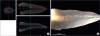
Specimens were numbered and scanned with a μ-CT (Skyscan 1076, Bruker, Kontich, Belgium). The scanning parameters for each scan were kept constant: 65 kV (10 W, 154 µA), 0.5 mm aluminum filter, 160 milllisecond exposure time, 0.70° rotation step, and isotropic voxel size of 35 µm. The volume percentage of the residual sealer divided by the total volume of the root canal system was calculated using an image analyzing program (CT-An, Bruker) (Figure 1A). CT-An software was utilized for the volume measurement of the residual sealer. To calculate the percentage of the total remaining remnant after the removal procedure, rendering was applied to the readily detected Ca(OH)2 paste. The region of interest (ROI) was set to the inside of the root canal system boundary and the volume of the residual paste was measured within that area. The vertical boundary of the measurement was set to the lowest level of the cemento-enemel junction of each sample. The volume of residual paste was constructed in CT-An software and qualitatively evaluated using CTVol (Bruker). Paste that filled lateral or accessory canals was not considered in the analysis.
Stereomicroscopic analysis
Grooves were prepared on the teeth using a diamond disk and were parallel to the long axis of the buccal and lingual surfaces and split longitudinally into halves. The samples were examined under a stereomicroscope (MZ16FA, Leica Microsystems, Wetzlar, Germany) and the images were obtained at X12.5 magnification (Figure 1B). The percentage ratios of the Ca(OH)2-coated surface area to the total canal surface area were calculated using an image processing analysis program (Image J, National Institutes of Health [NIH], Bethesda, MD, USA).
Evaluation of radiopacity
To verify whether the medicaments were noticeable in μ-CT images and how the difference of radiopacity might affect the results obtained by μ-CT analysis, the radiopacity of the tested materials was evaluated using the method recommended by ISO 6876:2012. Briefly, the tested paste was placed into a Teflon mold (1 mm in thickness and 10 mm in diameter) and the mold was placed on occlusal X-ray film (Kodak Insight, Rochester, NY, USA) along with an aluminum (99.5% pure) step wedge with step heights ranging from 1 to 10 mm in increments of 1 mm (n = 5). A Kodak-2200 X-ray machine (Kodak Insight) operating at 70 kV, 10 mA, 18 pulses/s, and with a focus-sensor distance of 30 cm was used. After the films were developed, they were converted into digital images at a resolution of 300 dpi using a scanner. The radiographic images were then analyzed using a densitometer (GS-800, Bio-Rad, Hercules, CA, USA). In brief, we created a calibration curve for the aluminum step wedge. The optical density of each specimen was then expressed in terms of the equivalent thickness of the wedge in accordance with the following formula:
y, optical density
x, thickness of aluminum
‘a’ and ‘b’, coefficients
ln, natural log value
Observation of precipitate formation
To examine how much of the vehicles remained solid ingredients in the suspension, the precipitate formation was evaluated. We placed the tested pastes (0.5 g) in transparent glass bottles containing distilled water (10 mL). Then, we vortexed the bottles until the paste was dissolved completely and observed the turbidity after 24 hours.
Agar overlay assay
To assess the cytotoxicity of the tested paste, an agar overlay assay was performed in accordance with ISO 10993-5:2009. The L929 cells from the Korean Cell Line Bank (Seoul, Korea) were seeded onto 100 mm Petri dishes (Nunc, Roskilde, Denmark) and incubated at 37°C and 5% CO2 until the concentrations of the cells exceeded 3 × 10⁵/mL. The agar media was created from 3% agar and minimum essential medium-α (HyClone Laboratories, Logan, UT, USA) containing 5% fetal bovine serum (HyClone Laboratories) in the ratio of 1:1. The agar medium was stained with a 0.01% neutral red solution (Sigma-Aldrich, St. Louis, MO, USA) for 2 hours. After removing the stain solution, we placed the Teflon rings with an internal diameter of 10 mm and height of 1 mm on the agar prior to inserting the material. We then inserted each tested material into the ring. A paper disc absorbing the distilled water was considered as a control. Each Petri dish was subsequently incubated at 37°C and 5% CO2 for 24 hours. The diameter of the decolorized area was measured using digital calipers (Absolute Digimatic, Mitutoyo, Kawasaki, Japan) and recorded. The test was repeated 3 times to ensure reliability.
Cell viability test
To evaluate the direct cytotoxic effect of the vehicles including NMP, PEG, and PG, cell viability was evaluated. L929 cells were seeded into a 24-well tissue culture plate (SPL Life Sciences, Pocheon, Korea) at a density of 2 × 10⁴ cells per well and pre-incubated in growth medium for 24 hours (n = 5). The cells were then treated with NMP, PEG, or PG (5 μL/mL each) for 48 hours. Cell viability was measured using the 3-(4,5-dimethylthiazol-2-yl)-2,5-diphenyltetrazolium bromide (MTT) assay. In brief, 200 mL of MTT solution (0.5 mg/mL) was added to each well, and the wells were incubated for 2 hours. Subsequently, 200 mL of dimethyl sulfoxide (Amresco, Solon, OH, USA) was added to each well. The plates were then shaken until the crystals had dissolved, and the solution in each well was transferred to a 96-well tissue culture plate. The reduced MTT was then measured spectrophotometrically at 540 nm in a dual-beam microtiter plate reader (SPECTROstar Nano, BMG Labtech, Ortenberg, Germany).
Statistical analysis
The data were statistically analyzed using Kolmogorov-Smirnov test for determination of normal distribution, and then by one-way analysis of variance (ANOVA) and Tukey's tests to detect any significance (p < 0.05). These analyses were performed with the SPSS software (SPSS 12.0K for Windows, SPSS Inc., Chicago, IL, USA).
RESULTS
Removal efficacy
In μ-CT analysis, as shown in Figure 2A, the cleaniCal group exhibited less residual volume percentage than the ApexCal and Calcipex II groups (p < 0.05). There was no significant difference between the ApexCal and Calcipex II groups (p > 0.05). Similarly, in the stereomicroscopic analysis, the cleaniCal group showed less area percentage of residual paste than the other groups (p < 0.05, Figure 2B). Furthermore, cleaniCal exhibited significantly higher radiopacity compared to Calcipex II (p < 0.05) and a similar value to ApexCal (Figure 3C and 3D). Therefore, it is shown that the radiopacity did not affect the results which demonstrated a higher removal efficacy of cleaniCal since it possessed higher or at least similar radiopaque sensitivity compared to the other groups. The distilled water in which cleaniCal was dissolved showed higher turbidity compared to the other groups, which means that more precipitate formed in ApexCal and Calcipex II than cleaniCal (Figure 3C).
 | Figure 2Removal efficacy and radiopacity of the tested materials. Mean percentage and standard deviation of the remaining volume (A) and remaining area (B) of the remnants in micro-computed tomographic (μ-CT) and stereomicroscopic assessments, respectively.
*A significant difference was determined at p < 0.05.
CL, cleaniCal; AC, ApexCal; CP, Calcipex II.
|
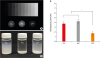 | Figure 3(A) A radiograph showing the radiopacity of each material and its equivalence to that of the aluminum step wedge. (B) Relative radiographic density of each material in comparison with that of a 10-step aluminum step wedge. (C) An image showing the turbidity of the suspension in which the pastes were dissolved. Note that white precipitate formed after sedimentation.
*A significant difference was determined at p < 0.05.
CL, cleaniCal; AC, ApexCal; CP, Calcipex II.
|
Cytotoxicity
In the agar overlay assay, cleaniCal, which uses NMP as a vehicle, showed a higher cytotoxicity compared to the other groups (p < 0.05, Figure 4A and 4B). The MTT assay showed that the cell viability of NMP is significantly lower than that of PEG and PG (p < 0.05, Figure 4C).
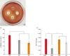 | Figure 4(A) A representative image showing the inhibition zone of each material in the agar overlay assay. (B) Mean and standard deviation of the inhibition zone of each material. (C) Cell viability of the vehicle for each product.
*A significant difference was determined at p < 0.05.
CON, control; CL, cleaniCal; AC, ApexCal; CP, Calcipex II; NMP, N-2-methyl-pyrrolidone; PEG, polyethylene glycol; PG, propylene glycol.
|
DISCUSSION
Intracanal medicaments such as Ca(OH)2 used in clinical practice should be removed from the root canal system because the remnants may have adverse effects on the outcomes of endodontic treatment. Recently, a new Ca(OH)2 product, cleaniCal, which contains NMP as a vehicle, was introduced into the endodontic field with the anticipation of promoting removal efficacy because of its potent solubility. In this study, we investigated the removal efficacy of cleaniCal in comparison with ApexCal and Calcipex II, which use different vehicles such as PEG and PG, respectively. As shown in Figure 2A, in the evaluation with μ-CT, the remaining volume percentage of cleaniCal in the root canal system space was significantly lower than those of ApexCal and Calcipex II (p < 0.05). Similarly, in the stereomicroscopic evaluation, the remaining area percentage of the cleaniCal group was significantly lower compared to the other groups (p < 0.05, Figure 2B). Therefore, it was demonstrated both times that the removal efficacy of cleaniCal, the NMP-based Ca(OH)2 paste, was higher than the other pastes that used PEG or PG as vehicles. NMP is known to possess potent solubility on organic substances and a cleansing effect for various industrial products such as semiconductors. Furthermore, there have been several studies which showed that NMP had better solubilizing efficiency than PEG or PG for poorly soluble compounds [161819].
Commercially available Ca(OH)2 pastes are a kind of colloid in which a substrate of microscopically dispersed insoluble particles including Ca(OH)2 and a radiopacifier are suspended throughout another substance such as a vehicle. The remnants from the Ca(OH)2 paste usually present as a form of precipitate which tends to stick onto the root canal wall and is difficult to remove through the irrigation procedure alone. Consequently, the more precipitates form, the greater amount of remnants remains on the surfaces of the root canal system. Therefore, the use of a vehicle that can dissolve more solid ingredients and generate less precipitates is recommended. In this study, we assumed that the solubility of Ca(OH)2 in NMP was higher than other vehicles because it contained more dissolved solid constituents. We then evaluated the degree of precipitate formation by measuring the turbidity of the supernatant in which the pastes were dissolved. In our results, cleaniCal exhibited significantly higher turbidity than ApexCal and Calcipex II (Figure 3C). This finding suggests that more Ca(OH)2 remained in the suspension of cleaniCal and less precipitate was formed after time-dependent sedimentation. This result may explain the reason why the removal efficacy of cleaniCal was higher than the other products.
NMP has been considered as a safe small chemical molecule [20] and is used as an approved solvent for pharmaceutical products especially implants with acceptable toxicological profiles [21]. NMP is a biodegradable Food and Drug Administration (FDA)-approved solvent listed as generally recognized as safe (GRAS) [22]. Agrawal et al. [23] reported that NMP contained in a drug delivery system for Leuprolide did not exhibit significant toxicity in mice. Recently, it was found that NMP has some biologic activity on the pulp-dentin complex as well as bone metabolism [24], and it is suggested as a promising vehicle for bone morphogenic protein (BMP) and enhances the bone regeneration effect [25]. Nevertheless, there are some controversies regarding the cytotoxicity of NMP. There have been several reports that NMP has certain toxic effects on humans as well as animals [2627]. In addition, the cytotoxicity of NMP as a vehicle for endodontic materials has yet to be evaluated. In this respect, the evaluation of the cytotoxicity of the NMP-based Ca(OH)2 paste is required. In the agar overlay assay, cleaniCal exhibited more cytotoxicity compared to the other products (p < 0.05, Figure 4A and 4B). We also assessed the cell viability of the vehicles including NMP, PEG, and PG, to verify if the vehicles were responsible for the cytotoxicity conducted by the agar overlay assay. As a result, NMP showed a significantly lower cell viability than the other vehicles (p < 0.05, Figure 4C). Therefore, we speculated that the higher cytotoxicity of cleaniCal shown in the agar overlay assay was induced mainly by the toxicity of NMP. Indeed, the cytotoxicity of intracanal medicaments may be an important concern especially when the material is extruded beyond the apical foramen. Kim et al. [28] demonstrated that extruded Ca(OH)2 pastes were widely dispersed into the periodontal tissues and resulted in foreign body granulomas. Kim et al. [29] also showed that overfilling of the root canal system with Ca(OH)2 paste resulted in a chronic foreign body granuloma in the periapex area which propagated to maxillary sinusitis. They indicated that the vehicle such as PG was not easily resorbed and was the causative element of the periapical granuloma and sinusitis. In this respect, the use of a bioinert vehicle is necessary to avoid possible pathological problems when accidental extrusion occurs. In this sense, NMP cannot be considered to be the safer substance as a vehicle compared to PEG or PG.
In this study, we measured the radiopacity of the tested materials and used 2 different methods to evaluate the removal efficacy: 1) μ-CT evaluation and 2) stereomicroscopic observation of the sectioned samples. The μ-CT is considered a useful tool for the evaluation of the remaining amount of root canal filling material. It has been shown that μ-CT gives a highly accurate, non-destructive, and 3-dimensional view of the internal structure of the root [30]. Therefore, there have been many studies using μ-CT for the assessment of the remaining amount of Ca(OH)2 paste [313233]. However, μ-CT has shortcomings that should be taken into account in evaluating the removal efficacy of Ca(OH)2 paste distinct from other root canal filling materials such as gutta-percha or sealer. Generally, the radiopacity of Ca(OH)2 paste, except for iodoform-based material, is lower than that of gutta-percha or sealer. Consequently, the removal efficacy of less radiopaque material can be exaggerated since the remaining material may not be detected in scanned images. This issue should be raised especially when the removal efficacy of the mainly tested material was higher than the compared materials. Notably, in this study, the removal efficacy of cleaniCal which is the protagonist of this study was higher than that of the other supporting materials. Therefore, it was strongly suggested that the radiopacity of the materials be measured. Here, the radiopacity of cleaniCal was significantly higher than or at least similar with that of Calcipex and ApexCal (Figure 3B). Therefore, we can state that the results obtained from the μ-CT evaluation were not influenced by the difference in radiopacity. Furthermore, we double-checked the result by assessing the sectioned specimen stereomicroscopically and the results were comparable to the μ-CT method. Therefore, we believe that the verifications make the results more reliable and predictable.
CONCLUSIONS
cleaniCal, the NMP-based Ca(OH)2 paste, exhibited better removal efficacy compared to ApexCal and Calcipex II which use PEG and PG as vehicles, respectively. This favorable effect may be due to the higher solubilizing efficiency of NMP compared to PEG or PG. However, the cytotoxicity of cleaniCal and its vehicle, NMP, was higher than the other products and vehicles. The null hypotheses of this study were rejected. Considering the distinct advantage in removal efficacy of the material, clinicians should be aware of the higher cytotoxicity of the NMP-based material and take into account its possible adverse effects to periradicular tissue when it is extruded beyond the apical foramen.
 XML Download