

 PDF
PDF ePub
ePub Citation
 Print
Print


Introduction
The determination of correct working length is essential for effective cleaning and shaping and it influences the clinical success of root canal treatment.1,2 Both radiographic and electronic methods have been used for correct working length determination. However, periapical radiographs have many limitations in determining the working length.3 Therefore, in search of more accurate working length measurements, electronic apex locators (EALs) have developed and nowadays are regarded as important tools in clinical endodontic practice and currently used in several clinical conditions.4 Although the function of EALs is consistent in 97% of cases, various factors (root canal pre-enlargement, different endodontic solutions, file size, stage of apex formation, retreatment procedures) affect the accuracy of different EALs.5,6
Although instrumentation procedures have improved considerably over the years, none of the existing techniques can completely clean the root canal system.1 Therefore, besides instrumentation, intracanal medication has been recommended in order to disinfect the root canal system and to improve the success of the root canal treatment, especially in multiple-visit cases.7 Calcium hydroxide (CH) mixed with an appropriate vehicle has become established as the most frequently used endodontic intracanal medicament owing to its good antimicrobial properties against the vast majority of endodontically relevant pathogens and its good biocompatibility.8,9 However, if this medication is not completely removed from the root canal, its presence on the dentin walls could compromise the endodontic treatment.10,11 Hence, CH dressing removal prior to the permanent root canal filling is required.12 The removal of CH is usually accomplished through several irrigation regimens in conjunction with different instrumentation techniques.12,13,14 During the removal, the most frequently described method is instrumentation of the root canal using a master apical file (MAF) and copious irrigation.13,15 However, it has been reported that the removal of CH from the root canal wall is difficult, because instrumentation and irrigation alone cannot completely clean the entire root canal.12,13,15,16
Moreover, no information is available regarding the influence of residual CH on the accuracy of EALs. Thus, the aim of this ex vivo study was to compare the ability of several techniques to remove CH from the root canal and to determine the influence of CH residues on the accuracy of the EAL.
Materials and Methods
Ninety maxillary lateral incisors with straight roots were used. Each tooth was decoronated at approximately the cemento-enamel junction (CEJ) to provide a flat horizontal surface to serve as a stable and unequivocal reference for all measurements. Root canal patency was evaluated using a size 10 K-file to discard any teeth with canal obstructions. After access preparation, a size 15 K-file was inserted into each canal until the tip of the file became visible through the foramen under microscope (×12). The file was then withdrawn until its tip lied tangential to the apical foramen. The silicone stop was adjusted to the nearest flat anatomical tooth landmark chosen as reference for root canal measurement. The distance from the base of the silicone stop to the file tip was measured under ×4.5 magnification with a millimeter ruler to the nearest 0.25 mm. Then 0.5 mm was subtracted from the measurement. Each measurement was repeated three times and the mean value calculated and computed. This value was recorded for each tooth as the reference length and registered as the true working length (TWL).17,18
The root canals were prepared using ProTaper rotary instruments (Dentsply-Maillefer, Ballaigues, Switzerland) up to master apical file F3 (size 30). Between each instrument, the canals were irrigated with 2 mL of 5.25% sodium hypochlorite (NaOCl), using a syringe and a 27 gauge-needle that was placed 2 mm short of the TWL. Prepared root canals were rinsed with 5 mL 17% ethylenediamine tetraacetic acid (EDTA, Vista Dental Products, Racine, WI, USA), followed by a final rinse of 5 mL distilled water, and were dried using paper points. Then, the canals were filled with an injectable CH paste (SurePaste, SureDent Co., Ltd., Seongnam, Korea). Then, radiographs were taken in mesiodistal and bucco-lingual directions to confirm the complete filling of the canals. The access cavities were temporarily sealed with cotton pellet and temporary filling material (Cavit, 3M ESPE, Seefeld, Germany). They were then stored at 37℃ and 100% relative humidity for 7 days. The teeth were randomly distributed amongst six experimental groups (n = 14) and a control group (n = 6) according to CH removal techniques (Table 1).
Electronic measurement was achieved using EAL (Root-ZX, J. Morita Corp., Tokyo, Japan) after performing CH removal techniques described in Table 1. Then, for the electronic working length (EWL) measurement, the test tubes were filled with 0.9% physiologic saline solution as an electrolyte such that the apical third of the roots was immersed into the liquid.19,20 Only one specialist (experienced in the use of EALs) measured the actual and also electronic lengths.17 To avoid bias, the measurements were taken by randomizing the order of the teeth. For the electronic measurements, the same sized (size 30) and the same tapered master apical Ni-Ti file was used with after removal of CH. The termination point used in this study was the red line on the meter designated by the manufacturer as the 'APEX' and measurements were done between 'APEX' and 1.0 mm points (green area). The silicone stop was adjusted to the nearest flat anatomical tooth landmark again and the distance from the base of the silicone stop to the file tip was measured under ×4.5 magnification with a caliper to the nearest 0.25 mm and registered the measured length as EWL. Each measurement was repeated three times and the mean value calculated and computed. The TWL was compared with the EWL. Differences were calculated (TWL - EWL), and tolerance limits of ± 0.5 and ± 1.0 mm were taken.21 A negative difference indicated that EWL was larger and file tip had crossed the foramen. A positive difference indicated that tip was short of foramen. The accuracy of Root-ZX was evaluated in terms of percentages of acceptable measurements (tolerance limit of ± 0.5 and ± 1.0 mm). Chi-square tests were used to compare the percentages (p < 0.05).
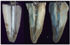
Thereafter, grooves were prepared with a water-cooled diamond bur on the buccal and lingual surfaces and the teeth were split along their long axis in a bucco-lingual direction using a surgical chisel.13 Both halves of the root canal were evaluated under a stereomicroscope (Olympus Corporation, Taichung, Taiwan) at ×40 magnification and photographed digitally. Digital images were imported into image analyzer software (Comef 4.3, OEG Messtechnik, Frankfurt, Germany), and the area occupied by the residual CH on the canal walls was measured (mm2). One-way ANOVA with post-hoc least significant difference (LSD) test was used for statistical analysis of collected data at a significance level of 5%. Pearson's correlation test was done to analyze the correlation between the amount of CH residues and EAL accuracy.
Results
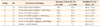
The calculated Root-ZX accuracy (%) and the amount of residual CH (%) are presented in Table 1. There was statistically significant effect of different removal techniques tested on the accuracy of Root-ZX for tolerance limits of ± 0.5 and ± 1.0 mm (χ2 test, p < 0.05). Within ± 0.5 mm accuracy, control group's measurements was statistically different from experimental groups and within ± 1.0 mm accuracy, control and 5.25% NaOCl + MAF groups' measurements were significantly different from other groups (χ2 test, p < 0.05). When the 17% EDTA + MAF and 5.25% NaOCl + MAF were used, EAL was accurate more than 85% of the time to ± 1.0 mm from the apical foramen (Table 1). However, the EAL accuracy was 28.6% of the time to ± 0.5 mm for saline and EDTA without MAF groups. In control group, EWL measurements were always shorter than TWL even when the file extruded from apical foramen.
The 17% EDTA and MAF group and 5.25% NaOCl and MAF group showed better removal efficiency than other groups (p < 0.05, Figure 1). MAF (hand filing) improved the removal efficiency of irrigation solutions (p < 0.05). Control group showed complete coverage of the canal walls with CH remnants densely packed in the canals (Figure 1).
Pearson's correlation coefficient (r) showed that there was strong negative correlation between amount of CH residues and EAL accuracy (r = -0.800 for ± 0.5 mm; r = -0.940 for ± 1.0 mm).
Discussion
The present study was designed to assess the efficacy of different techniques for CH removal by means of a sectioning technique and to evaluate the influence of CH remnants in root canal on the accuracy of Root-ZX apex locator, in vitro. A number of methods for investigating EALs have been used in which extracted teeth were immersed in various media with electrical resistance similar to the periodontium. The advantages of these in vitro models were their simplicity, ease of use and the ability to have strict control over the experimental conditions tested. Furthermore, a greater number of canals can be tested over a shorter period of time than could have been achieved by clinical means.22 The removal of CH from radicular dentin is essential in order to maintain the integrity of the root canal seal.10,23
In the present study, the complete removal of CH from the canal walls was not obtained with any protocol tested. This result is similar to the findings of previous studies tried to achieve the best protocol to remove CH medicament before root canal filling and showed considerable amounts of CH remaining on the canal walls, notwithstanding the removal technique used.14,15,24 Compared with the NaOCl and EDTA-only groups, the combined use of NaOCl and EDTA with MAF (hand filing) improved the removal efficiency. This is in agreement with the results of a previous study, which showed the importance of recapitulation using MAF to improve the removal of CH.25 On the other hand, irrigation only with 17% EDTA performed significantly better results than those of saline and 5.25% NaOCl. This can be explained by the ability of EDTA to dissolve inorganic substances such as calcium.24 Calt and Serper reported complete removal of CH from the root canal after irrigation with EDTA and NaOCl in comparison with NaOCl alone.23 However, a previous study using the same irrigation regime (EDTA and NaOCl) could not confirm these results and still found extensive remnants of CH.15 Similarly, in the present study, although the most successful method of CH removal was EDTA + MAF combination, there is no evidence that EDTA can completely remove CH from the root canal. This difference could be a result of the variable dimensions of the root canal system and their subsequent preparation size and taper, which affects the irrigant penetration.
High reliability index in readings and more precise measurements compared with other apex locators in previous studies were the reasons to use Root-ZX Apex locater in the present study.18,26,27 McDonald recommended the use of files with sizes comparable with the root canal diameter claiming that this would result in more accurate readings.28 Hence, in the present study, the electronic measurements were performed with the same sized and the same tapered master apical Ni-Ti file after removal of CH, while the TWL measurement was done with size 15 stainless-steel K-file. Manufacturers of new-generation EALs claim that these new devices are not affected from the irrigation solutions, and work even in the presence of electrolytes without calibration.29,30 Previous studies reported that with either 5% NaOCl or 14% to 17% EDTA, no interference in detecting the apical foramen was observed.31,32 Goldberg et al. reported that the Root-ZX to be 62.7 to 68.0% accurate within ± 0.5 mm, while Dunlap et al. reported that 82% of electronic values recorded with the Root-ZX were accurate within ± 0.5 mm of the apical constriction when 2.5% NaOCl was used.33,34 In this study, accuracy values of EAL were 50.0 to 64.3% within ± 0.5 mm, when 5.25% NaOCl was used. Against those results, Haffner et al. compared some EALs under in vivo conditions and found Root-ZX more accurate when the root canals were dried with paper points.35 According to the present results, 5.25% NaOCl showed most accurate results amongst the irrigants. Moreover, EAL accuracy were enhanced when MAF combined with irrigation procedure in all groups, except for the control group within ± 0.5 and also ± 1.0 mm. Although CH residues were less in 17% EDTA and MAF group than in 5.25% NaOCl and MAF group, electronic measurements were significantly more accurate in latter group than other MAF combined groups. This can be explained with highly electro-conductive property of NaOCl, resulting in the reduction of electrical impedance of the root canal wall and the better electrical contact with the apical tissues as well.36
Conclusions
Within the limitations of this study none of the irrigants or their combination with MAF filing removed the CH completely. Nevertheless, the results of this study suggest that the addition of hand instrumentation to irrigation is more effective in removing CH from the root canal than irrigation alone. The CH dressing residues affected the accuracy of EAL adversely.
 XML Download