

 PDF
PDF ePub
ePub Citation
 Print
Print


Introduction
The major goal of root canal treatment is to eliminate microorganisms and remove inflamed or necrotic organic tissue.1 Several studies have shown that mechanical instrumentation alone is not able to reduce the microbial population in the root canal system.234 Furthermore, a nonhomogeneous structure, called the smear layer, is formed on dentin surface by the mechanical action of the instruments.5 This layer is comprised of inorganic and organic substances such as fragments of odontoblastic processes, microorganisms, and necrotic tissue.6 The removal of the smear layer could contribute to the disinfection of the root canal system and improve the seal of the root canal filling.7 Therefore, to effectively clean and disinfect the root canal system, irrigation is an essential part of root canal treatment.8
Sodium hypochlorite (NaOCl) solutions are the most widely recommended irrigants in endodontics with regards to their good antimicrobial efficacy and unique capacity to dissolve organic tissue remnants.8 On the other hand, NaOCl is cytotoxic and can cause local tissue necrosis in case of extrusion to the periapical area.9 Therefore, there have been attempts to identify alternative irrigants with the same efficacy as NaOCl, but less toxicity.10 Calcium hypochlorite (Ca(OCl)2) is a powder that is chemically similar to NaOCl, and it is a chlorine source, which forms hypochlorous acid as twice as NaOCl and calcium hydroxide (Ca(OH)2) when mixed with water. Ca(OCl)2 is as widely used as NaOCl for water treatment and as a bleaching agent. To date, only one study evaluated its application in endodontics, and it was found to be as efficient as NaOCl for dissolving organic tissue.11 However, since the smear layer has an inorganic component as well, calcium chelators are used with NaOCl in root canal treatment to ensure its complete removal.12 Ethylenediaminetetraacetic acid (EDTA) has a long-standing history as a chelator agent in endodontics. However, researchers have reported its potential to cause irritation.1314 In addition, several reports have indicated that the sequential use of EDTA and NaOCl may lead to dentinal erosion on the root canal wall.1516 A copolymer of acrylic acid and maleic acid (poly[AA-co-MA]) is a chelator that is obtained by radical copolymerization of maleic anhydride (MA) with acrylic comonomers. In the literature, many data can be found regarding the application of MA copolymers in the medical and pharmaceutical fields. This copolymer is considered to be biocompatible with good chelating properties, and it has the following formula:17
The purposes of this study were to evaluate poly(AA-co-MA) and Ca(OCl)2 in removing the smear layer and debris and to investigate their erosive effects on the root canal wall using scanning electron microscope (SEM).
Materials and Methods
Preparation of the materials
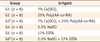
The materials used were 7% Ca(OCl)2 (Sigma-Aldrich Chemie GmbH, Steinheim, Germany), 25% Poly(AA-co-MA) (Aldrich Chemical Co., Milwaukee, WI, USA), 17% EDTA (Vista Dental Products, Racine, WI, USA) and and 2.5% NaOCl. Ca(OCl)2 solution was freshly prepared by dissolving 25 g Ca(OCl)2 granules in 75 mL of deionized water, and then gravimetrically analyzed to determine the exact mass ratio. This stock solution was used after dilution to 7% Ca(OCl)2. Poly(AA-co-MA) (50 wt% solution in water, pH = 1) was diluted to 25% with distilled water.
Selection and preparation of the specimens
This study was revised and approved by the Ethics Committee of Hacettepe University (Project No., GO 13/364). Twenty-four single-rooted human teeth with a single canal were used in this study. The teeth were decoronated at the cementoenamel junction with a diamond bur. Each root canal was negotiated with a size 10 K-file (Dentsply Maillefer, Ballaigues, Switzerland) and the working length (WL) was calculated by subtracting 0.5 mm from this length. Size 15 and 20 K-files (Dentsply Maillefer) were used at the WL and any root with an apical constriction diameter wider than a size 20 file was excluded. Thereafter, ProTaper rotary instruments (Dentsply Tulsa Dental Products, Tulsa, OK, USA) were used in continuous clockwise rotation using a gentle in-and-out motion. SX was used at two thirds of the WL, then Shaping Files 1 and 2 (S1 and S2) and the Finishing Files 1 through 3 (F1 - F3) at the WL with circumferential filing to touch all surfaces. Each set of instruments was used to enlarge 2 canals only. All preparations were performed by one operator. The root canals were irrigated with distilled water between each instrument. After instrumentation, the apical and coronal thirds of each root were removed with carborundum discs. Two longitudinal grooves were prepared on the buccal and lingual surfaces without exposing the root canals. Thereafter, the 5 mm middle third sections were split into two equal pieces with a hammer and chisel.
Distribution of specimens and final irrigation
Forty-eight samples were randomly divided into 6 groups and subjected to the irrigants as described in Table 1. A total volume of 2 mL of each irrigant was used for each specimen. A final flush was done with 2 mL of distilled water in all samples. In combination groups, 2 mL mid-irrigation with distilled water was applied to prevent possible interaction between the solutions. Each irrigant was applied for 5 minutes.
SEM examination
After irrigation procedures, the specimens were kept in an incubator at 37℃, then mounted on stubs, gold-sputtered and examined with SEM (JSM-6400, JEOL Ltd., Tokyo, Japan) at magnification of ×300, ×1,000 and ×3,000. It is well known that SEM evaluation is subjective and qualitative, as the selection of the observation area is directly operator-dependent. Thus, to standardize the examined area for each sample, we applied the technique described by Paque et al.18 For this purpose, the central beam of the SEM was directed to the center of the specimen under ×10 magnification, and the magnification was increased gradually to ×3,000. The area of the canal wall captured on the screen of the SEM was used for scoring the sample.
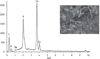
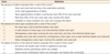
The scores and scales of the presence of debris, smear layer and degree of erosion of dentinal tubules were presented in Table 2.1920 The scoring was done by two independent examiners. When a difference occurred in the scoring of an image, the two examiners reevaluated the image and discussed it until an agreement was obtained. One image was analyzed with energy dispersive spectroscopy (EDS) on suspicion of precipitate formation.
Results
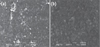
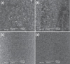
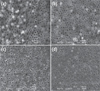
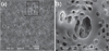
The smear layer, debris and erosion scores are presented in Table 3. G1 and G4 showed the presence of debris, and heavy smear layer and there was no significant difference between them (Figure 1). The smear layer and debris were totally removed in G2, G3, G5 and G6 (Figure 2). The results indicated that G1 and G4 were statistically different from the remaining groups in removing debris and smear layer (p < 0.05). In G1 and G4, scoring for erosion could not be done because of the presence of debris and heavy smear layer. G2, G3 and G5 did not show any erosion, and there was no significant difference between them (Figure 3). G6 showed severe erosion and was statistically different from G2, G3 and G5 (p < 0.05, Figure 4). EDS microanalysis of a sample from G3 showed the presence of Na, P, and Ca elements on the surface of suspected precipitate (Figure 5).
Discussion
While there are no evidence-based studies that provide outcomes of treatment based solely on the removal of the smear layer, it seems reasonable to suggest that removal of the smear layer can result in a more thorough disinfection of the root canal system and the dentinal tubules, which would ensure a better adaptation between the obturation materials and the root canal walls.7 Several studies have shown that the use of a combination of sodium hypochlorite (2.5 - 5%) and EDTA (10 - 17%) is particularly effective in the removal of smear layer and debris.152122 However, recent research seems to demonstrate an excessively aggressive effect of this combination on the root canal walls that could cause too much erosion and degradation of the dentin.71523 Although its effect on the long-term success of endodontic treatment remains unclear, erosion could result in an alteration of mechanical properties of dentin and create more difficulties in the adaptation of the root filling materials to the canal walls.2425
It is a well-established point that the smear layer is created only where the endodontic instruments cut the dentinal walls effectively.26 On the other hand, with both rotary nickel-titanium instruments and traditional stainless-steel hand instruments almost half of the root canal walls are left uninstrumented.1827 This can become an issue when examining a surface that belongs to a smear-free area. The reason of this issue is that the smear-free area could be produced by the chelating solution or it may merely indicate an uninstrumented area.26 This situation may cause inaccurate results in SEM examinations. In order to eliminate any potential problems that may develop due to uninstrumented areas, we chose to use 5 mm mid-roots in the present study, because it was reported previously that there is less untouched surface in the middle third of the root after root canal preparation.28 Furthermore, this laboratory study was aimed to analyze the effects of two novel irrigants on dentin structures. Therefore, to ensure a uniform and direct contact of each irrigant with the root canal walls, final irrigations were performed after 5 mm mid-roots were divided into two pieces longitudinally. Although this approach does not represent the clinical situation, it is a useful method when evaluating the effects of a potential irrigant on dentin surface regardless of any canal variations.
A review of irrigation in endodontics revealed that optimal irrigation is commonly achieved by combining two or more irrigant solutions in a specific sequence.8 However, there is a concern that combined irrigation regimens may cause undesirable interactions between irrigants such as creation of erosion, loss of activity and development of potentially toxic by-products.29 To avoid such interactions, we used distilled water between two irrigants in each of the combination groups. Nevertheless, we found that EDTA and NaOCl caused erosion around dentinal tubules in 5 minutes when used sequentially, whereas no erosion was observed in the combination group of novel irrigants. Although we used distilled water between irrigants, it was found that some precipitate can occur even when distilled water is used between the specific irrigants.29 Therefore we considered this fact in the present study, as the tested materials were novel irrigants, and only one specimen from the combination group of novel irrigants showed an unusual view on its surface and was analyzed further with EDS. According to the analysis Na, P, and Ca elements which are the components of dentin were detected in the area indicating no chemical precipitate. More studies are required to examine whether a precipitate occurs in the contact of these two novel irrigants.
Poly(AA-co-MA) is a polymer with numerous applications for biomedical purposes. Their applications as drugs, drug-conjugates, enzyme-conjugates or gene delivery systems are also reported.17 It was reported that in pharmaceutical forms, poly(AA-co-MA) was able to improve mucosal permeability by its chelating abilities similar to EDTA.30 According to our results, this copolymer demonstrated promising results as a novel and potential irrigant. Poly(AA-co-MA) produced entirely clean surfaces without damaging the dentinal surface when it was used alone or combined with Ca(OCl)2. Our results also showed that Ca(OCl)2 was ineffective in removing the smear layer, which was also the same for NaOCl. The inability of NaOCl to dissolve the inorganic structures has been well documented in the literature.8 Considering the similar chemistry of these two chlorine products, our study also indicated that Ca(OCl)2 was ineffective on inorganic structures. On the other hand, a recent study concluded that Ca(OCl)2 was as effective as NaOCl in dissolving organic tissues, but with a slower rate which could contribute less tissue irritation in cases of periapical extrusion.11 When NaOCl and Ca(OCl)2 are mixed with water the following reactions occur:
Ca(OH)2 and the higher generation of hypochlorous acid may enhance the antimicrobial efficacy of Ca(OCl)2. Furthermore, its effect on organic tissue makes it a potential root canal irrigant.11 However, many aspects of this irrigant, such as its antimicrobial effectiveness, cytotoxicity and the interactions with other root canal irrigants should be investigated.
Conclusions
Poly(AA-co-MA) is effective in removing the smear layer and debris without causing erosion either alone or with Ca(OCl)2. Both irrigants have the potential to be used as irrigants in endodontics. More studies are necessary to further investigate the properties of these two novel irrigants.

 XML Download