

 PDF
PDF Citation
 Print
Print


INTRODUCTION
Dysphagia after stroke is a common disabling problem and is estimated to occur in 22%–78% depending on the method used and timing after stroke [1234]. Dysphagia is one of the significant risk factors for aspiration pneumonia in stroke patients and aspiration pneumonia increases mortality and the length of hospitalization [4]. Therefore, early detection of post-stroke dysphagia can decrease the incidence of aspiration pneumonia, which is an important part of acute stroke management [5].
Dysphagia can be detected with various diagnostic methods, which include various bedside swallowing screening tests (BSST), a formal instrumental test such as videofluoroscopic swallowing study (VFSS), and fiberoptic endoscopic evaluation of swallowing (FEES).
Bedside tests are safe and easily repeatable test [6], but has variable sensitivity (42%–92%), specificity (59%–91%), and inter-rater reliability [7]. Meanwhile, VFSS is a more reliable test and still a standard test for detecting dysphagia [89]. During VFSS, an examiner can trace the process of swallowing from the oropharyngeal to esophageal phases with various food consistencies and subject posture. The examiner can also determine the management plans such as optimal food consistency and posture for patients. Aspiration detected during VFSS in the early stage of stroke is an important risk factor for developing subsequent aspiration pneumonia during the subacute stage of stroke [10].
Although VFSS has advantages over bedside tests in terms of test accuracy and in determining a management plan, it also has drawbacks. For VFSS, patients should be transferred to a room equipped with videofluoroscopy and have the ability to sit up and be cooperative during the procedure, which might be difficult for a patient in the very early stage of stroke. An example would be when a patient is in a stroke unit. This limitation can force physicians to decide on a post-stroke dysphagia management plan based on BSST alone. As a result, physicians tend to be more conservative in determining the diet level than in a determination based on VFSS, which is especially true for high-risk patients.
There is no established guideline for reassessment of swallowing function in post-acute period. The need for reassessment was highlighted in a recent study [11]. Heckert et al. [11] retrospectively reviewed the medical records of 146 stroke cases admitted to their inpatient rehabilitation facility, and compared the swallowing function during post-acute stroke rehabilitation care with initial swallowing assessment during acute care setting. The authors reported that 11% of subjects were newly identified as having dysphagia and, following reassessment, 12% required more conservative diets than prescribed in the acute stage. However, the necessity for swallowing reassessment as a part of routine evaluation during post-acute stroke rehabilitation care is not fully established yet, and the authors proposed prospective observation to establish a standard.
Previous prospective studies comparing bedside tests and VFSS were small sized with a heterogeneous stage and reassessment interval [1213] or performed within the short term which could not reflect the practical interval between acute and sub-acute phase of the stroke treatment process [1415]. Therefore, the purpose of this study was to prospectively compare the test outcome of a standardized bedside swallowing screening test in the acute period with that of VFSS in the sub-acute period with a fixed reassessment interval.
We hypothesized that the need for changing the diet modification plan would still exist after reassessment, justifying the necessity of routine post-acute reassessment of swallowing function with VFSS.
MATERIALS AND METHODS
Patients
This was a prospective observational study of consecutive patients with first-ever ischemic or hemorrhagic stroke admitted to our stroke center located in a tertiary university hospital from May 2009 to May 2010. The diagnosis of stroke was confirmed by brain magnetic resonance imaging (MRI) in all cases. A total of 550 stroke patients were admitted during the study period. Of these, 207 patients who had previous stroke and 91 who needed intensive medical care such as mechanical ventilation due to unstable medical conditions were excluded from the study. Two hundred fifty-two patients underwent the initial BSST. Forty-nine subjects were further excluded because they were discharged early from the hospital or lost to follow up (n = 47), or died (n = 2) before VFSS evaluation. Thirteen patients refused to undergo a confirming VFSS after the initial BSST, and four patients could not finish VFSS due to poor cooperation or inability to tolerate the procedure. Finally, 186 patients were analyzed in this study. Among them, 182 patients undertook VFSS during hospital stay, and the remaining four patients revisited for a VFSS after being discharged (Fig. 1). General characteristics of the stroke patients are summarized in Table 1.
Fig. 1
Flow chart of stroke patients eligible and included for analysis. A total of 550 stroke patients were admitted to stroke center. Among them, 186 patients underwent both BSST and VFSS.
BSST, bedside swallowing screening test; VFSS, videofluoroscopic swallowing study.
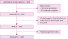
Table 1
General characteristics of the 186 stroke patients
*Refer to the Supplementary Data for details.
BSST, bedside swallowing screening test; SD, standard deviation; n, number.
Initial BSST
Our stroke center’s protocol is to perform initial BSST within 48 hours of admission. Initial BSST was performed by consulting rehabilitation physicians using a standardized protocol (see Supplementary Data for detailed protocol). Until initial BSST, all patients were kept nil per os (NPO).
All the patients analyzed in this study fulfilled our protocol. Briefly, maintaining nasogastric tube or commencing diet were decided based on flow diagram assessment, which sequentially evaluated patients’ mental state, as well as dry and water swallowing ability. Patient risk was classified as low, medium, and high according to the results of wet swallowing test (see Supplementary Data). In detail, subjects in this study were instructed to swallow 20 mL of water and following 4 factors were checked: 1) delayed swallowing (> 2 seconds) or swallowing not possible, 2) cough response (until 3 minutes later), 3) drooling, 4) wet voice change. Number of factors observed was used for risk categorization (no factor: low risk, one factor: medium risk, 2 or more factors: high risk). Only low and medium risk group patients were allowed to have oral diet before formal VFSS. Other patients were kept on nasogastric tube until a formal VFSS was conducted for a dysphagia management plan. Risk classification of patients is summarized in Table 1. BSST was not possible in 10.2% of the patients due to poor mental status and 3.2% of patients were kept on nasogastric tube because they failed the dry swallow test. Patients at high, medium, and low risk were 10.8%, 13.4%, and 62.4%, respectively.
VFSS
VFSS was performed 9.2 ± 7.2 days after initial BSST. All patients were reassessed within 3 weeks, and 86% were re-evaluated with VFSS within 2 weeks after initial BSST. VFSS was performed according to a modification of a published protocol [16]. Patients were seated upright in a chair for the duration of the study and were given 2 mL and 5 mL of diluted barium (35% weight/volume), honey and nectar-like thickened liquid, curd-type yogurt as a liquid diet, and boiled and normal rice as solid diet. When it was considered safe to do so, patients were given a cup with 30 mL of diluted barium through uninterrupted swallows. When the examiner found the subglottic aspiration on diluted barium or thickened fluid, VFSS was suspended without further testing of solid diet. The penetrating voltage of the X-ray was set at 40 kV peak to facilitate viewing of the soft tissues of the oropharyngeal structures. Images were recorded as a digital movie file for later review. The results of VFSS was analyzed using the penetration-aspiration scale (PAS) [17]. The results compatible with PAS score 5 which means ‘Contrast contacts vocal folds; visible residue remains’ were defined as penetration. Based on the same criteria, patients were classified as being aspirated if the PAS score was over 5 which means ‘Contrast passes glottis.’
Between BSST and VFSS, all patients were managed according to our hospital’s routine dysphagia management protocol, which includes diet modification, posture education, oropharyngeal exercise, and pharyngeal electrical stimulation when applicable. The study protocol was approved by the Institutional Review Board of (our blinded) hospital.
Statistical analysis
We examined change in diet prescription according to findings of VFSS reassessment. Using a χ2 goodness-of-fit test, we compared actual change with the predicted change, which reflected expected recovery estimate, based on the published literature concerning the natural recovery of dysphagia [18] and the results of a post-acute reassessment [11].
RESULTS
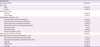
Table 2 summarizes dysphagia identification at initial BSST and follow-up VFSS. At initial BSST, 70 patients (38%) were considered to have dysphagia. At follow-up VFSS, aspiration was detected in 46 patients (25%). Among them, 16% (16/97) were new aspirators who were not considered to have dysphagia at initial BSST. In other words, 14% (16/116) of patients who passed the initial BSST revealed new aspiration during follow-up VFSS. Meanwhile, among the patients who were considered to have dysphagia at initial BSST, 24% (17/70) showed a near-normal finding at follow-up VFSS, suggesting spontaneous recovery due to time delay in the assessment from BSST to VFSS or improvement in dysphagia, or mismatch between the two tests.
Table 2
Dysphagia at initial BSST and follow-up VFSS

| Follow-up VFSS | |||||
|---|---|---|---|---|---|
| Dysphagia (+) | Dysphagia (−) | Total | |||
| Aspiration | Penetration/residue | ||||
| Initial BSST | Dysphagia (+) | 30 | 23 | 17 | 70 |
| Dysphagia (−) | 16 | 28 | 72 | 116 | |
| Total | 46 | 51 | 89 | 186 | |
In the present study, the sensitivity, specificity, positive predictive value (PPV), negative predictive value (NPV), and positive likelihood ratio of BSST for detecting aspiration were 65.2% (30/46), 71.4% (100/140), 42.9% (30/70), 86.2% (100/116), and 2.28, respectively. The respective values for identifying dysphagia (considering for both aspiration and penetration/food residue) were 54.6% (53/97), 80.9% (72/89), 75.7% (53/70), 62.1% (72/116) and 2.86.
Changes in recommendations for solid diet and fluid consistency after VFSS as compared to initial BSST are summarized in Tables 3 and 4.
Table 3
Changes in solid diet recommendation after VFSS

Terminology was adopted from ‘Australian standardized labels and definitions’ [30].
VFSS, videofluoroscopic swallowing study; BSST, bedside swallowing screening test.
Table 4
Changes in recommendation for fluid consistency after VFSS

Terminology was adopted from by Logemann [16].
VFSS, videofluoroscopic swallowing study; BSST, bedside swallowing screening test.
Agreement in prescribed diet between BSST and VFSS occurred in only 47.8% of patients (59.1% for solid diet recommendation and 50.5% for recommendation for fluid consistency). Whereas diet level was more conservatively downgraded in 28.0% of patients (19.4% for solid diet, 26.3% for fluid), the diet level was upgraded in 23.1% of patients (21.5% for solid diet, 23.1% for fluid) after follow-up VFSS (Table 5).
Table 5
Changes in diet recommendation after VFSS

| Changes in diet recommendation | Upgrade | Downgrade | Same | Other | |
|---|---|---|---|---|---|
| Changes according to diet category | |||||
| Solid diet, n (%) | 40 (21.5) | 36 (19.4) | 110 (59.1) | 0 (0.0) | |
| Fluid, n (%) | 43 (23.1) | 49 (26.3) | 94 (50.5) | 0 (0.0) | |
| Overall, n (%) | 43 (23.1) | 52 (28.0) | 89 (47.8) | 2 (1.1) | |
| Changes in previous studies | |||||
| Heckert et al. [11], n (%) | 53 (36.3) | 18 (12.3) | 71 (48.6) | 4 (2.7) | |
| Finestone and Greene-Finestone [18], (%) | (36.0) | (0.0) | (64.0) | (0.0) | |
DISCUSSION
The main finding of this study is that many patients who successfully pass the initial BSST still show aspiration in subsequent VFSS (14%), or need to change diet to more conservative level after VFSS (28%). The findings justify the necessity of VFSS as a routine standard evaluation of post-stroke dysphagia, even though patients have already undergone BSST during the acute stage of stroke.
In this study, the incidence of dysphagia detected by VFSS at first onset stroke was 52%, which was comparable to previously published data [119]. VFSS performed at median time of 8 days after onset identified aspiration in 24.7% and penetration/food residue in 27.4% of stroke patients. A previous report involving patients who underwent videofluoroscopy within a median time of 2 days after stroke onset reported comparable results of aspiration (22%), while penetration was reported in 46% of patients [2]. In another study, in which 95% of patients were examined within 2 weeks of onset, 26% of patients were judged to have aspiration [14].
Mann et al. [20] prospectively studied 128 stroke patients with clinical and videofluoroscopic swallowing assessment within a median time of 3–10 days after stroke onset. Clinical swallowing assessment identified aspiration in 50% of patients and videofluoroscopy identified aspiration in 22% and penetration in 45% of patients [20]. In our study, 25% of patients were identified as having aspiration in VFSS, which is comparable to their report, although our prevalence of penetration/residue (27%) was lower than their report. The relative high false positivity (17/70, 24%) in our BSST was probably due to the fact that we regarded patients with drowsy mentality as having dysphagia without testing water swallowing function.
In terms of clinical assessment, dysphagia was observed in 37.6% (70/186) of patients within 2 days from onset. These results were also comparable with previous studies (39%–41%) in which clinical assessments were performed 1–7 days from the onset [221].
Previous studies used “failure on the 50 mL (or 85 mL) water swallowing” as an accurate indicator for aspiration [1422]. In our BSST, we used a combination of clinical findings as an indicator for dysphagia; pass or fail to 20 mL and/or 3 mL thin liquid swallowing test, presence of wet/gurgly voice, drooling, and cough. We used 20 mL fluid test rather than 50 mL, because we felt 50 mL was too challenging for patients immediately after stroke onset.
McCullough et al. [14] previously evaluated the clinical swallowing assessment tool, which is similar to the BSST. They suggested that the clinical assessment had a relatively low sensitivity as compared with specificity, and that too many screening assessment combinations would render the bedside screening test difficult in ruling out aspiration. The result would be a tendency to recommend a more conservative diet strategy.
Two studies surveyed clinician’s decision-making practices based on clinical and instrumental assessment for dysphagia [2324]. In one study, 89% of clinicians felt that an instrumental evaluation, mostly VFSS, was needed when clinical signs of aspiration were noted during commencement of an oral diet, and 78% of clinicians felt the need for VFSS when clinical findings of oro-pharyngeal swallowing dysfunction without definite aspiration symptoms were observed. More experienced clinicians were less apt to recommend an instrumental test with marginal significance. Given that 14% of non-aspirators at initial BSST were subsequently revealed as an aspirator at follow-up VFSS in our study, the necessity for undertaking VFSS, even after skilled BSST, is justified.
Heckert et al. [11] reviewed the medical records of stroke patients in a rehabilitation program who had been referred from an acute care hospital. They retrospectively compared initial dysphagia assessment in the acute care setting with dysphagia reassessment results in the rehabilitation setting. Although they did not detail the dysphagia as aspiration or penetration, the proportion of dysphagia in BSST (38%) and follow-up VFSS (52%) in our study were less than their report; in acute care (66%) and in post-acute phase reassessment (64%). They indirectly judged the presence of dysphagia in the acute setting by retrospective medical record review on previously prescribed diet, rather than using the VFSS or BSST reports. This could explain the discrepancy in prevalence of dysphagia at acute phase between two observations.
In our study, the percentage of patients in whom diet recommendation was downgraded towards a more conservative way was higher and diet upgrades was lower than in the study of Heckert et al., considering comparable age, days from screening to VFSS between two studies, suggesting we were more conservative in terms of diet management plan (Table 5) [311]. One way χ2 Goodness-of-fit tests showed statistically significant disagreement between our and the results of Heckert et al. [11] at the level of α = 0.01 of significance. Heckert et al. [11] recommended diet upgrades in 36.3% of patients, which seemed to be attributable to spontaneous recovery of post-stroke dysphagia during the days from onset to admission to their rehabilitation facility, judging from previously published data regarding spontaneous recovery of post-stroke dysphagia [18].
Given the dichotomies between studies, the question of when is the optimal time for formal VFSS remains open. It would be reasonable to screen swallowing function with VFSS as soon as VFSS is feasible and the patient can tolerate the procedure. In a prospective follow-up study after stroke, 13.4% of patients developed aspiration pneumonia and all of those patients experienced the first episode of pneumonia within the first month after stroke onset [25].
Wilkinson et al. [26] suggested that clinicians should consider insertion of a percutaneous endoscopic gastronomy (PEG) tube when patients cannot tolerate spoon-thick fluids or a puree diet 14 days after stroke. They reported that chance of needing a PEG insertion rose from 29% on day 7 to 50% by day 14 when patient could not tolerate a puree diet.
This study was designed as a prospective study and all first-stroke patients who could bear the BSST were tested. However, we could not reassess 25% of the patients using VFSS who were initially evaluated with BSST because they were discharged earlier than VFSS arrangement or refused to enroll. Patients who dropped out were graded as low risk (n = 53), medium risk (n = 1), high risk (n = 2), not in good state (n = 3), and a mild drowsy state (n = 1) upon initial BSST. If we included these patients, specificity and NPV would have been improved.
In our study VFSS evaluators were not totally blinded to the initial BSST results, which might have biased the interpretation of VFSS findings, and test items were limited to water because our screening sheet was designed to briefly identify whether dysphagia was present or not. A screening test using various consistencies/textures of liquids and solids, such as Gugging Swallowing Screening [27] could render more sophisticated diet management strategies [28] at the expense of more time [29].
CONCLUSION
Dysphagia is one of leading causes of morbidity after stroke and early detection of dysphagia may prevent the consequent aspiration pneumonia. To avoid complications due to undetected aspiration or unnecessary dietary restriction for fear of complication, a tailored management plan in an appropriate time is essential. However, a standardized evaluation pathway along the course of stroke recovery has yet to be established. BSST is an initial gateway to identify dysphagia, but there are still discrepancies between the BBST and VFSS. In our study, in 14% of patients who passed initial BSST, aspiration was still detected in follow-up VFSS, and diet recommendation was changed in 51% of patients following VFSS. The present findings support the need for reassessing swallowing function using VFSS, even if the initial BSST was performed during the acute period of stroke.
 XML Download