Journal List > Brain Neurorehabil > v.9(2) > 1094167

Yu, Kim, Seo, and Cho: Brain Plasticity and Neurorestoration by Environmental Enrichment

Abstract

In the adult mammalian brain, neural-lineage cells are continuously generated in the subventricular zone (SVZ) and dentate gyrus of the hippocampus. These cells in vivo arising from the adult SVZ may be regulated by environmental enrichment (EE). EE is a method of raising animals in a huge cage containing novel objects, running wheels and social interaction with a complex combination of physical, cognitive, and social stimulations. EE can affect neural plasticity via overexpression of growth factors such as brain-derived neurotrophic factor (BDNF), vascular endothelial growth factor (VEGF), insulin-like growth factor-1 (IGF-1), fibroblast growth factor-2 (FGF-2), and synaptic activity-regulating genes. EE also have advanced effects on brain functions including the enhancement of motor and cognitive functions in normal and pathological states. Additionally, behavioral changes by EE are related with molecular changes including neurogenesis, gliogenesis, angiogenesis, axonal sprouting, and dendritic branching in the adult brain. In this review, we focus on brain plasticity and neurorestoration associated with molecular changes of neurotrophic growth factors such as BDNF, VEGF, IGF-1, FGF-2 and synaptic activity-regulating genes that occurs in interaction to EE.

Highlights

  • Environmental enrichment (EE) is a method of raising animals in a huge cage containing novel objects, running wheels and social interaction.

  • EE can affect neural plasticity via overexpression of neurotrophic growth factors and synaptic activity-regulating genes.

  • We focus on brain plasticity and neurorestoration associated with molecular changes that occurs in interaction to EE.

INTRODUCTION

Rearing animals specially in rodents have generally been treated by environmental enrichment (EE) to stimulate the effects of a complex combination of physical, cognitive, and social interaction [12]. EE is designed for enhancing social interactions, sensory and motor stimulation and learning and memory in a larger cage than standard cage, and this cage contains a variety of toys such as tunnels, platforms, and running wheels [34] (Fig. 1).
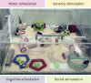
Fig. 1
Representative finding of environmental enrichment.
An enriched environment was provided in a huge cage (86 × 76 × 31 cm3) containing various objects, such as tunnels, shelters, toys, and running wheels for voluntary exercise, and allowed for enriched social interaction (approximately 10–15 mice/cage) with a complex combination of motor, sensory, cognitive, and social stimulations.
bn-9-e2-g001
Sensory, motor, and cognitive stimulation, interaction by EE are important elements in enhancing and adapting the neuronal system for the normal brain function [5]. Biochemical facilitation and morphological changes can promote functions in animals with brain injury [167]. Previous studies have been shown that the surrounding environment causes and stimulate many plastic responses in the adult brain, varying from biochemical parameters to dendritic arborization, gliogenesis, and neurogenesis [189]. EE significantly stimulates neuronal components such as cell body and size of nucleus, dendritic branching, dendritic spine density, and the amount of synapses [10111213].
Lately, researches have been focused on neural stem cells and neurogenesis because the neuronal replacement is the key point of brain reformation in the adult vertebrates [14]. Since EE supports the survival rate of the progeny cells of neuronal precursor in the hippocampus, the volume of the hippocampus expends in an animal model by increasing the number of neurons in the hippocampus [15]. An adult hippocampal neurogenesis is an active progression that can be induced by physiological and pathological causes. EE- or injury-induced hippocampal neurogenesis suggests common pathways of regulation by lesion and by environmental interventions [16].
EE can affect neural plasticity via overexpression of neurotrophic factors and neurotransmitter receptors [171819]. Several studies have shown that, in a brain lesion, brain-derived neurotrophic factor (BDNF), vascular endothelial growth factor (VEGF), insulin-like growth factor-1 (IGF-1), or fibroblast growth factor-2 (FGF-2) mediate the neurogenesis and angiogenesis promoting effects of EE [5710202122]. These growth factors are essential mediators of the effects of physical activities and EE in brain plasticity [23]. Moreover, EE have been reported as the treatment that can improve neurological functions in several animal models such as hypoxic–ischemia brain injury [7], and traumatic brain injury [24], Alzheimer’s disease [25], and Parkinson’s disease [2627]. Therefore, EE is considered as an advanced treatment to counteract cognitive and somatosensory deficits in neurological disorders with following brain injury [28]. Similarly, EE is known to have advanced effects on brain functions including the enhancement of motor and cognitive functions in normal and pathological states. The behavioral changes are related with these molecular changes [2930].
In this review, we will explain the brain plasticity and neurorestoration associated with molecular changes of neurotrophic growth factors such as BDNF, VEGF, IGF-1, FGF-2 and synaptic activity-regulating genes that occurs in interaction to EE.

BRAIN-DERIVED NEUROTROPHIC FACTOR (BDNF)

Function of BDNF is important in synaptic plasticity [3132] and previous studies report that its expression is changed following EE [533]. Physical exercise exposure has been shown to increase object recognition memory and to encourage BDNF expression in rats [3034], and the relations of EE and exercise on sensory gating and spatial memory has been formerly reported in rodents [3035]. In rodents, EE can improve memory and long-term potentiation (LTP) [4]. Another study described that BDNF is increased following EE, and this EE-induced BDNF goes with the enhancement of the object recognition memory and the improvement of LTP [5]. BDNF has been found to play a key role in the late phase of LTP, the best known cellular plasticity occurrence in the brain [363738], which is thought to be one of the plastic occurrence that underlies learning and memory [394041]. Especially, the hippocampus appears to be a specifically sensitive region to EE [42]: enriched animals show enhanced LTP [434445], improved learning and memory [464748], recovered behavioral functions after ischemia [49] and rescued memory deficits related to interruption of N-methyl-D-aspartate (NMDA) receptors [4546].
BDNF is also required for EE-mediated improvements of hippocampal neurogenesis [50]. Exposure of rodents to EE is an useful method for increasing both the adult hippocampal neurogenesis [155152] and the survival ratio of new-born neurons in the dentate gyrus [5354]. Since several studies described that neurogenesis and the survival of new-born neurons are connected to synaptic plasticity and memory function through BDNF/TrkB signaling [555657], it is likely that effects of EE on synaptic plasticity and cognition are enabled by these actions [5].

VASCULAR ENDOTHELIAL GROWTH FACTOR (VEGF)

The function of VEGF in the central nervous system (CNS) is comprehensive. Apart from its angiogenic effect in developmental and pathological angiogenesis [58], it has been described to have neuroprotective, neurotrophic and neurogenic effects [59606162]. Among these effects, angiogenesis is importantly associated with functional outcomes. At the same time, angiogenesis is essential for the repair of ischemic brain and functional recovery because it encourages blood flow and metabolism in the peri-ischemic region [2863]. Moreover, EE stimulated angiogenesis on cerebral cortex in an ischemic rat model. EE also encouraged the recovery of motor coordination and motor skills, and improved performances of learning and memory [28].
In addition, numerous studies have been reported that VEGF, as a direct mediator, improved cognition function in rodents [646566] and induced long-term synaptic enhancement in hippocampal neurons [67]. VEGF encourages hippocampus-dependent memory independently of its effects on neurogenesis and angiogenesis, by increasing the synaptic strength [6869]. Recent studies have been reported that short-term exposure to EE is also enough to alter synaptic plasticity and molecular markers related to the cognitive function [4]. EE-facilitated LTP was dependent upon NMDA receptors. These changes in synaptic plasticity occurred with raised levels of phosphorylated cAMP response element-binding protein (CREB) and VEGF [4].
Another study reported that hippocampal neurogenesis is responsive to VEGF expression by environmental inducements with enriched shelter and performance in a maze for the hippocampus-dependent test. This potential mechanism was confirmed by inhibiting VEGF expression by RNA interference, which entirely obstructed the environment induced neurogenesis [1664].

INSULIN-LIKE GROWTH FACTOR-1 (IGF-1)

IGF-1 has functions such as the control of cell proliferation, gliogenesis, neurogenesis, neuron survival, differentiation, synaptogenesis, and myelination in prenatal and postnatal CNS development [70717273]. Lately, neuroprotective effects of physical activities including exercise and EE support IGF-1 to regulate neuronal death [747576], the enhancement in hippocampal plasticity, and learning and memory in the adult [73777879]. The function of IGF-1 on both synaptic plasticity and hippocampal neuronal replacement seems mainly relevant to display such cognitive reserve. Like this, serum IGF-1 is necessary in the adult brain not only for both basal hippocampal neurogenesis and exercise and EE-induced increases of neurogenesis [22], but also for synaptic plasticity and cognition [23]. Recent study described serum IGF-1 has a direct role in cognition, although some long-term behavioral effects of exercise are IGF-1 independent at the same time [80].
Running also stimulates uptake of IGF-1 by certain groups of neurons enhancing electrical activity [76] and increases IGF-1 expression in the hippocampus [79]. Remarkably, EE has been shown to upregulate IGF-1 receptor gene in the adult hippocampus and sensorimotor cortex [81]. Especially, early exposure to EE could increase IGF-1 and BDNF levels in the brain, thus affecting hippocampal glucocorticoid receptor expression and stress response in adult brain [82].
In addition, IGF-1 increases astrocyte intercellular gap junctional communication [83], promotes dendritic arborization [84], regulates the rate of neurogenesis [23], and increases oligodendrogenesis [85]. Many studies described that learning and memory respond to IGF-1 after experience- or activity-induced events such as physical exercise and EE. All these actions, together with the above-mentioned evidences about the function of IGF-1 regulating diverse membrane channels, many neurotransmitter receptors, and neurotransmitter release, point to a control of critical aspects of neuronal excitability and, therefore, of the neuronal integrative capacities [2386].

FIBROBLAST GROWTH FACTOR-2 (FGF-2)

FGF-2 is existed throughout the CNS, and many previous studies reported that FGF-2 is important for the survival, reproduction, and differentiation of cells [878889]. The function of FGF-2 as a mediator on the brain is supported by demonstrations that FGF-2 is not only a solid proangiogenic factor [90] but also a neurotrophic factor which is generally produced by astrocytes in the adult brain [21919293]. Previous study reported that expression of FGF-2 was expressively raised in mice exposed to EE compared with exposed to standard cage, causing that the upregulation of FGF-2 might be a key factor in functional recovery after hypoxic-ischemic brain injury [7]. In addition, increasing FGF-2 protein by intraventricular infusions induced increased hippocampal neurogenesis [9495]. The studies showing a FGF-2 dependent neurogenic response in the dentate gyrus and subventricular zone (SVZ) from ischemia are according to a condition for injury and/or inflammation facilitating FGF2-mediated neurogenesis [72196].
The elevated expression of FGF-2 induced by EE improved neurogenesis and angiogenesis in animal models of chronic hypoxic-ischemic brain injury [721]. Physical activities including exercise and EE are interesting to speculate that these factors act at least in part by stimulating FGF signaling [97]. The previous study described that FGFs directly affected on hippocampal stem cells [94] and that the decreased neurogenesis with age is not only due to a decreased ability of stem cells to generate new neurons or respond to FGF signals, but also a decrease in the availability of ligands in their environment [97]. In addition, FGF-2 have a potent ability of chemotactic factor for endothelial cells [98], and it showed the function of regulating recovery from cerebral brain injury [99100]. FGF-2 improves sensorimotor deficits and to decrease infarct size following cerebral ischemic brain injury in adult rats [101], and neutralizing antibodies to FGF-2 blocks recovery from motor cortex lesions [102].
Since FGF-2 is initially essential for migration of neural progenitors [103], neuronal recruitment into the striatum may have been potentiated by increased FGF-2 [21]. Moreover, neurogenic repair processes mostly depend on FGF-2, which is largely expressed by reactive astrocytes in the injured brain [721]. Although both risky and beneficial activities have been attributed to reactive astrocytes, recent studies have shown functions for these cells in limiting inflammation and protecting neurons and oligodendrocytes in brain lesions [104105]. EE has been shown to increase overall expression of astrocyte marker, glial fibrillary acidic protein (GFAP) similar to the pattern of FGF-2 released from activated astrocytes [106], which suggests that astrocyte activation can mediate both neurogenesis and angiogenesis, consequently resulting in functional improvement through a FGF-2 dependent mechanism [721] (Fig. 2).
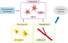
Fig. 2
Brain plasticity and neurorestoration by environmental enrichment.
Environmental enrichment has been shown to increase overall expression of astrocyte marker GFAP similar to the pattern of FGF-2 released from activated astrocytes, which suggests that astrocyte activation can mediate both neurogenesis and angiogenesis, consequently resulting in functional improvement through a FGF-2 dependent mechanism.
GFAP, glial fibrillary acidic protein; FGF-2, fibroblast growth factor-2.
bn-9-e2-g002

SYNAPTIC ACTIVITY-REGULATING GENES

Experience-driven changes in brain include both modifications of the synaptic connectivity of the circuits in a local synapse-specific manner [107], and the induction of activity-dependent gene expression [108]. An increasing number of molecules and genes have been involved in activity-induced plasticity [23]. Previous studies [1109] reported that the expressions of genes related with neuronal structure, synaptic plasticity, and transmission were significantly changed by short-term and long-term exposure to EE. When microarray-based determination of gene expression patterns has been used to find pathways that are affected by EE [1109110], and recent study [1] specifically described that the gene sets expression related with various synaptic changes including chemical synaptic transmission, postsynaptic neurotransmitter receptor binding and G protein activation, kainate receptor activation, the opening of the presynaptic calcium channel, and acetylcholine release were up-regulated, while those associated with neurotransmitter transporters such as dopamine transporters were down-regulated [19].
Above all described things, G protein activation is important for transducing transmitter attachment into postsynaptic responses [111]. Specially, synaptic plasticity is controlled by G protein coupled receptors whose signal pathways aim the NMDA subtype of excitatory glutamate receptor in hippocampus [112]. The recent study [1] exhibited that the expression of G protein-coupled purinergic receptor P2RY12 increased in the hippocampus, where the proteins were mainly spread, regulating the signaling functions in neurotransmission and regeneration [113114]. It is probable that the upregulation of P2RY12 might be related with improved memory function [1].
Activation of kainate receptor, a non-NMDA glutamate receptor, also has a function in synaptic plasticity, affective the postsynaptic cell firing and the presynaptic neurotransmitter release underlying regulation of excitatory synaptic transmission in the hippocampus [115]. The molecular mechanisms underlying these alterations in synaptic features have been traced to NMDA receptor-dependent synaptic plasticity [116] and rises in postsynaptic [Ca2+] [117]. In this way, it is remarkable that the physical stimulation including EE is a physiological inducement delivering the brain with peripheral trophic sustenance [123].
In addition, EE-directed improvement of motor and cognitive functions is not only related to growth factors but also related to the modification of synaptic activity-regulating genes [1]. These genes are related with postsynaptic receptor activity, and the genes are modulated in the presynaptic neurotransmitter transporters that potentially leading to the more efficient use of neurotransmitters and enhanced synaptic plasticity. For examples, dopaminergic receptors are up-regulated in the postsynaptic area, and on the other hands, dopamine transporters are down-regulated in the presynaptic area, consequently resulting in the increase of the dopamine neurotransmitter in the synaptic level [119].

CONCLUSION

EE enhanced neural plasticity such as neurogenesis, gliogenesis, angiogenesis, and neurobehavioral functions. Additionally, EE can affect neural plasticity via overexpression of various neurotrophic growth factors such as BDNF, VEGF, IGF-1, FGF-2 and synaptic activity-regulating genes such as Na+/Cl-dependent neurotransmitter transporters including dopamine transporter. Therefore, EE is considered as an advanced treatment to counteract neurological disorders with cognitive and somatosensory deficits following brain injury. We reviewed the brain plasticity and neurorestoration associated with molecular changes of neurotrophic growth factors such as BDNF, VEGF, IGF-1, FGF-2 and synaptic activity-regulating genes in interaction to EE. Taken together, these findings suggest that EE has beneficial effects on the brain plasticity and neurorestoration.

Notes

Funding This study was supported by grants from the National Research Foundation of Korea (NRF-2014R1A2A1A11052042, NRF-2015M3A9B4067068), the Ministry of Science and Technology, Republic of Korea, and the Korean Health Technology R & D Project (HI16C1013; HI16C1012), Ministry of Health and Welfare, Republic of Korea.

Conflict of Interest The authors have no potential conflicts of interest to disclose.

References

1. Lee MY, Yu JH, Kim JY, Seo JH, Park ES, Kim CH, et al. Alteration of synaptic activity-regulating genes underlying functional improvement by long-term exposure to an enriched environment in the adult brain. Neurorehabil Neural Repair. 2013; 27:561–574.
crossref
2. Rosenzweig MR, Bennett EL, Hebert M, Morimoto H. Social grouping cannot account for cerebral effects of enriched environments. Brain Res. 1978; 153:563–576.
crossref
3. Mora F, Segovia G, del Arco A. Aging, plasticity and environmental enrichment: structural changes and neurotransmitter dynamics in several areas of the brain. Brain Res Brain Res Rev. 2007; 55:78–88.
crossref
4. Stein LR, O’Dell KA, Funatsu M, Zorumski CF, Izumi Y. Short-term environmental enrichment enhances synaptic plasticity in hippocampal slices from aged rats. Neuroscience. 2016; 329:294–305.
crossref
5. Novkovic T, Mittmann T, Manahan-Vaughan D. BDNF contributes to the facilitation of hippocampal synaptic plasticity and learning enabled by environmental enrichment. Hippocampus. 2015; 25:1–15.
crossref
6. Will B, Galani R, Kelche C, Rosenzweig MR. Recovery from brain injury in animals: relative efficacy of environmental enrichment, physical exercise or formal training (1990-2002). Prog Neurobiol. 2004; 72:167–182.
crossref
7. Seo JH, Yu JH, Suh H, Kim MS, Cho SR. Fibroblast growth factor-2 induced by enriched environment enhances angiogenesis and motor function in chronic hypoxic-ischemic brain injury. PLoS One. 2013; 8:e74405.
crossref
8. Cummins RA, Walsh RN, Budtz-Olsen OE, Konstantinos T, Horsfall CR. Environmentally-induced changes in the brains of elderly rats. Nature. 1973; 243:516–518.
crossref
9. Holloway RL Jr. Dendritic branching: some preliminary results of training and complexity in rat visual cortex. Brain Res. 1966; 2:393–396.
crossref
10. Komitova M, Johansson BB, Eriksson PS. On neural plasticity, new neurons and the postischemic milieu: an integrated view on experimental rehabilitation. Exp Neurol. 2006; 199:42–55.
crossref
11. Sirevaag AM, Greenough WT. Differential rearing effects on rat visual cortex synapses. II. Synaptic morphometry. Brain Res. 1985; 351:215–226.
crossref
12. Sirevaag AM, Greenough WT. Differential rearing effects on rat visual cortex synapses. III. Neuronal and glial nuclei, boutons, dendrites, and capillaries. Brain Res. 1987; 424:320–332.
13. Walsh RN, Cummins RA. Changes in hippocampal neuronal nuclei in response to environmental stimulation. Int J Neurosci. 1979; 9:209–212.
crossref
14. Parent JM. Adult neurogenesis in the intact and epileptic dentate gyrus. Prog Brain Res. 2007; 163:529–540.
crossref
15. Kempermann G, Kuhn HG, Gage FH. More hippocampal neurons in adult mice living in an enriched environment. Nature. 1997; 386:493–495.
crossref
16. Yu JH, Seo JH, Lee JY, Lee MY, Cho SR. Induction of neurorestoration from endogenous stem cells. Cell Transplant. 2016; 25:863–882.
crossref
17. Zhu SW, Yee BK, Nyffeler M, Winblad B, Feldon J, Mohammed AH. Influence of differential housing on emotional behaviour and neurotrophin levels in mice. Behav Brain Res. 2006; 169:10–20.
crossref
18. Mlynarik M, Johansson BB, Jezova D. Enriched environment influences adrenocortical response to immune challenge and glutamate receptor gene expression in rat hippocampus. Ann N Y Acad Sci. 2004; 1018:273–280.
crossref
19. Kim MS, Yu JH, Kim CH, Choi JY, Seo JH, Lee MY, Yi CH, Choi TH, Ryu YH, Lee JE, et al. Environmental enrichment enhances synaptic plasticity by internalization of striatal dopamine transporters. J Cereb Blood Flow Metab. 2015; 0271678X15613525.
crossref
20. Fabel K, Fabel K, Tam B, Kaufer D, Baiker A, Simmons N, et al. VEGF is necessary for exercise-induced adult hippocampal neurogenesis. Eur J Neurosci. 2003; 18:2803–2812.
crossref
21. Seo JH, Kim H, Park ES, Lee JE, Kim DW, Kim HO, et al. Environmental enrichment synergistically improves functional recovery by transplanted adipose stem cells in chronic hypoxic-ischemic brain injury. Cell Transplant. 2013; 22:1553–1568.
crossref
22. Trejo JL, Carro E, Torres-Aleman I. Circulating insulin-like growth factor I mediates exercise-induced increases in the number of new neurons in the adult hippocampus. J Neurosci. 2001; 21:1628–1634.
crossref
23. Llorens-Martín M, Torres-Alemán I, Trejo JL. Mechanisms mediating brain plasticity: IGF1 and adult hippocampal neurogenesis. Neuroscientist. 2009; 15:134–148.
crossref
24. de Witt BW, Ehrenberg KM, McAloon RL, Panos AH, Shaw KE, Raghavan PV, et al. Abbreviated environmental enrichment enhances neurobehavioral recovery comparably to continuous exposure after traumatic brain injury. Neurorehabil Neural Repair. 2011; 25:343–350.
crossref
25. Jankowsky JL, Melnikova T, Fadale DJ, Xu GM, Slunt HH, Gonzales V, et al. Environmental enrichment mitigates cognitive deficits in a mouse model of Alzheimer’s disease. J Neurosci. 2005; 25:5217–5224.
crossref
26. Jadavji NM, Kolb B, Metz GA. Enriched environment improves motor function in intact and unilateral dopamine-depleted rats. Neuroscience. 2006; 140:1127–1138.
crossref
27. Rojas JJ, Deniz BF, Miguel PM, Diaz R, Hermel EE, Achaval M, et al. Effects of daily environmental enrichment on behavior and dendritic spine density in hippocampus following neonatal hypoxia-ischemia in the rat. Exp Neurol. 2013; 241:25–33.
crossref
28. Yu K, Wu Y, Zhang Q, Xie H, Liu G, Guo Z, et al. Enriched environment induces angiogenesis and improves neural function outcomes in rat stroke model. J Neurol Sci. 2014; 347:275–280.
crossref
29. Rosenzweig MR. Environmental complexity, cerebral change, and behavior. Am Psychol. 1966; 21:321–332.
crossref
30. Hendershott TR, Cronin ME, Langella S, McGuinness PS, Basu AC. Effects of environmental enrichment on anxiety-like behavior, sociability, sensory gating, and spatial learning in male and female C57BL/6J mice. Behav Brain Res. 2016; 314:215–225.
crossref
31. Kang H, Schuman EM. Long-lasting neurotrophin-induced enhancement of synaptic transmission in the adult hippocampus. Science. 1995; 267:1658–1662.
crossref
32. Poo MM. Neurotrophins as synaptic modulators. Nat Rev Neurosci. 2001; 2:24–32.
crossref
33. Gobbo OL, O’Mara SM. Impact of enriched-environment housing on brain-derived neurotrophic factor and on cognitive performance after a transient global ischemia. Behav Brain Res. 2004; 152:231–241.
crossref
34. Bechara RG, Kelly AM. Exercise improves object recognition memory and induces BDNF expression and cell proliferation in cognitively enriched rats. Behav Brain Res. 2013; 245:96–100.
crossref
35. Pietropaolo S, Feldon J, Alleva E, Cirulli F, Yee BK. The role of voluntary exercise in enriched rearing: a behavioral analysis. Behav Neurosci. 2006; 120:787–803.
crossref
36. Raymond CR. LTP forms 1, 2 and 3: different mechanisms for the “long” in long-term potentiation. Trends Neurosci. 2007; 30:167–175.
crossref
37. Minichiello L. TrkB signalling pathways in LTP and learning. Nat Rev Neurosci. 2009; 10:850–860.
crossref
38. Bramham CR. Local protein synthesis, actin dynamics, and LTP consolidation. Curr Opin Neurobiol. 2008; 18:524–531.
crossref
39. Whitlock JR, Heynen AJ, Shuler MG, Bear MF. Learning induces long-term potentiation in the hippocampus. Science. 2006; 313:1093–1097.
crossref
40. Pastalkova E, Serrano P, Pinkhasova D, Wallace E, Fenton AA, Sacktor TC. Storage of spatial information by the maintenance mechanism of LTP. Science. 2006; 313:1141–1144.
crossref
41. Bekinschtein P, Oomen CA, Saksida LM, Bussey TJ. Effects of environmental enrichment and voluntary exercise on neurogenesis, learning and memory, and pattern separation: BDNF as a critical variable? Semin Cell Dev Biol. 2011; 22:536–542.
crossref
42. Teather LA, Magnusson JE, Chow CM, Wurtman RJ. Environmental conditions influence hippocampus-dependent behaviours and brain levels of amyloid precursor protein in rats. Eur J Neurosci. 2002; 16:2405–2415.
crossref
43. Duffy SN, Craddock KJ, Abel T, Nguyen PV. Environmental enrichment modifies the PKA-dependence of hippocampal LTP and improves hippocampus-dependent memory. Learn Mem. 2001; 8:26–34.
crossref
44. Foster TC, Dumas TC. Mechanism for increased hippocampal synaptic strength following differential experience. J Neurophysiol. 2001; 85:1377–1383.
crossref
45. Artola A, von Frijtag JC, Fermont PC, Gispen WH, Schrama LH, Kamal A, et al. Long-lasting modulation of the induction of LTD and LTP in rat hippocampal CA1 by behavioural stress and environmental enrichment. Eur J Neurosci. 2006; 23:261–272.
crossref
46. Rampon C, Tang YP, Goodhouse J, Shimizu E, Kyin M, Tsien JZ. Enrichment induces structural changes and recovery from nonspatial memory deficits in CA1 NMDAR1-knockout mice. Nat Neurosci. 2000; 3:238–244.
crossref
47. Tang YP, Wang H, Feng R, Kyin M, Tsien JZ. Differential effects of enrichment on learning and memory function in NR2B transgenic mice. Neuropharmacology. 2001; 41:779–790.
crossref
48. Lee EH, Hsu WL, Ma YL, Lee PJ, Chao CC. Enrichment enhances the expression of sgk, a glucocorticoid-induced gene, and facilitates spatial learning through glutamate AMPA receptor mediation. Eur J Neurosci. 2003; 18:2842–2852.
crossref
49. Dahlqvist P, Zhao L, Johansson IM, Mattsson B, Johansson BB, Seckl JR, et al. Environmental enrichment alters nerve growth factor-induced gene A and glucocorticoid receptor messenger RNA expression after middle cerebral artery occlusion in rats. Neuroscience. 1999; 93:527–535.
crossref
50. Rossi C, Angelucci A, Costantin L, Braschi C, Mazzantini M, Babbini F, et al. Brain-derived neurotrophic factor (BDNF) is required for the enhancement of hippocampal neurogenesis following environmental enrichment. Eur J Neurosci. 2006; 24:1850–1856.
crossref
51. Auvergne R, Leré C, El Bahh B, Arthaud S, Lespinet V, Rougier A, et al. Delayed kindling epileptogenesis and increased neurogenesis in adult rats housed in an enriched environment. Brain Res. 2002; 954:277–285.
crossref
52. Bruel-Jungerman E, Laroche S, Rampon C. New neurons in the dentate gyrus are involved in the expression of enhanced long-term memory following environmental enrichment. Eur J Neurosci. 2005; 21:513–521.
crossref
53. Tashiro A, Makino H, Gage FH. Experience-specific functional modification of the dentate gyrus through adult neurogenesis: a critical period during an immature stage. J Neurosci. 2007; 27:3252–3259.
crossref
54. Ben M’Barek K, Pla P, Orvoen S, Benstaali C, Godin JD, Gardier AM, et al. Huntingtin mediates anxiety/depression-related behaviors and hippocampal neurogenesis. J Neurosci. 2013; 33:8608–8620.
crossref
55. Snyder JS, Kee N, Wojtowicz JM. Effects of adult neurogenesis on synaptic plasticity in the rat dentate gyrus. J Neurophysiol. 2001; 85:2423–2431.
crossref
56. Bruel-Jungerman E, Rampon C, Laroche S. Adult hippocampal neurogenesis, synaptic plasticity and memory: facts and hypotheses. Rev Neurosci. 2007; 18:93–114.
crossref
57. Sisti HM, Glass AL, Shors TJ. Neurogenesis and the spacing effect: learning over time enhances memory and the survival of new neurons. Learn Mem. 2007; 14:368–375.
crossref
58. Ferrara N, Gerber HP, LeCouter J. The biology of VEGF and its receptors. Nat Med. 2003; 9:669–676.
crossref
59. Jin K, Zhu Y, Sun Y, Mao XO, Xie L, Greenberg DA. Vascular endothelial growth factor (VEGF) stimulates neurogenesis in vitro and in vivo. Proc Natl Acad Sci USA. 2002; 99:11946–11950.
crossref
60. Rosenstein JM, Krum JM. New roles for VEGF in nervous tissue--beyond blood vessels. Exp Neurol. 2004; 187:246–253.
crossref
61. Storkebaum E, Lambrechts D, Carmeliet P. VEGF: once regarded as a specific angiogenic factor, now implicated in neuroprotection. BioEssays. 2004; 26:943–954.
crossref
62. Kaya D, Gürsoy-Ozdemir Y, Yemisci M, Tuncer N, Aktan S, Dalkara T. VEGF protects brain against focal ischemia without increasing blood--brain permeability when administered intracerebroventricularly. J Cereb Blood Flow Metab. 2005; 25:1111–1118.
crossref
63. Zhang ZG, Chopp M. Neurorestorative therapies for stroke: underlying mechanisms and translation to the clinic. Lancet Neurol. 2009; 8:491–500.
crossref
64. Cao L, Jiao X, Zuzga DS, Liu Y, Fong DM, Young D, et al. VEGF links hippocampal activity with neurogenesis, learning and memory. Nat Genet. 2004; 36:827–835.
crossref
65. During MJ, Cao L. VEGF, a mediator of the effect of experience on hippocampal neurogenesis. Curr Alzheimer Res. 2006; 3:29–33.
crossref
66. Plaschke K, Staub J, Ernst E, Marti HH. VEGF overexpression improves mice cognitive abilities after unilateral common carotid artery occlusion. Exp Neurol. 2008; 214:285–292.
crossref
67. Kim BW, Choi M, Kim YS, Park H, Lee HR, Yun CO, et al. Vascular endothelial growth factor (VEGF) signaling regulates hippocampal neurons by elevation of intracellular calcium and activation of calcium/calmodulin protein kinase II and mammalian target of rapamycin. Cell Signal. 2008; 20:714–725.
crossref
68. Licht T, Goshen I, Avital A, Kreisel T, Zubedat S, Eavri R, et al. Reversible modulations of neuronal plasticity by VEGF. Proc Natl Acad Sci USA. 2011; 108:5081–5086.
crossref
69. Ortuzar N, Rico-Barrio I, Bengoetxea H, Argandoña EG, Lafuente JV. VEGF reverts the cognitive impairment induced by a focal traumatic brain injury during the development of rats raised under environmental enrichment. Behav Brain Res. 2013; 246:36–46.
crossref
70. D’Ercole AJ, Ye P, Calikoglu AS, Gutierrez-Ospina G. The role of the insulin-like growth factors in the central nervous system. Mol Neurobiol. 1996; 13:227–255.
crossref
71. D’Ercole AJ, Ye P, O’Kusky JR. Mutant mouse models of insulin-like growth factor actions in the central nervous system. Neuropeptides. 2002; 36:209–220.
crossref
72. O’Kusky JR, Ye P, D’Ercole AJ. Insulin-like growth factor-I promotes neurogenesis and synaptogenesis in the hippocampal dentate gyrus during postnatal development. J Neurosci. 2000; 20:8435–8442.
crossref
73. Aberg ND, Brywe KG, Isgaard J. Aspects of growth hormone and insulin-like growth factor-I related to neuroprotection, regeneration, and functional plasticity in the adult brain. ScientificWorldJournal. 2006; 6:53–80.
crossref
74. Carro E, Trejo JL, Busiguina S, Torres-Aleman I. Circulating insulin-like growth factor I mediates the protective effects of physical exercise against brain insults of different etiology and anatomy. J Neurosci. 2001; 21:5678–5684.
crossref
75. Koopmans GC, Brans M, Gómez-Pinilla F, Duis S, Gispen WH, Torres-Aleman I, et al. Circulating insulin-like growth factor I and functional recovery from spinal cord injury under enriched housing conditions. Eur J Neurosci. 2006; 23:1035–1046.
crossref
76. Carro E, Nuñez A, Busiguina S, Torres-Aleman I. Circulating insulin-like growth factor I mediates effects of exercise on the brain. J Neurosci. 2000; 20:2926–2933.
crossref
77. Markowska AL, Mooney M, Sonntag WE. Insulin-like growth factor-1 ameliorates age-related behavioral deficits. Neuroscience. 1998; 87:559–569.
crossref
78. Cotman CW, Berchtold NC. Exercise: a behavioral intervention to enhance brain health and plasticity. Trends Neurosci. 2002; 25:295–301.
crossref
79. Ding Q, Vaynman S, Akhavan M, Ying Z, Gomez-Pinilla F. Insulin-like growth factor I interfaces with brain-derived neurotrophic factor-mediated synaptic plasticity to modulate aspects of exercise-induced cognitive function. Neuroscience. 2006; 140:823–833.
crossref
80. Trejo JL, Piriz J, Llorens-Martin MV, Fernandez AM, Bolós M, LeRoith D, et al. Central actions of liver-derived insulin-like growth factor I underlying its pro-cognitive effects. Mol Psychiatry. 2007; 12:1118–1128.
crossref
81. Keyvani K, Sachser N, Witte OW, Paulus W. Gene expression profiling in the intact and injured brain following environmental enrichment. J Neuropathol Exp Neurol. 2004; 63:598–609.
crossref
82. Ciucci F, Putignano E, Baroncelli L, Landi S, Berardi N, Maffei L. Insulin-like growth factor 1 (IGF-1) mediates the effects of enriched environment (EE) on visual cortical development. PLoS One. 2007; 2:e475.
crossref
83. Aberg ND, Blomstrand F, Aberg MA, Björklund U, Carlsson B, Carlsson-Skwirut C, et al. Insulin-like growth factor-I increases astrocyte intercellular gap junctional communication and connexin43 expression in vitro. J Neurosci Res. 2003; 74:12–22.
crossref
84. Cheng CM, Mervis RF, Niu SL, Salem N Jr, Witters LA, Tseng V, et al. Insulin-like growth factor 1 is essential for normal dendritic growth. J Neurosci Res. 2003; 73:1–9.
crossref
85. Aberg ND, Johansson UE, Aberg MA, Hellström NA, Lind J, Bull C, et al. Peripheral infusion of insulin-like growth factor-I increases the number of newborn oligodendrocytes in the cerebral cortex of adult hypophysectomized rats. Endocrinology. 2007; 148:3765–3772.
crossref
86. Davila D, Piriz J, Trejo JL, Nunez A, Torres-Aleman I. Insulin and insulin-like growth factor I signalling in neurons. Front Biosci. 2007; 12:3194–3202.
crossref
87. Woodbury ME, Ikezu T. Fibroblast growth factor-2 signaling in neurogenesis and neurodegeneration. J Neuroimmune Pharmacol. 2014; 9:92–101.
crossref
88. Eckenstein FP. Fibroblast growth factors in the nervous system. J Neurobiol. 1994; 25:1467–1480.
89. Çelik Y, Atıcı A, Beydağı H, Reşitoğlu B, Yılmaz N, Ün İ, et al. The effects of fibroblast growth factor-2 and pluripotent astrocytic stem cells on cognitive function in a rat model of neonatal hypoxic-ischemic brain injury. J Matern Fetal Neonatal Med. 2016; 29:2199–2204.
crossref
90. Folkman J, Klagsbrun M. Angiogenic factors. Science. 1987; 235:442–447.
crossref
91. Baird A. Fibroblast growth factors: activities and significance of non-neurotrophin neurotrophic growth factors. Curr Opin Neurobiol. 1994; 4:78–86.
crossref
92. Reuss B. von Bohlen und Halbach O. Fibroblast growth factors and their receptors in the central nervous system. Cell Tissue Res. 2003; 313:139–157.
crossref
93. Walicke PA. Basic and acidic fibroblast growth factors have trophic effects on neurons from multiple CNS regions. J Neurosci. 1988; 8:2618–2627.
crossref
94. Jin K, Sun Y, Xie L, Batteur S, Mao XO, Smelick C, et al. Neurogenesis and aging: FGF-2 and HB-EGF restore neurogenesis in hippocampus and subventricular zone of aged mice. Aging Cell. 2003; 2:175–183.
crossref
95. Rai KS, Hattiangady B, Shetty AK. Enhanced production and dendritic growth of new dentate granule cells in the middle-aged hippocampus following intracerebroventricular FGF-2 infusions. Eur J Neurosci. 2007; 26:1765–1779.
crossref
96. Yoshimura S, Takagi Y, Harada J, Teramoto T, Thomas SS, Waeber C, et al. FGF-2 regulation of neurogenesis in adult hippocampus after brain injury. Proc Natl Acad Sci USA. 2001; 98:5874–5879.
crossref
97. Kang W, Hébert JM. FGF Signaling Is Necessary for Neurogenesis in Young Mice and Sufficient to Reverse Its Decline in Old Mice. J Neurosci. 2015; 35:10217–10223.
crossref
98. Bikfalvi A, Klein S, Pintucci G, Rifkin DB. Biological roles of fibroblast growth factor-2. Endocr Rev. 1997; 18:26–45.
crossref
99. Comeau WL, Hastings E, Kolb B. Pre- and postnatal FGF-2 both facilitate recovery and alter cortical morphology following early medial prefrontal cortical injury. Behav Brain Res. 2007; 180:18–27.
crossref
100. Monfils MH, Driscoll I, Kamitakahara H, Wilson B, Flynn C, Teskey GC, et al. FGF-2-induced cell proliferation stimulates anatomical, neurophysiological and functional recovery from neonatal motor cortex injury. Eur J Neurosci. 2006; 24:739–749.
crossref
101. Li Q, Stephenson D. Postischemic administration of basic fibroblast growth factor improves sensorimotor function and reduces infarct size following permanent focal cerebral ischemia in the rat. Exp Neurol. 2002; 177:531–537.
crossref
102. Rowntree S, Kolb B. Blockade of basic fibroblast growth factor retards recovery from motor cortex injury in rats. Eur J Neurosci. 1997; 9:2432–2441.
crossref
103. Zhang H, Vutskits L, Pepper MS, Kiss JZ. VEGF is a chemoattractant for FGF-2-stimulated neural progenitors. J Cell Biol. 2003; 163:1375–1384.
crossref
104. Sofroniew MV. Reactive astrocytes in neural repair and protection. Neuroscientist. 2005; 11:400–407.
crossref
105. Azin M, Mirnajafi-Zadeh J, Javan M. Fibroblast growth factor-2 enhanced the recruitment of progenitor cells and myelin repair in experimental demyelination of rat hippocampal formations. Cell J. 2015; 17:540–456.
106. Briones TL, Woods J, Wadowska M, Rogozinska M, Nguyen M. Astrocytic changes in the hippocampus and functional recovery after cerebral ischemia are facilitated by rehabilitation training. Behav Brain Res. 2006; 171:17–25.
crossref
107. Malinow R, Malenka RC. AMPA receptor trafficking and synaptic plasticity. Annu Rev Neurosci. 2002; 25:103–126.
crossref
108. Flavell SW, Greenberg ME. Signaling mechanisms linking neuronal activity to gene expression and plasticity of the nervous system. Annu Rev Neurosci. 2008; 31:563–590.
crossref
109. Rampon C, Jiang CH, Dong H, Tang YP, Lockhart DJ, Schultz PG, et al. Effects of environmental enrichment on gene expression in the brain. Proc Natl Acad Sci USA. 2000; 97:12880–12884.
crossref
110. Li C, Niu W, Jiang CH, Hu Y. Effects of enriched environment on gene expression and signal pathways in cortex of hippocampal CA1 specific NMDAR1 knockout mice. Brain Res Bull. 2007; 71:568–577.
crossref
111. Neves SR, Ram PT, Iyengar R. G protein pathways. Science. 2002; 296:1636–1639.
crossref
112. MacDonald JF, Jackson MF, Beazely MA. G protein-coupled receptors control NMDARs and metaplasticity in the hippocampus. Biochim Biophys Acta. 2007; 1768:941–951.
crossref
113. Burnstock G. Potential therapeutic targets in the rapidly expanding field of purinergic signalling. Clin Med (Lond). 2002; 2:45–53.
crossref
114. Sasaki Y, Hoshi M, Akazawa C, Nakamura Y, Tsuzuki H, Inoue K, et al. Selective expression of Gi/o-coupled ATP receptor P2Y12 in microglia in rat brain. Glia. 2003; 44:242–250.
crossref
115. Contractor A, Swanson GT, Sailer A, O’Gorman S, Heinemann SF. Identification of the kainate receptor subunits underlying modulation of excitatory synaptic transmission in the CA3 region of the hippocampus. J Neurosci. 2000; 20:8269–8278.
crossref
116. Bortolotto ZA, Bashir ZI, Davies CH, Collingridge GL. A molecular switch activated by metabotropic glutamate receptors regulates induction of long-term potentiation. Nature. 1994; 368:740–743.
crossref
117. Abraham WC, Bear MF. Metaplasticity: the plasticity of synaptic plasticity. Trends Neurosci. 1996; 19:126–130.
crossref
TOOLS
Similar articles