

 PDF
PDF Citation
 Print
Print


INTRODUCTION
The evaluation of recovery following a stroke is key for both treatment and research. Despite severe disabilities and neurological impairments during the early post-stroke period, most stroke patients achieve some degree of recovery over time [12]. For example, some stroke patients show early motor function recovery, which occurs primarily within the first few months [3]. Although the degree of paralysis is a primary predictor, it cannot be used to predict the rate of motor recovery accurately during the subacute stage based on the patient's initial condition [3]. Improvement in motor function in the lower body is observed in –65% of patients with initial motor deficits [3]; however, the probability of recovery of normal functioning in the upper limbs is much lower (< 15%) [4]. Additionally, the rate of clinical recovery is relatively rapid during the first few weeks after a stroke, and then decelerates 1–3 months later [56]. Between 3 and 6 months after stroke, most previous studies report little or no observable recovery [57]. In contrast, Lee et al. [6] showed a small but significant improvement in all recovery variables 3–6 months after a stroke, with the exception of lower-body motor function, indicating that recovery had not yet plateaued. In addition, it is known that most of the recovery of gait and motor function generally occurs within 6 months after a stroke [89]. Although the change in recovery varies after stroke, the recovery process does not differ markedly [10]. For this reason, the analysis of recovery profiles is important because this information can help to provide more specific plans for stroke rehabilitation [11]. Several investigators have studied clinical post-stroke recovery, mostly in terms of upper and lower motor function, gait, balance, cognition, trunk control, and activities of daily living (ADL) [67912131415161718192021]. Our knowledge of the details of stroke recovery remains limited, and there are few validated predictors of clinical recovery and generally insufficient data on the degree of recovery that can be achieved.
CORRELATION BETWEEN BRAIN LESION AND MOTOR RECOVERY
Clarification of the cause of motor weakness is essential for prognosis and for adoption of scientific strategies for successful rehabilitation [2223242526]. Recovery following a stroke is typically classified into neurological recovery and functional recovery; functional recovery is influenced by the external environment, continuity of rehabilitation, and motivation, whereas neurological recovery varies according to stroke pathogenesis and lesion site [27]. Severe paralysis has been found to be a valid predictor of poor functional recovery [28].
A process for motor recovery in stroke rehabilitation was suggested in a review by Cauraugh and Summers [4]: 1) ipsilesional compensatory changes by functional reorganization of normal tissues in the peri-infarct area [29]; 2) activation of the contralesional motor area and ipsilateral corticospinal tract (CST) [30]; and 3) increased activation of non-motor areas, such as the supplementary motor area, inferior parietal cortex, cingulate, insula, and cerebellum [313233]. Furthermore, Jang [11] suggested a motor recovery mechanism as follows: 1) an ipsilateral pathway from the unaffected cortex; 2) peri-lesional reorganization; 3) recovery of the damaged lateral CST; and 4) contributions from other motor areas. As mentioned in these reviews, key factors in motor recovery were the degrees of sparing in the CST and sensorimotor cortex [343536].
In the human brain, the neural tracts for motor function are classified as CST and non-CST. The CST and the corticoreticulospinal tract have been regarded as important neural tracts for the mediation of voluntary movements [37]. The main function of the CST is control of movements of the distal extremities, particularly fine-motor movements of the hands [3738], whereas the corticoreticulospinal tract, consisting of the corticoreticular pathway (CRP) and the reticulospinal tract, innervates the proximal extremities and axial muscles. Thus, it is involved in postural control and locomotor function [243940414243]. Therefore, determining the state of the CST and CRP is important for determining the cause of motor weakness in stroke patients. Although it lacked statistical power, a study by Yoo et al. [44] suggested that patients with injury to the CST, which is closely related to hand function, showed worse hand function than did patients with injury to the CRP, and patients with injury to the CRP, which is more closely related to poor recovery of gait function. The CRP originates primarily from the premotor cortex (PMC) and terminates at the pontomedullary reticular formation (PMRF) [43]. As mentioned by Nudo [45], injury to the CST in the primary cortex (M1) could be compensated for by spared motor cortex areas, such as the PMC. The corticoreticulospinal tract is one of the extrapyramidal motor pathways in the human brain [41]. A previous study found that the CRP descends through the corona radiata and the posterior limb of the internal capsule anterior to the CST. In the midbrain and pons, it passes through the tegmentum and terminates at the PMRF [46]. Freund et al. [474849] reported an association between PMC lesions and proximal muscle weakness (shoulder and hip muscles). In addition, the intact PMC showed better recovery in terms of mobility and motor function of the hip [50]. Motor functions of patients with injury to both the CST and CRP were significantly poorer than those in patients with injury to either the CST or CRP [44]. Putaminal hemorrhage frequently accompanies injuries to both the CST and CRP, and the CRP appears to be more vulnerable than the CST. These findings suggest the need for evaluating both the CRP and the CST in patients with putaminal hemorrhage [44].
NEUROIMAGING TECHNIQUES TO INVESTIGATE BRAIN DAMAGE AND STROKE RECOVERY
Many studies have investigated the mechanism of motor recovery after stroke using transcranial magnetic stimulation (TMS), functional magnetic resonance imaging (fMRI), and diffusion-tensor imaging (DTI) [11]. To establish relationships between functional recovery and brain anatomy using lesion-mapping approach, it is essential to have information of the location and extent of a brain injury. Recently introduced imaging methods made it possible to identify regions that are commonly involved in many individuals with similar deficit. Furthermore, group analysis allowed higher accuracy of such identification despite of different shapes, sizes, and structures of the individuals by transforming each brain into a standard stereotaxic space [50].
There are several issues that need to be considered for using the lesion method. This technique assumes that discrete anatomical modules are involved in different functions, which is different in reality as many brain functions work in a distributed network rather than in a single anatomical module [51]. In addition, brain damages are heterogeneous, since they depend rather on the vascular supply but not solely on its functional unit. As a result, disruption of the blood supply can bring a wide range of deficits, however, on the other hand, some small lesions may not reveal any obvious functional problem due to the redundancy of the human brain [52].
Identification of a critical area for a certain function by superimposing individual lesions is based on the assumption that these functional components are in the same locations in different individuals. Specifically, functional compensation may occur at different sites [53]. Another assumption of this technique is that the intact regions of the brain continue to function in the same manner as prior to the injury. Some areas of the cortex are particularly prone to damage by stroke [54]. Locations of brain damages are not randomly distributed and the extents of those damages vary depending on the blood supply specific to the location. Hence it makes interpretation of lesion overlay plots challenging. Additionally, brain damage may result in diaschisis, i.e., a functionally disabled region that is still structurally intact. The functions of these regions that appear to be structurally healthy may be impaired by de-afferentation. Conventional structural imaging techniques such as computed tomography (CT) and MRI make it possible to demonstrate whether such structurally intact region is functionally normal.
The lesion method has other drawbacks. When testing participants in the acute stage of their illness, it is difficult to accurately identify all the impaired brain regions since patients having relatively small lesions can present with temporary malfunctions in large regions of their brain due to disrupted perfusion. However, after waiting for these initial problems to resolve, the changes associated with brain plasticity will become more pronounced. Unfortunately, the lesion method does not explain the precise time course of information processing. Thus, the lesion method when used for temporal resolution is especially poor. The lesion method also has several problems in terms of differential vulnerability, plasticity, and disconnection.
Functional MRI measures neuronal activity indirectly by obtaining hemodynamic response function using blood oxygenation level dependent signal, which can provide information of task-related brain activity with good time resolution. However, there still are challenges in interpreting brain activation studies and has its own limitations [55]. We can put togetherevidence that a particular task is correlated with activation in a particular brain region. However, it is not clear whether this region is necessary to perform this task, and it cannot be concluded that an area that shows no change in blood flow during a task is not involved in that task. Furthermore, it is not possible to use the technique in a patient with poor motor function [11]. Because brain injuries such as stroke, can severely disrupt regional blood flow, fMRI may fail in detecting prominent activity in neurological patients for 2 reasons. First, brain injury can result in reduced metabolism (nutrient not absorbed by damaged region), which result in the surrounding intact areas receiving more blood supply than needed for normal demands (a situation known as ‘luxury perfusion’). Second, the surviving arteries can exhibitsustained dilation to compensate for compromised arteries. Either of these effects can reduce the task-related dilation and constriction of blood flow that would be measured with fMRI as the correlate of brain activity. Consequently, a perfectly well functioning region may appear unresponsive during a standard fMRI analysis.
DTI, an extension of diffusion weighted imaging (DWI), takes advantages of the brain's limited perpendicular water motion to fiber tracts but less constrained motion in the direction of fiber tracts. DTI can help to identify disconnected regions and determine the extent of white matter damage post stroke [56]. DTI is advantageous when it comes tovisualization and estimation of the CST [57]. DTI provides imaging of axonal pathways in the living brain and provides information about tissue microstructure by measuring fractional anisotropy (FA) [58].
Both the lesion method and neuroimaging techniques have clear limitations. However, it should be noted that some of the strengths and weaknesses of these tools are complementary. Although some limitations of lesion method are inherent to the technique itself, some of its weaknesses are being addressed by recent technical innovations. In particular, new imaging protocols and improved analysis of lesion data may reform its shortcomings and improve the ability in measuring functional extent of brain damage. In addition, it may assist in refining our understanding of the relationship between brain anatomy and function.
LESION NORMALIZATION TO ALIGN BRAINS FROM DIFFERENT INDIVIDUALS
The quality of normalization is crucial; if different individuals' brains are not matched accurately, same regions of the brains are not compared thus reducing the statistical power of these tests. Even in healthy individuals, normalization is a complex process where an ideal solution is yet to be found. Further challenges are faced when working with elderly stroke patients. These patients typically have larger and more variable ventricles due to both age and injury related atrophy. Furthermore, automated normalization techniques face difficulty with injured brain region as the location may be distorted in comparison to the template image.
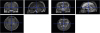
It was necessary to turn each individual's T1 scan, an anatomical image, and lesion images on a T2 scan into a lesion map, and the pathological scan was the scan used to draw lesions [5960]. The first step is re-aligning the T1 image to standard space. This approximately locates the anterior commissure, which aids in the subsequent normalization. The next step is to coregister the T2 scan to the space of the T1 scan. The T2 image is moved and rotated until it is closely aligned to T1 (Figs. 1 and 2).
Fig. 1
Lesion normalization using Statistical Parametric Mapping (SPM; Wellcome Trust Centre for Neuroimaging, University College London, London, UK). Images shown are individual anatomical images (T1) realigned to a standard template image. The left shows a mean T1 image from 152 people, and the right shows the individual's T1 image. The 2 images are now roughly aligned to each other. This is not a perfect match, but it is a good starting estimate for subsequent analyses.
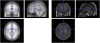
Fig. 2
Lesion normalization using Statistical Parametric Mapping (SPM; Wellcome Trust Centre for Neuroimaging, University College London, London, UK). Images shown are co-registered pathological (T2) and anatomical images (T1). The T2 image is moved and rotated until it is closely aligned to the T1.
Currently, two types of scan are commonly used for MRI lesion studies. T1 weighted scans offer good discrimination of anatomical landmarks by sharp contrast between gray and white matter. Meanwhile, T2-weighted scans, which usually highlight regions of damage, provide good pathological information. However, therestill are limitations in assessing acute infarct lesions with T1- and T2-weighted MRI scans. First, conventional T1 and T2 scans often fail to detect acute strokes, even when the behavior pathology is severe enough that emergent clinical intervention is necessary. Second, these sequences do not always determine whether an area is functioning normally or not. Techniques such as DWI enable strokes to be visualized at an early stage [57]. T2-weighted fluid-attenuated inversion recovery (FLAIR) imaging sequences can be used for delineating the lesion extent in acute cerebral infarcts.[616263].
STATISTICAL ANALYSIS OF LESION DATA
In lesion analysis, patients are typically grouped either by lesion or by behavior. In the lesion-defined approach, the behavioral performance of a group of patients with a common area of injury (for example, dorsolateral prefrontal cortex) is compared with that of a control group or another patient group. This method is valuable for assessing the functional roles of particular regions of interest (ROIs), but can lose information if an ROI contains multiple subregions that each contribute to behavior. Additionally, regions outside the ROI that are part of a distributed functional network may be overlooked. In the behavior-defined approach, patients are grouped according to a specific behavioral deficit. Lesion reconstructions from patients with the deficit are overlaid to find common area(s) of infarction, and these are compared to lesion overlays from patients without the deficit. These contrasting overlays, or subtracted images, are effective in identifying brain regions that may contribute to a cognitive skill, but in situations where the behavioral data are continuous rather than binary, a cutoff must be applied, and information reflecting varying degrees of performance can be lost [64].
Simple overlay plots for patients who have a disorder can be misleading, as the regions that they highlight might reflect increased vulnerability of certain regions to injury because of their vasculature [55]. A control group of neurological patients who do not exhibit the deficit of interest is thus indispensable for valid anatomical conclusions. In order to investigate a brain lesion related to a functional impairment, a group of control patients who do not show any definite impairment is needed. These control patients should have brain lesions in the same hemisphere and must be similar to the patients of interest with respect to other variables. Thus, this approach uses a traditional overlay plot with lesion subtraction and analyzes contrasts in lesion contrast between a study group of patients (a lesion overlay with positive value) and a control group (a lesion overlay with negative values; Fig. 3) [65]. There are, however, 2 primary problems with this approach [66]. First, it is not an objective test; any random collection of patient lesions will necessarily show some region of maximal overlap, regardless of whether this location has any influence on symptoms or behavior. Second, brain injury is not random; because of the vasculature and architecture of the brain, certain regions of the brain are particularly vulnerable to injury. Thus, specific brain regions are commonly injured regardless of their influence on symptoms.
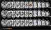
Fig. 3
Subtraction analysis using the MRIcron program (Chris Rorden, University of South Carolina, Columbia, SC, USA). The top 2 figures (A, B) were overlaid from patients with mass synergic movement recovery and with isolated movement recovery. The bottom (C) shows a subtraction analysis where an overlay of patients with isolated movement recovery was subtracted from an overlay of those with mass synergic movement recovery [65].
Because both the lesion size and location could affect functional recovery, lesion location should be significantly different between study patients and a matched lesion volume in a control group. Large individual variation substantially reduces the power of anatomical studies. One solution is to conduct analyses on a large number of patients. A second solution is to restrict analysis to patients with focal lesions. Ideally, we want to know whether differences in lesion frequency (for example, between patients who show the disorder of interest and patients who do not) might be due to chance or are reliable predictors of behavior. Naturally occurring lesions vary greatly in location and in the extent of the damage, adding a huge degree of variability to an analysis. Voxel-based lesion-symptom mapping (VLSM) estimates statistical parameters on a voxel-by-voxel basis, allowing fairly high spatial precision. With VLSM, similarity between statistical maps can be assessed by calculating the correlation between t-scores on 2 tasks, treating voxels as subjects [64]. To work effectively, brain images from different patients must be aligned in a common stereotaxic space. This method reduces the tendency to detect areas that are correlated with large lesions but not specifically with the deficit being investigated.
Statistical lesion analyses are conducted by examining damage to predefined anatomical ROIs or on a voxel-wise basis, with an independent test computed for every 3-dimensional pixel of the brain image. Region-of-interest studies can offer better statistical power because fewer tests are computed simultaneously, requiring less correction for multiple comparisons. However, voxel-wise analysis can offer much better spatial resolution, and it is typically better suited when there is no strong a priori prediction regarding the spatial location of the critical lesion. The basic question underlying lesion analyses is whether the spatial location of the brain lesion can predict the individual's behavioral symptoms. The choice of a test for lesion analysis is driven by the nature of the symptoms. Binomial tests are performed when the symptoms are binary (e.g., the presence or absence of a disorder), whereas the t‑test is traditionally used if the severity of symptoms lies on a continuum, with patients exhibiting a graded range of performance. Additionally, all patients included should be investigated at the same time point after stroke onset; otherwise, differences in behavior may reflect different stages of recovery rather than differences due to different brain lesion locations.
CONCLUSION
Many studies have examined the relationship between brain damage and motor recovery using brain images and the lesion method. For brain imaging studies, the statuses of the CST and CRP are important for determining the cause of motor weakness in stroke patients. Also, with the lesion method and overlapping brain data, involvement of the putamen, internal capsule, thalamus, corona radiata, and premotor cortex is related to poor motor recovery after a stroke. This may be because the CRP and CST descend through the corona radiata and the posterior limb of the internal capsule. However, in lesion studies, an area may emerge as relevant because it has a direct causal role or because of a diaschitic effect, involving correlated lesions some distance away. The lesion method has much to offer neuroscience, despite its limitations. The strengths and weaknesses of these tools are complementary. Indeed, some brain functions might be impossible to determine using only the lesion method or functional neuroimaging alone. Because other techniques, such as event-related potential (ERP), magnetoencephalography (MEG), and TMS, also have important complementary strengths, we could solve some problems by combining these techniques. New imaging techniques to improve statistical power will provide a better understanding of the total extent of disruption after brain injury. These tools have clear implications for improving patient care, but will also improve our theoretical understanding of brain function. Advances in imaging have been complemented by new tools for analyzing lesion data.
 XML Download