

 PDF
PDF Citation
 Print
Print


INTRODUCTION
Alzheimer's disease (AD) is one of the most common degenerative diseases. As the elderly population increases, the prevalence and concern of AD are growing. Moreover, it is dreadful that there has been no disease modifying treatment for this disease. Currently, only pharmacological treatment that improves cognitive function is used as a symptomatic treatment. This treatment has a limited effect, and its effect decreases over time, and even if it has side effects, it could not be used [1]. Therefore, due to the limitations of currently available pharmacological treatments, alternative therapies area required.
Transcranial direct current stimulation (tDCS) is one of the brain stimulation techniques, noninvasive, safe, painless, and relatively easy to use [2]. To date, tDCS has been shown to have beneficial effect in numerous diseases including major depressive disorder [3], pain [45], stroke [67], drug addiction [8], refractory epilepsy [9], focal hand dystonia [10], and other degenerative disease [11].
Since tDCS enhances neural function by modulating cortical excitability and synaptic plasticity, it was showed the favorable effects of cognitive function in previous healthy human studies with tDCS [12131415]. This effects may contribute to the treatment of AD [16], therefore brain stimulation is considered as an alternative treatment for AD.
In this review, we briefly introduced what tDCS was, summarized the pathogenesis of AD in terms of neural circuit and discussed possible mechanisms of tDCS that could be applied to the treatment of AD.
BASIC PRINCIPLES OF tDCS
tDCS is one of the noninvasive brain stimulation methods and it is a technique to control the neuronal transmembrane potential by flowing a weak current of 1–2 mA to the scalp [17]. tDCS regulates spontaneous neuronal network activity through polarization of the resting membrane potential, rather than causing neuronal firing by suprathreshold neuronal membrane depolarization. Two small electrodes with an area of 25 to 35 cm2 are put on the scalp and current is passed through it, the anodal or cathodal stimulation is determined according to the direction of the electric flow between the 2 electrodes. Although the skull has a high resistance, a considerable amount of the electrical current reaches the brain. Previously, it was questionable whether the current of tDCS would reach the brain, however recent studies have shown that appropriate electrode placement and montage influence electrical current reaching the cortex during stimulation [18]. In addition, since the conductivity of the skull is low, the current density is homogenously transmitted to the brain below the site where the electrode is located [19].
The effect of tDCS depends on the direction of current polarity of the electrodes, anodal stimulation increases cortical activity and excitability, while cathodal stimulation decreases. The effects of tDCS are observed not only during stimulation but also after the end of stimulation (after-effect) [20]. The factors affecting stimulation are duration, intensity, polarity of stimulation and baseline cortical excitability state [21]. The tDCS stimulation is usually performed for 20–30 minutes, and after-effects are observed to last for more than 1 hour. While the effect during stimulation is caused by changes in spontaneous neuronal firing such as neuronal depolarization or hyperpolarization, the after-effect is provoked by altering the synaptic microenvironment, such as altering N-methyl-D-aspartate (NMDA) receptor dependent activity [22].
PATHOPHYSIOLOGY OF AD: FROM THE PERSPECTIVE OF NEURAL CIRCUIT
Amyloid beta deposition
Amyloid beta in the brain parenchyma influenced loss of synapse, neurodegeneration, and alteration in neuronal activity. These changes impaired neural circuits, which led to widespread network dysfunction and cognitive decline. Amyloid beta protein had unfavorable effects on neurons and other types of brain cells [23]. Oligomeric amyloid beta directly stimulated neuronal apoptosis through interaction with cell-surface receptors. Additionally, long-term accumulation of toxic amyloid beta in parenchyma led to oxidative damage of deoxyribonucleic acid (DNA) and protein, physical injury of cellular organelle, and dysregulation of intracellular calcium level, resulting in cell death [24].
Impairment of synapses and destabilization of circuits
Amyloid beta induced deficits in synaptic plasticity, circuit function, and cognition initiated before cell loss occurred [25]. Meanwhile, synaptic activity could also promote the accumulation of amyloid beta in the brain parenchyma by affecting amyloid beta metabolism [26]. Excitatory activity promoted amyloid precursor protein proteolysis and released it into the extracellular space [27].
The effect of amyloid beta on synapse activity varied with the extracellular concentration. Low levels of amyloid beta enhanced excitatory activity and higher levels inhibited it [2829]. A slight increase in amyloid beta contributed to activity through presynaptic acetylcholine receptors, which increased internal calcium concentration to induce the release of glutamate [30]. Postsynaptic excitation further increased amyloid beta and synaptic excitability through positive feedback, however increasingly high levels of amyloid beta reduced synapse activity by modifying the synapse strength through several mechanisms such as internalization of glutamate receptors [31]. Acute increases in synaptic amyloid beta impaired the long-term potentiation (LTP) of synaptic strength and increased the depression of synaptic activity [282932]. Moreover, chronic elevations of amyloid beta weakened connectivity, altered dynamics of dendritic spines and increased synapse loss [3233]. Loss of dendritic spines due to amyloid beta induced neuron hyperexcitibility and made it more easily stimulated [34]. Amyloid beta also affected the inhibitory interneurons, altering the balance of excitatory activity and inhibitory activity. Loss of synaptic inhibition through the inhibitory interneuron deficit, including the downregulation of cell-surface voltage-gated sodium channels, led to modification of network activity and cognitive dysfunction [35].
Network susceptibility
Amyloid deposition altered neural circuit connectivity and network activity. In AD, the default mode network, which was a functionally connected region that was activated during passive thinking, remembering and planning, coincided with the deposition of amyloid beta [36]. Additionally, in previous studies, the areas where the cerebral blood flow (CBF) increased when performing memory-related activities were consistent with the default mode network [3738]. In functional magnetic resonance imaging (MRI) study, the medial temporal lobe activity was also correlated with spontaneous thought process with less attention, which mainly activates the default mode network [39]. These studies suggested that memory processes might be linked to the default mode and that involvement of memory networks in default state could explain why memory was predominantly vulnerable in AD. In AD patients, amyloid deposition had little change with disease progression [40], whereas progression of default mode network dysfunction was associated with clinical deterioration. In early stage of disease, the areas of the posterior default mode network, such as posterior cingulate/precuneus cluster, started to be disrupted. As the disease progresses, connectivity within the ventral and anterior systems of default mode network were weakened [41].
Intracellular pathophysiology
Tau protein
Dynamic binding of tau attributed to the stability and functionality of the microtubule, which influenced neuronal activity and circuit connectivity by engaging in connections between long-distance cells [4243]. Phosphorylation weakened the microtubule-binding capacity of tau, releasing tau into the cytosol, decreasing solubility to cause aggregation inside the cell forming paired helical filaments and neurofibrillary tangles [4445]. Tau was found in the trans-entorhinal cortex in the earliest stages of the disease. Then it spread to limbic area and neocortex, sequentially [46]. And their spreading correlated with the degree of cognitive decline and neurodegeneration [42]. When tau aggregates were released into the extracellular space, it induced abnormal changes in neurons adjacent to it, leading to cell-to-cell propagation that could explain the widespread distribution of tau in AD [47]. Presynaptic excitatory neuronal activity increased tau release and accelerated propagation of tauopathy through circuits [47]. Neuronal circuit hyperactivity and calcium influx also triggered aberrant tau phosphorylation [48]. Hyperphosphorylated tau might be toxic by itself [49], moreover accumulation of hyperphosphorylated tau in the dendritic spines resulted in a disruption of synaptic function by damaging the glutamate receptors [50].
Lysosomal degradation
Lysosomal degradation through autophagy was an intracellular degradation process that cleared proteins and organelles from the cytoplasm [51]. The defect of autophagy in neuron was one of the pathogenesis of AD. Lysosomal degradation occurred by using clathrin-mediated endocytosis in the pre- and post-synaptic terminal. It played an important role in controlling intracellular and extracellular environments to influence synaptic activity and plasticity. Amyloid beta itself and genetic susceptibility could disrupt clathrin-mediated endocytosis that contributed to neurodegeneration in AD [52].
Neuroinflammation
Microglia, the phagocytic cells of the central nervous system (CNS), played an important role in maintaining the extracellular environment by taking up the amyloid beta oligomers. The loss of phagocytic function of microglia to oligomers led to long-term depression (LTD) and adverse effects on synapses. It might cause the inefficient synaptic communication and circuit dysfunction before amyloid plaque formation [53]. In addition to microglia, astrocytes, oligodendrocytes, and endothelial cells played an important role in maintaining circuit function and worked on neuronal activity [23].
Brain-derived neurotrophic factor (BDNF)
BDNF is an important mediator for neuronal survival, synaptic plasticity, and cellular differentiation. It acted on cell survival function as well as cognitive activity such as learning, behavior, and memory [54]. In mice, the lack of neuronal BDNF induced the impairment of LTP in the hippocampus, and when BDNF was injected, the LTP was brought back [55]. BDNF induced the secretion of acetylcholine by enhancing the differentiation and survival of cholinergic neurons in the basal forebrain [56]. BDNF might play an important role in the pathogenesis of AD through neurotrophic effects on basal cholinergic neurons. Lack of BDNF synthesis might also be associated with neuronal dysfunction in AD.
MECHANISMS OF tDCS FOR AD PATIENTS
Depolarization of neuronal resting membrane potentials
tDCS induced the changes in plasticity, anodal stimulation increased cortical excitability, and cathodal stimulation decreased. In addition, the after-effect that excitability changes persisted for several minutes after cessation of current stimulation, might be due to polarity-driven alterations of resting membrane potential by tDCS [22]. The intensity and duration of the after-effects are also adjusted according to the current intensity and duration [21]. The tDCS stimulation induced migration of the transmembrane protein, which affected the propagation of neuronal activity and non-synaptic plasticity by changing the properties and number of ion channels. tDCS also caused water electrolysis, which affected the membrane, receptor, and cell function by changing the acid-base balance [57].
Alteration of synaptic plasticity
According to pharmacological studies, during short-lasting tDCS, membrane polarization occurred, leading to cortical excitability alteration, which regulated the conductance of sodium and calcium channels [58]. On the other hand, the long-lasting after-effects of tDCS were mostly caused by NMDA receptor-dependent neuroplastic changes [2258]. tDCS led to LTP and LTD-like effects, the duration of which depended on duration and intensity of stimulation, the origin of the cerebral cortex, and the prolonged effect depended on NMDA-receptor activity [59]. In addition, the modulation mediated by glutamatergic and gamma-aminobutyric acid-ergic (GABAergic) neurons in the neocortex resulted in LTP and LTD-like changes. In particular, anodal stimulation caused excitatory effects due to reduction of GABAergic inhibition as well as NMDA receptor dependency, and cathodal stimulation induced inhibitory effects mediated by reduction of excitatory glutamatergic neurotransmission [60].
When anodal stimulation activated the NMDA receptor, the intracellular Ca2+ of the postsynaptic neuron was increased. Depending on the degree of NMDA receptor activation, the extent of Ca2+ uptake was different and had distinct effects on synaptic modulation. A small increase in postsynaptic Ca2+ induced LTD-like changes, moderate increase induced no synaptic modulation, and greater increase led to LTP-like changes [261].
Modulation of astrocytes
Anodal stimulation increased the resistibility of neurons to amyloid beta toxicity and modulated astrocytes. Activated astrocytes are neurotoxic, and tDCS reduced expression of inflammatory factors by less activating astrocytes [66].
Alterations in CBF
Although both anodal and cathodal stimulation increased CBF, anodal stimulation was 3 times greater than cathodal one. Also, CBF increased linearly with an increase in anodal current strength [67]. Compared to sham stimulation, anodal and cathodal stimulations increased and decreased CBF respectively, in the cortical and subcortical areas [68]. CBF reflected regional neuronal activity indirectly, which means that tDCS caused sustained and widespread changes in regional neuronal activity.
Modulation of functional connectivity
In an animal study, anodal stimulation on frontal cortex induced neuronal activation in the frontal cortex and its associated brain regions [69]. In human studies, when anodal stimulation was applied to dorsolateral prefrontal cortex, the synchrony in the anticorrelated network components increased and the synchrony in the default mode network components decreased, which means that the intrinsic brain activity network reconfiguration occurred after tDCS [70]. tDCS also caused changes in brain synchronization and topological functional organization [71]. Because cognitive impairment in AD was related to abnormal neural synchronization, changes in brain synchronization due to tDCS might be helpful in improving cognitive function.
CLINICAL TRIALS OF tDCS IN PATIENTS WITH AD
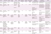
There were a total of 8 tDCS studies for AD, 1 for case report and 7 for clinical trials. Ferrucci et al. [72] reported 10 patients with anodal or cathodal stimulation in the bilateral temporo-parietal area for one 15-minute period. After 30 minutes, word recognition memory was measured. Anodal stimulation showed improvement and cathodal stimulation showed worsening of word recognition memory in this study. Boggio et al. [73] studied 10 patients with AD. Anodal stimulation was given to the left temporal area or left dorsolateral prefrontal cortex for 30 minutes, and visual recognition memory was measured during stimulation. Visual recognition memory was improved during stimulation for each of the 2 sites. Boggio et al. [74] had shown in a subsequent study that this improvement in visual recognition memory lasted up to 4 weeks after the end of the stimulation.
Thereafter, a parallel-group clinical trial of the effects of tDCS was conducted. Khedr et al. [75] performed a double-blind, sham-controlled, parallel-group clinical trial in 34 patients with AD. The left dorsolateral prefrontal cortex was subjected to anodal or cathodal stimulation 10 times for 25 minutes and P300, event-related potential, was measured as well as cognitive function. In this study, both anodal and cathodal stimulation showed a change in cognitive function improvement and shortening of P300 latency. However, in other randomized parallel-group clinical trials, tDCS stimulation did not show significant effects on enhancement of cognitive function [767778] (Table 1). Most of these previous clinical studies were small, and the characteristics of subjects, conditions of stimuli and outcome measures were different from study to study. Therefore, it is difficult to pinpoint the effect of tDCS on AD through these studies.
Table 1
Clinical trials of tDCS in AD
| Articles | Study design | Participant | Sample size | Anode | Cathode | Stimulation intensity, mA | Electrode area, cm2 | Session number/duration | Methods of outcome measures | Time point of outcome measures | Results |
|---|---|---|---|---|---|---|---|---|---|---|---|
| Ferrucci et al. [72] | Randomized-controlled cross over | MMSE > 20 | 10 | Bilateral temporoparietal areas (P3-T5 + P6-T4) | Right deltoid muscle | 1.5 | 25 | 1/15 min | WRT (recognition memory) | Prestimulation, 30 min after tDCS ended | atDCS: improved WRT |
| VAT (attention cue task) | ctDCS: worsened WRT | ||||||||||
| No differences of VAT | |||||||||||
| Boggio et al. [73] | Randomized-controlled cross over | MMSE 12–25 | 10 | Left temporal cortex (T7) | Right supraorbital region | 2.0 | 35 | 1/30 min | Visual recognition memory | During stimulation (test started 10 min after stimulation onset and lasted until the end of stimulation) | Visual recognition memory enhanced after tDCS of both active stimulation |
| Left DLPFC | Stroop test (selective attention) | Stroop test, digit span did not show significant differences | |||||||||
| Digit span test (working memory) | |||||||||||
| Boggio et al. [74] | Randomized-controlled cross over | MMSE > 15 | 15 | Bilateral temporal lobes (T3 + T4) | Right deltoid muscle | 2.0 | 35 | 5/30 min | MMSE | Baseline | VRT improved after anodal stimulation |
| ADAS-Cog | End of treatment day 5 | Improvement persisted for 4 wk after stimulation ended | |||||||||
| VRT | After 1 wk | ||||||||||
| VAT | After 4 wk | ||||||||||
| Khedr et al. [75] | Randomized-controlled parallel-group (1:1:1) | MMSE 12–23 | 34 (anodal 11, cathodal 12, sham 11) | Left DLPFC | Right supraorbital region | 2.0 | 24/100 (anodal/cathodal) | 10 (10 day)/25 min | Cognitive measure: MMSE, WAIS-III | Baseline | MMSE and WAIS were improved after active stimulation |
| Neuropsychological measure: P300, rMT, aMT, CSP | At the end of 10th session | atDCS or ctDCS reduce the P300 latency | |||||||||
| 1 mon after the end of the sessions | |||||||||||
| 2 mon after the end of the sessions | |||||||||||
| Cotelli et al. [76] | Randomized-controlled parallel-group (1:1:1) (active + memory training active + motor training sham + memory training) | MMSE 20–22 | 36 (anodal 12, cathodal 12, sham 12) | Left DLPFC | Right deltoid muscle | 2.0 | 25/60 (anodal/cathodal) | 10 (2 wk)/25 min | Face-naming association memory task | Baseline | Memory training improved the cognitive function |
| Neuropsychological scale | After 2 wk of treatment | Stimulation effect was not existed | |||||||||
| Functional scale | 3 mon after the beginning of treatment | ||||||||||
| 6 mon after the beginning of treatment | |||||||||||
| Suemoto et al. [77] | Randomized-controlled parallel-group (1:1) | MMSE 10–20 with apathy | 40 (anodal 20, sham 20) | Left DLPFC | Right orbit | 2.0 | 35 | 6 (2 wk)/20 min | NPI | Baseline | No differences |
| ADAS-Cog | At the end of the 6th session | ||||||||||
| Depression scale | After 1 wk | ||||||||||
| Bystad et al. [78] | Randomized-controlled parallel-group (1:1) | MMSE > 17 | 25 (active 12, placebo 13) | Left temporal area (T3) | Right frontal lobe (Fp2) | 2.0 | 35 | 6 (10 day)/30 min | CVLT-II | Baseline | No differences |
| MMSE | At the end of 6th session | ||||||||||
| Clock-drawing test | |||||||||||
| TMT A and B |
tDCS, transcranial direct current stimulation; AD, Alzheimer's disease; MMSE, mini-mental state examination; WRT, word recognition task; VRT, visual recognition task; atDCS, anodal tDCS; ctDCS, cathodal tDCS; DLPFC, dorsolateral prefrontal cortex; ADAS-Cog, Alzheimer's disease assessment scale-cognitive subscale; VAT, visual attention task; WAIS-III, Wechsler adult intelligence scale-3rd edition; P300, event-related potential; rMT, resting motor thresholds; aMT, active motor thresholds; CSP, cortical silent periods; NPI, neuropsychiatry inventory; CVLT-II, California verbal learning test-2nd edition; TMT, trail making test.
![]()
CONCLUSION
As tDCS is non-invasive and easy to use, it could be alternative therapeutic method for AD. The mechanisms of tDCS are based on modulation of neuronal resting membrane potentials, synaptic plasticity, cortical neurotransmitters, astrocytes, CBF, and functional connectivity, which may restore cognitive impairment in AD. However, there are inconsistent results in clinical studies in patients with AD. Since most clinical studies so far are pilot studies with small sample sizes, future clinical studies should be more systematic with extensive scale.
 XML Download