

 PDF
PDF ePub
ePub Citation
 Print
Print


INTRODUCTION
While metal-ceramic restorations have favorable characteristics, several problems can be encountered with this treatment modality. The opaque structure of the metal framework, reflection of the metal in the gingiva, and allergy and corrosion related to non-precious metals are some of these problems that often direct the clinician and patient to use all-ceramic restorations.
Due to their high flexural strength, chemical stability, and biocompatibility, zirconia frameworks can also be used in posterior restorations, and have been shown to have properties superior to those of other dental ceramics.
Fabrication procedures for zirconia restorations resemble those for metal-ceramic restorations. Zirconia frameworks are also veneered with low-fusing dental ceramic having a compatible thermal expansion coefficient to achieve better esthetics. Thus, the restoration looks like the natural tooth and shares its esthetic properties.1 The bond strength between the zirconia framework and the ceramic veneer is vital for a successful long-term performance. However, recent clinical trials have shown that the most common failure in zirconia-supported ceramic restorations is due to fracture occurring in veneering ceramic (chipping).2,3,4
Many factors can affect the bond strength between a zirconia framework and the veneering ceramic. Although these factors have been identified, their exact mechanisms have not yet been defined.5 The veneering surface of the framework and the mechanical retention of this surface, compatibility of thermal expansion coefficients, and volumetric shrinkage of the veneering ceramic, viscosity, and wettability - all affects bond strength. Mechanical properties of these materials should also be compatible for good bonding.6
Zirconia frameworks are more esthetically acceptable compared with metallic frameworks but their opaque and whitish appearance remains a handicap. Thus, colored zirconia frameworks were introduced to obtain a more natural-looking color match. The main advantage of colored zirconia ceramics is that they enable color to be reflected from the inner layer, as in the dentin and enamel structure of natural teeth.
Color shading of zirconia frameworks can be accomplished by different techniques, such as the addition of metallic pigments to the initial zirconia powder before or after pressing the milling blocks, the dipping of milled frameworks into the dissolved coloring agents, or the application of liner material to sintered white frameworks.3
In a review of the literature, it was found that there are few studies evaluating the effects of color-shading procedures on the structure of zirconia-based restorations. In some studies, it has been shown that the color-shading procedure affects the structure of the zirconia framework.7,8,9 In the same way, bond strength between colored zirconia frameworks and veneering ceramic has also been examined.10,11 However, only one type of coloring liquid and one length of dipping time were used in these studies.10,11
The aim of the present study was to evaluate whether subjecting zirconia frameworks to different coloring liquids and different lengths of dipping time affects the bond strength between zirconia frameworks and veneering ceramic. The null hypothesis was that the application of different coloring liquids and different dipping times to the zirconia framework does not affect the bond strength between the framework and the ceramic veneer.
MATERIALS AND METHODS
Yttrium partially stabilized zirconia dioxide blocks (ICE Zirkon, Zirkonzahn, South Tyrol, Italy) were cut into discs by means of a low-speed diamond saw (Struers Ltd, Lanarkshire, United Kingdom). In total, 132 discs, 15 × 12 × 1.6 mm, were used as test samples. The samples were divided into six groups: Vita A3, B1, C4, D2, D4 shades, and a non-colored control group. Each group was then divided in half, since each shade group would be subjected to two different (3 or 60 seconds) dipping times.
Test samples (except for the control group) were dipped in the coloring liquid (Zirkonzahn) with plastic holders, held there for 3 or 60 seconds, and dried under a warming lamp (Zirkonlampe 250, Zirkonzahn) for 45 minutes. After the shading procedure, samples were sintered in a sintering oven (Zirkonzahn) according to the manufacturer's instructions (Table 1).
After being sintered, all samples were sandblasted with 50 µm aluminum oxide particles (Al2O3) from a distance of 10 mm for 20 seconds under 3.5 bar pressure, to increase surface roughness and enhance bonding strength. All samples were then cleaned in an ultrasonic cleaner (Quantrex 90, L & R Ultrasonics, Kearny, NJ, USA) for 10 minutes, rinsed, and air-dried.
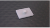
The translucent porcelain (CZR, Noritake Co., Kiza, Nagoya, Japan) was used to veneer zirconia samples. To standardize the veneering procedure, a metal template with a hole corresponding to the center of the zirconia disc was used. The metal template enabled us to apply a standard layer of veneering ceramic (3.5 mm in diameter and 3 mm thick) to the zirconia disc (Fig. 1). The ceramic veneer was then sintered according to the manufacturer's instructions (Table 2).
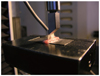
Zirconia discs with ceramic veneers were then placed in the Universal Testing Machine (TSTM 02500 Elista Ltd., Istanbul, Turkey), and shear bond strengths (SBS) of the samples were evaluated at a speed of 1 mm per minute (Fig. 2). The load cell used was a Z-type one with a measuring capability of 5,000 N.
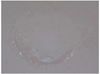
After the SBS test, fracture modes were examined by optical microscopy (Olympus SZ4045 TRPT) at magnifications of 10× and 20×, to determine the types of failure. Photographs of the surfaces were also taken, and fracture types were classified as "cohesive", "adhesive", (Fig. 3).
Statistical analysis of the data was performed by the 'SPSS 15.0 for Windows' package program. Statistical analysis was performed applying repeated measuments ANOVA followed by Tukey's HSDs at a significance level set at P<.05. The paired-samples t-test was used for binary comparisons of variations in the same color after the 3- and 60-second dipping times.
RESULTS
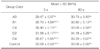
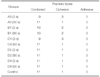
SBSs of the samples according to different color shades and coloring times are given in Table 3. Results show that the bonding strength of zirconia and veneering ceramic can be as high as 36.4 MPa (C4, 3 seconds) and as low as 29.47 MPa (A3, 3 seconds).
When the 3-seconds color-shaded groups were considered statistically significant differences were observed between groups A3 and C4 (P<.05, while there were no statistically significant differences among the other groups (P>.05). No statistically significant differences were observed among the 60-seconds color-shaded groups (P>.05). Color groups were also compared according to shading times (3 seconds vs. 60 seconds), and no statistically significant difference was obtained except for color B1 (P=.036).
Fracture types were classified as "adhesive", "cohesive", or "combined". Fracture types between zirconia framework and veneering ceramic were observed as cohesive and combined, while no adhesive type was observed (Table 4).
DISCUSSION
In the present study, the effects of different coloring liquids and two dipping times (3 seconds and prolonged 60 seconds) on the bond strength between zirconia and veneering ceramic were evaluated. The null hypothesis that different coloring liquids and different dipping times do not affect the bond strength between zirconia framework and veneering porcelain was not accepted. Results show that coloring liquids can affect the bond strength between zirconia framework and veneering ceramic.
It has been shown that bond strength between metal and ceramic should be at least 25 MPa for metal-ceramic restorations to resist fracture stresses.12 In the present study, the bond strengths obtained between zirconia frameworks and veneering ceramics for all groups were between 29.47 and 36.4 MPa. These results were higher than the standard bond strengths of metal-ceramic restorations in terms of durability and clinical performance.
There are studies that compared the SBSs metal-ceramic and zirconia-based ceramic restorations which have showed no significant differences between them13,14 or showed lower bond strengths for zirconia-based restorations than metal-ceramic restorations.15,16,17 The bonding mechanism between metal and layering ceramic may be the reason for this difference.18 However, the bonding mechanism between zirconia and ceramic veneer is still not fully understood.
The studies evaluating the bond strength between zirconia frameworks and veneering ceramics reported values between 16.8 and 48.8 Mpa.3,5,11,19,20,21,22,23,24,25,26,27,28 The mean bond strengths obtained in our study (29.47 MPa-36.4 MPa) for all color groups and time periods are compatible with those reported in the studies mentioned above.
The effect of coloring procedure on the bond strength between zirconia frameworks and veneering porcelain was also investigated. Mosharraf et al.11 evaluated the effects of colored zirconia frameworks and different surface treatments on bond strength and concluded that using colored frameworks had no effect on the bond strength between zirconia and ceramic, while surface treatment had a statistically significant effect. They reported bond strengths between 21.33 and 30.83 MPa. Aboushelib et al.10 investigated the effects of different surface treatments on the bond strength between pre-colored and non-colored zirconia frameworks and layering ceramic (Cercon white and yellow, Lava white and yellow, Procera zirconia). They indicated that using pre-colored or non-colored zirconia frameworks had a statistically significant effect on the bond strength between zirconia and ceramic.
Hjerppe et al.7 examined the effects of different coloring solutions (A3, B1, C4, D2, D4) and dipping periods on the fracture resistance of zirconia by using the same coloring liquids as in the present study and found that the coloring procedure had a negative effect on the fracture strength of zirconia. Those researchers reported that the fracture resistance of zirconia frameworks gradually decreased with prolonged dipping time, and that a 60 seconds dipping time generated a lower fracture resistance for all groups when compared with that of the 3-seconds groups. The present study also showed negative results for prolonged dipping time of 60 seconds, except for the A3 and D2 groups which displayed higher shear bond strength with no statistically significant difference. A prolonged dipping time decreased the bond strength between zirconia frameworks and veneering ceramic. The highest bond strength measurements occurred within the 3-second groups (Table 3). Hjerppe et al.7 also found a statistically significant difference between colored groups and the non-colored control group. However, in the present study, the control group had an average bond strength value of 33 MPa which is similar to that of other groups.
Examining fracture types under a microscope is the most important tool for understanding the mechanism of brittle fracture of materials such as dental ceramics. The location of crack onset, sizes, and types shows how cracks spread, reaching fractures of macroscopic dimensions.29 Combined and cohesive fracture types are the most frequently reported by most studies.13,14,16,22,26,27 In the present study, the fracture surfaces of the test samples were also observed, and it was found that they were composed of cohesive and combined types (combined dominantly), while no adhesive type was observed. In most of the combined-type fractures, a broad layer of ceramic veneer was observed on the fracture surfaces of the samples and also showed that the fractures began as a cohesive type at the side of load application, and terminated as an adhesive type on the opposite side. Choi et al.16 reported similar types of fractures in their study. The initiation of the fracture within the ceramic veneer, and its propagation in this layer, indicates that the bond strength between zirconia framework and veneering ceramic is greater than the fracture resistance of the ceramic veneer.
CONCLUSION
According to the results of the present study different coloring procedures can affect the bonding strength between the zirconia framework and the ceramic veneer. Results also indicated that the bond strength between zirconia frameworks and ceramic veneers is clinically acceptable, even in the weakest group. Combined and cohesive type of fractures observed on the samples showed that bond strength between zirconia framework and ceramic veneer may not be the cause of ceramic veneer delamination.


 XML Download