

 PDF
PDF ePub
ePub Citation
 Print
Print


INTRODUCTION
Geographic tongue (lingua geographica), also known as benign migratory glossitis or annulus migrans, is an inflammatory disorder of unknown etiology caused by local loss of filiform. The condition usually presents as asymptomatic erythematous patches with serpiginous borders. The patches are irregular and sharply demarcated, resembling a map1. Candidiasis of the tongue may resemble or may be associated with geographic tongue.
CASE REPORT
A 63-year-old male patient came to our outpatient clinic with painful white lesions on his tongue. He explained that his lesions had occurred approximately one year prior to arrival. He used topical corticosteroid and antiinflammatory agents, but his lesions did not respond to those therapies. He had likely been evaulated for geographic tongue before coming to our clinic.
There was no history of immunosuppressive drug usage in our patient. On laboratory investigation, there was no evidence of anemia or diabetes mellitus in the complete blood cell count and biochemistry tests. The iron, total iron binding capacity, vit-B12, folate, and ferritin levels were in normal range. Sedimentation rate, posteroanterior chest radiography, abdominopelvic ultrasonography, and tumor markers were also found to be normal. Thus, we did not detect any immunosuppressive condition in the patient.
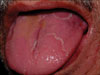
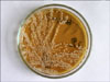
Dermatological examination revealed annular white plaques on the left part of the tongue (Fig. 1). There were no other lesions in the mouth or other parts of the body. Clinically, the diagnosis was thought to be geographic tongue and/or candidiasis. A sample of the lesion from the tongue was obtained with a sterile cotton swab. Direct mycologic examination was performed with potassium hydroxide 15%, and yeast cells with blastoconidia and pseudohyphae were seen microscopically (Fig. 2). The sample was also cultured at 37℃ on Sabouraud's dextrose agar (SDA), and typical yeast colonies were determined after 72 hours (Fig. 3). Growth was identified as C. albicans by germ-tube production, and further examination revealed pseudohyphae with clusters of blastoconidia at the septa and terminal chlamydospores on cornmeal-Tween 80 agar after a passage from SDA using the Dalmau method.
We prescribed fluconazole 150 mg once a week for two weeks and asked the patient to gargle with nystatin three times a day for ten days. With that topical and systemic antifungal therapy, complete clinical improvement was observed. As a result, we determined that the patient was more likely to have candidiasis than geographic tongue.
DISCUSSION
Geographic tongue is also known as lingua geographica, transitory benign plaques of the tongue, glossitis areata exfoliativa, and benign migratory glossitis. In some patients, it is a manifestation of atopy or psoriasis, but generally it is an isolated finding. The dorsal surface of the tongue is the site that is usually affected. Geographic tongue begins with a depression on the lateral border or tip of the tongue, smoother and redder than the rest of the tongue surface. It spreads peripherally, with the formation of sharply circumscribed ringed or gyrate red patches, each with a narrow yellowish white border, making the tongue resemble a map. The appearance changes from day to day. Patients occasionally complain of glossodynia1. There is no race or gender predilection for geographic tongue, which is seen in approximately 2% of the population. It appears to be more common among children. The etiology is unknown2. Zargari investigated the prevalence and significance of fissured tongue and geographical tongue in psoriatic patients and suggested that geographical tongue is common in early onset psoriasis and may be an indicator of disease severity3.
Two clinical variants of geographic tongue have been described. In one type, discrete, annular, "bald" patches of glistening, erythematous mucosa with absent or atrophic filiform papillae are noted. Another type shows prominent circinate or annular white raised lines that vary in width up to 2 mm1,4.
Candidal infection of the oral mucosa may resemble or may be associated with geographic tongue. Infection of the oral mucosa with candida may present with different clinical symptoms5. Acute pseudomembraneous candidiasis or thrush is the most common form of oral candidiasis. Predisposing factors include diabetes mellitus, systemic steroid use, antibiotic use, pernicious anemia, malignancies, radiotherapy to the head and neck, and cell-mediated immunodeficiency6. In pseudomembranous candidiasis, the characteristic sign of this condition is a sharply defined patch of creamy, crumbly, curd-like white pseudomembrane, which, when removed, leaves an underlying erythematous base. It may be seen in healthy infants or secondary to immunsupression. Acute erythematous candidosis (acute atrophic oral candidiasis) is another form. In that condition, there is marked soreness and denuded atrophic erythematous mucous membranes on the dorsum of the tongue. The condition is associated with broad-spectrum antibiotic therapy, glucocorticoid use, and human immunodeficiency virus infection. There is both an asymptomatic and symptomatic variant, the latter of which is characterized by burning or pain7. Chronic atrophic candidiasis (denture stomatitis) is a common form of oral candidiasis seen in 24 to 60 percent of those wearing dentures. Female patients are affected more commonly than males. Clinical findings of chronic erythema and edema of the palatal mucosa that contacts the dentures as well as angular cheilitis are present. Candidal cheilosis (angular cheilitis), so-called perleche, is characterized by erythema, fissuring, maceration, and soreness at the angles of the mouth. That condition is commonly encountered in habitual lip lickers, usually in the young, and in elderly patients with sagging skin at the oral comissures. It is often associated with chronic atrophic candidiasis due to denture wear6. The other clinical form of oral candidiasis is chronic hyperplastic candidiasis with a single fixed plaque, falling into the clinical spectrum of leukoplakia8.
In the literature we did not find any case report of mucosal candidiasis with sharply circumscribed annular lesions, except a case of candida keratitis with annular infiltration9. Thus, it should be kept in mind that mucosal lesions of mycotic infections may appear in annular configuration, such as in dermatophyte infections of the skin.
With this interesting case, we discussed the clinical forms of oral candidiasis and suggested that clinical presentation of mucosal candidiasis may vary according to the stage of infection and individual factors.

 XML Download