

 PDF
PDF ePub
ePub Citation
 Print
Print


INTRODUCTION
The supporting tissues of the periodontium are vulnerable to the physiological variations in the levels of circulating steroid hormones in both males and females. Several pathways exist for estrogens and progestins, as well as androgens to exacerbate gingival inflammation and promote periodontal disease.1 Periodontal alveolar bone loss could be due to systemic osteoporosis, but more so to periodontal disease.2 Several authors have suggested that the mandibular bone density may be indicative of the systemic bone density.3,4 There has been increasing interest in the relationship between systemic osteoporosis, oral bone loss, tooth loss and risk factors for these conditions. Changes in bone metabolism and decrease in the bone mineral component (BMC) of the skeleton especially the jaw may be a stimulating factor of periodontal disease.3,4 This relationship is difficult to establish, as the results may easily be confounded by other factors, such as gender, hormone intake, smoking, race and age.5
It has been speculated that osteopenia/osteoporosis plays a role in the progression of oral bone loss following the menopause3,6,7 Although osteoporosis is more common in women, it also occurs in men with hypogonadism of prepubertal onset.8,9 Hypergonadotropic hypogonadism (primary hypogonadism) is characterized by a low testosterone level, high levels of gonadotropins and elevated luteinizing hormone (LH) and follicle-stimulating hormone (FSH).10 Hypogonadism is a well-established cause of osteoporosis in men and hypogonadal osteoporosis is associated with increased bone resorption and decreased mineralization. The pathogenesis of bone loss in hypogonadal men remains uncertain, although it has been suggested that it may be due to the direct effects of androgen, estrogen deficiency, malabsorption of calcium and reduced circulating calcitonin levels.8
Recently, biochemical markers for bone metabolism have been developed and are expected to reflect minor changes of bone turnover.11 The rate of bone formation or degradation can be assessed either by measuring an enzymatic activity of the osteoblastic or osteoclastic cells, such as that of the alkaline and acid phosphatase or by measuring components of the bone matrix that are released into circulation during formation or resorption.12 The alkaline phosphatase (ALP) activity is the most commonly used marker of bone formation. ALP has been found in various periodontal tissues13,14 and its serum level can be used as an indicator of bone formation.12 Studies have revealed that ALP can often be measured in the gingival crevicular fluid (GCF) to examine its relations to periodontal conditions and disease activity.15-18 Ishikawa and Cimasoni19 demonstrated that enzyme levels in the GCF were 3 times higher than those in the serum and significant correlation was shown between the ALP concentration in the GCF and the pocket depth. Binder et al.15 performed a longitudinal study of 8 patients and demonstrated a positive relation of the GCF alkaline phosphatase concentration with attachment loss.
To date, the studies have focused on the relationship between periodontal disease and osteoporosis in relation to the hormonal changes in women.3,6,20 However, there have been a limited number of studies that show the effects of the hormonal changes in men on periodontal tissues.1 However, there is no information concerning the periodontal condition in men with hypergonadotropic hypogonadism. The aim of this preliminary study was to determine the possible relationship between ALP levels in the GCF and periodontal disease in men with hypergonadotropic hypogonadism.
MATERIALS AND METHODS
Study population
A total of 41 patients, divided into 4 groups, participated in this study. Twenty males with HH (18 to 20 years old; mean age 20.81 ± 1.33 years) were derived from an army population at the Gulhane Military Medical Academy (GATA). The patients in need of periodontal treatment were referred from the Endocrinology Department to the Periodontology Department. The study was approved by the Ethical Committee of the GATA, Sciences of Dentistry, Ankara, Turkey. The informed consent of all subjects was obtained and all procedures were fully explained before the study. After clinical and radiographic examinations, 9 males with gingivitis (G), with a mean age: 20.45 ± 0.20 years and 11 males with periodontitis (P), with a mean age: 21.44 ± 0.29 years, were selected from the HH patients.
There were 21 volunteers in the systemically healthy control (C) group (9 males with periodontitis (P), with a mean age: 20.33 ± 0.44 years and 12 males with periodontally healthy (H),with a mean age: 21.17 ± 0.36 years) were drawn from staff at the GATA. The periodontitis patients were selected based on the radiographical evidence of bone loss and the presence of four or more sites with pocket depths > 5 mm. The gingivitis patients had no evidence of bone loss but varying degrees of gingival inflammation. The periodontally healthy individuals were selected on the basis of no radiographic evidence of alveolar bone and attachment losses, with no probing depths greater than 3 mm and no significant bleeding on probing. The subjects had not received professional teeth cleaning or systemic medication, including androgen substitution drugs, during the 6 months prior to the study.
Periodontal examination
The clinical evaluation of patients was based on the following parameters: the plaque index (PI),21 gingival index (GI),22 probing depths (PD) and attachment level (AL). All parameters were measured with a Williams probe calibrated in millimeters. All clinical parameters were performed on 6 points per tooth (mesio-mid and disto-buccal; mesio-mid and disto-lingual). Clinical examinations were carried out by the same investigator.
GCF sampling and processing
In order to avoid blood contamination and possible stimulation of GCF flow during the clinical measurements, the GCF samples were collected before any other clinical recordings. The GCF sample of each subject was collected from the maxillary anterior area. Prior to GCF collection, the supragingival plaque was removed. The sample site was gently dried using an air syringe and isolated from saliva with cotton rolls. The GCF was collected using Whatman chromatography paper (2 × 8 mm). Paper strips with a notch, were inserted 1 mm into the gingival sulcus and left in place for 30 seconds.23 Care was taken to avoid any mechanical injury. Strips contaminated with blood or exudate were discarded. The amount of GCF on the strips was measured by weighing the accumulated fluid. The paper strips were placed into coded sealed plastic microcentrifuge tubes. To determine the amount of GCF, an electronic balance (Precisa 62 A, Precisa Instruments AG, CH-Dietikan, Zürich, Switzerland) was used for weighing the paper strips before and immediately after the collection. The strips were stored at -70℃ until assayed. The mass (mg) of the fluid on each strip was converted to a volume in milliliters (mL) by assuming a GCF density of 1.24
GCF and serum enzyme-linked immunosorbent assay (ELISA) analysis for ALP
The levels of ALP in the GCF were measured using an ELISA kit (Biomereux, SA, Marcy I'Etoile, France) according to the manufacturer's instructions. The ALP levels in the GCF samples were determined from samples on filter paper strips placed in 96-well microtiter plates. Two hundred and fifty µl of AMP buffer (pH 10.5 in amino-2 methyle-2 propanol-1 buffer) were dispensed into each well and the strips removed after soaking for 90 min at room temperature.
Fifty µL of 0.95 mM p-nitro phenylphosphate in buffer AMP was added to each well and the plate incubated for 45 min. Fifty µL of 1 M NaOH were added. Optical density readings of each well were measured at 405 nm. by using ELISA reader (Bio-Tek Instrument inc. Vinooski, VT, Vermont, USA). The final results were expressed as corrected optical densities or enzyme units, where 1 unit = 1 µmol p-nitro phenylphosphate converted to pnitro phenol plus inorganic phosphate per min at the pH and temperature indicated for each enzyme.
Serum samples were assayed using the protocol described for the GCF. The optical density was measured using a Technicon Dax-48 analyzer (Bayer Diagnostica, Leverkusen, Germany).
Statistical analysis
The mean values and standard errors of the measured parameters were assessed by a one-way analysis of variance (ANOVA). The significant differences were examined using the Duncan test.
The correlations between the GCF enzymes and clinical parameters in all groups were examined by a Pearson correlation analysis. All calculations were undertaken using a statistical software package (SPSS v.10.0 Inc., Chicago, IL, USA).
RESULTS
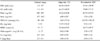
The biochemical parameters for diagnostic evaluation of the Hypergonadotropic hypogonadism patients are presented in Table 1.
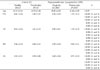
The clinical parameters data (mean ± SE) for all groups are presented in Table 2. When considering the mean age, there were no differences among the groups. No significant difference could be detected in the mean clinical parameter data between the P/HH and P/C groups (p > 0.05). When the PI, GI, PD and AL scores were considered, the periodontally healthy control group (H/C) exhibited significant differences compared to all the other periodontitis groups (p < 0.001). The periodontitis patients in both groups (P/C and P/HH) had higher mean probing depths than the H/C and G/HH patients (p < 0.001). There was no difference between the pocket depths of the G/HH and H/C patients.
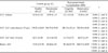
The mean GCF and serum ALP levels and GCF volume are summarized in Table 3. Periodontitis patients (P/HH and P/C) demonstrated significantly increased GCF volumes compared to healthy (H/C) and gingivitis (G/HH) patients (p < 0.01). In the P/HH group, the mean total ALP level was higher than in the H/C and G/HH groups. The difference was found to be statistically significant (p < 0.01), while that between the P/HH and P/C groups was not (p > 0.05). The healthy control group (H/C) presented lower ALP concentrations than the other patient groups. The differences between the H/C and P/C group and H/C and P/HH group were found to be statistically significant (p < 0.01). The P/HH group showed higher serum ALP than the other groups which were statistically significant (p < 0.001).
Table 4 shows the correlations between the clinical parameters and the GCF enzyme activity in all groups. In the P/HH group, the total ALP activity and ALP concentration presented negative and non-significant correlations with PI and GI but a positive correlation with the mean PD (p > 0.05). In the P/C group, the total ALP activity and ALP concentration exhibited weak-positive correlations with PI, GI and AL but strong-positive and significant correlations with PD (p < 0.05). There was a strong negative correlation between the GCF volume and GI scores in the P/HH group (p < 0.05).
DISCUSSION
Hypogonadism is one of the most frequent causes of osteoporosis in men.25,26 The diagnosis of hypogonadism may not always be clinically apparent and the routine measurements consider all definable causes for osteoporosis in men.27 In the present study, the patients were diagnosed randomly from an army population during routine laboratory testing. The diagnostic biochemical evaluations for osteoporosis in hypogonadism patients are presented Table 1. Low serum testosterone levels were seen in our hypogonadism patients and it's insufficiency is well known as a major risk factor for male osteoporosis.8,25
ALP has often been measured in the GCF to examine relations to periodontal conditions.14 The aim of this preliminary study was to assess the relationship between clinical periodontal conditions and alkaline phosphatase levels in men with hypergonadotropic hypogonadism.
No published report on the GCF ALP level in hypergonadotropic hypogonadism patients could be found, so our data could not be compared with similar studies. In the present study, no statistically significant differences could be detected between the two periodontitis groups (P/HH-P/C) with any of the clinically parameters. These results indicate that the degree of periodontal disease was similar in both groups. In the present study the level of supragingival plaque accumulation was found to be significantly different between the P/HH and G/HH, and the H/C and P/C groups. There is no doubt that bacterial plaque is a major cause of the initiation and maintenance of gingival inflammation.
Ishikawa and Cimasoni19 reported that ALP levels in the GCF harvested from periodontitis correlated positively and significantly with the pocket depth and the mean percentage of bone loss. Our findings are in accordance with those of previous studies.15,16,19 Total amounts and concentrations of GCF ALP were significantly and positively correlated with the probing depth in our P/C group. The present study also supports the findings of previous studies that reported increased GCF ALP levels in the presence of periodontal destruction.16 Ishikawa and Cimasoni19 demonstrated that the GCF ALP levels were significantly higher from those in the serum. In the present study, the ALP levels in the GCF from both the patients and control groups were found to be higher than those measured in the serum, which is contradictory to previous findings that indicate the local production of significant amounts of ALP by periodontal tissues or within the periodontal pocket. In addition the ALP within the GCF is probably a mixture of that from the serum, host tissue, polymorphonuclear leukocytes (PMNL) and bacterial origin.15
While periodontal disease is a local disease, hypergonadotropic hypogonadism is a systemic disease. Bone loss is a common feature of both diseases. Biochemical markers, including the ALP, have also been associated with bone loss from periodontal disease. We could suggest that systemic bone loss in these patients may lead to periodontitis with increased ALP levels.
Both the concentrations and total amounts of ALP were significantly higher in the periodontitis groups compared to the healthy and gingivitis groups (Table 3). However the mean total GCF ALP in the P/HH group was higher than that in the P/C group, but no significant difference was found between the two periodontitis groups. The total amount in the GCF correlated better rather than the enzyme concentrations.28 It is possible to suggest for the ALP, the data presented as total activity are more precise in reflecting the existing clinical periodontal status compared to the concentration expressed. The concentration of GCF ALP is dependent upon the GCF volume. The GCF volume is also influenced by many other factors such as the flow rate, gingival trauma and repeated sampling.29,30 It is logical to expect that the total level of this enzyme in gingival fluid will correlate better with the disease than the enzyme concentrations, as in the study of Chapple et al.17 According to our data, It has been shown that the GCF volume increases in the order healthy, gingivitis and periodontitis patients and reflects the presence of gingival inflammation.
The findings of this study suggest a role of HH in periodontal disease could be implicated as a contributing factor to the progress of periodontal disease. However, no data in the medical literature suggest an interaction between periodontal destruction and HH in men. Therefore additional studies are needed to define the relationship between hypergonadotropic hypogonadism in men, alveolar bone loss and periodontal disease.

 XML Download