

 PDF
PDF ePub
ePub Citation
 Print
Print


INTRODUCTION
Cerebrovascular disease is a leading cause of death and a major cause of permanent neurological and physical impairment in adults. In the United States, cerebrovascular disease is the fifth most common cause of death, with approximately 795,000 stokes occurring each year.1) Cerebrovascular disease is also the third leading cause of death in Korea; 48.2 persons per 100,000 die from cerebrovascular disease every year.2) Among several types of cerebrovascular disease, ischemic stroke is the most prevalent, with approximately 15–20% of all ischemic strokes occurring from atherosclerotic carotid artery stenosis, particularly in the internal artery. Significant carotid artery stenosis is observed in about 0.5% of patients aged 60–79 years old, while it is observed in about 10% of patients aged 80 years old and over. Most patients have no symptoms. Symptomatic carotid artery stenosis is defined as stenosis in the internal carotid artery with cerebral symptoms associated with ipsilateral carotid lesions. It is a significant cause of ischemic stroke, and patients with symptomatic carotid artery stenosis have increased risk of recurrent cerebrovascular events.
Carotid revascularization prevents recurrent ischemic stroke in patients with significant symptomatic carotid artery stenosis. Carotid endarterectomy has been the gold standard treatment for symptomatic significant carotid artery stenosis for more than 60 years.3) Carotid artery stenting (CAS) (or carotid artery stent implantation) has developed rapidly over the last 30 years, and its frequency is increasing because it is less invasive than carotid endarterectomy with a low risk of cardinal injury and fewer surgical complications.4) However, the value of CAS remains unclear in managing patients with significant carotid artery stenosis. In this review article, we discuss the basic concepts and procedural techniques of CAS.
STROKE AND ITS ASSOCIATION WITH CAROTID ARTERY STENOSIS
The estimated prevalence of stroke increases with age. In 2014, approximately 2.8% of the general population over 18 years of age had a history of stroke.1) Additionally, about 795,000 people had a new or recurrent stroke in each subsequent year, and ischemic stroke accounts for about 87% of the total stroke cases in the United States. There were 6.5 million stroke deaths worldwide,5) accounting for approximately one of every 20 deaths in 2014 in the United States. Moreover, 3% of males and 2% of females were disabled from stroke in 2014.4) In 2010 in the U.S., ischemic stroke-based loss of disability-adjusted life years was quantified at 39.4 million.6) In Korea, stroke is the second leading cause of death.7) From 2000 to 2010, the annual increase in stroke hospitalization was 6.4%, and health care expenditures increased by 13.2%.
There is a close association between stroke occurrence and carotid artery stenosis, which can cause cerebral embolization and transient ischemic attack (TIA) or thromboembolic stroke. TIA is a warning sign and is often followed by ischemic stroke, and carotid artery stenosis accounts for 20% of all ischemic stroke incidence.8) Carotid artery stenosis should be identified in patients with ischemic stroke or TIA, and it can usually be diagnosed by duplex ultrasonography with color Doppler imaging of the carotid arteries. Duplex ultrasonography is a widely available first-line imaging modality that can evaluate hemodynamic status. It is also useful in decision making.9) Ultrasound examination can be used to diagnose significant carotid artery stenosis if the peak systolic velocity is greater than 250 cm/sec or if the end diastolic velocity is greater than 120 cm/sec. Moreover, a lipid-rich or heterogeneous hemodynamic profile indicates the presence of high-risk plaques.10)11)
Other noninvasive imaging modalities include computed tomography (CT) angiogram and magnetic resonance imaging (MRI). Either of these modalities can be used if the carotid stenosis is far from the carotid bifurcation and cannot be detected by duplex ultrasonography.12)13) In cases of symptomatic significant stenosis (≥70%), carotid revascularization with endarterectomy or intervention are established treatments to reduce further stroke risk. Either magnetic resonance angiography or CT angiography should be utilized to evaluate aortic arch anatomy as well as carotid morphology in patients with planned CAS (class I B indication).14)
CAROTID ARTERY REVASCULARIZATION
History and clinical outcomes of carotid endarterectomy
The first carotid endarterectomy was performed in 1953, and has since been regarded as the gold standard treatment for symptomatic carotid artery stenosis.15) However, there have been concerns about complications and efficacy of the procedure, and large-scale multicenter studies have been conducted to address these issues. Among these studies, the European Carotid Surgery Trial (ESCT) and the North American Symptomatic Carotid Endarterectomy Trial (NASCET) showed that carotid endarterectomy is helpful in preventing ischemic stroke in patients with symptomatic carotid artery stenosis.16)17)18) The Asymptomatic Carotid Atherosclerosis Study (ACAS) and the Asymptomatic Carotid Surgery Trial (ASCT) are 2 representative studies evaluating the effects of carotid endarterectomy for asymptomatic carotid artery stenosis.19)20) Both studies demonstrated that carotid endarterectomy can be effective for the long-term prevention of ischemic stroke. Based on the NASCET and ESCT results, the number needed to treat (NNT) to prevent one stroke event was as low as 5 in patients over 75 years of age, and the time from the last event was less than 2 weeks.21)
However, there are still questions about the effects of carotid endarterectomy because patients with asymptomatic carotid artery stenosis have low annual stroke incidence, and there are complications and costs associated with the surgery. Moreover, the development of pharmacologic agents such as antiplatelet agents, statins, and angiotensin-converting-enzyme inhibitors can stabilize atherosclerotic plaque and reduce the incidence of ischemic stroke. Studies such as Stent Protected Angioplasty versus Carotid Endarterectomy (SPACE)-2 that compare surgery with stenting and best medical therapies are currently underway.22)
There are discrepancies between neurologists and surgeons in interpreting the results of clinical studies. In a meta-analysis of 51 studies conducted between 1980 and 1996, the stroke frequency evaluated by neurologists was significantly higher than the stroke frequency evaluated by surgeons (7.7% vs. 2.3%).23) In terms of the interpretation of clinical findings, it is important to consider that results depend on the skill of the surgeon performing the operation, the age and general condition of the patient, and the complexity of the lesion.
History and clinical outcomes of CAS
Intervention for symptomatic carotid artery stenosis was introduced in early 1980. CAS has increased dramatically over the last 30 years because of advantages including convenience, rapid recovery, low complications, and the introduction of devices to prevent embolic stroke. Since 2000, researchers have published large-scale clinical studies comparing carotid endarterectomy with CAS, including the Sapphire, Endarterectomy versus Angioplasty in Patients with Symptomatic Severe Carotid Stenosis (EVA-3S), SPACE, the International Carotid Stenting Study (ICSS), and the Carotid Revascularization Endarterectomy versus Stenting Trial (CREST).24)25)26)27)28) The EVA-3S was the first clinical study to demonstrate that CAS with a filter device is equivalent to carotid endarterectomy in the development of clinical events over 3 years.24) In fact, in each of the 3 large studies conducted in Europe, EVA-3S, SPACE, and ICSS, stent implantation for symptomatic carotid artery stenosis showed more clinical events than did carotid endarterectomy.20)23)25) Regardless, these studies have several limitations, including low practitioner proficiency in the CAS group and limited use of embolic protection devices (EPDs). The CREST was the largest clinical trial performed in the United States. In this trial, highly experienced interventionists performed CAS using EPDs in more than 95% of cases. The incidence of stroke in the stent group was 4.1%, which was significantly higher than that in the carotid endarterectomy group (2.3%). However, the rate of myocardial infarction was significantly lower (1.1% vs. 2.3%), showing that the frequency of clinical events between the 2 groups was similar.24) When we divided the total period of the CREST (from 2000 to 2008) into 2 stages, the rate of clinical events of the CAS group was 5.7% at the initial stage, but dropped to 1.1% at the late stage, suggesting that the improved skill of the interventionists from repeated procedures reduced the number of clinical events during the trial. In a recent publication on long-term CREST results, there were no differences in clinical outcomes including death, myocardial infarction, and stroke.29) Moreover, the incidence of post-procedural ipsilateral stroke did not differ between endarterectomy and stenting groups.
INDICATIONS FOR CAROTID ARTERY REPERFUSION AND SELECTION OF A REPERFUSION TECHNIQUE
Indications for carotid artery reperfusion
Because patients with more than 60% symptomatic carotid artery stenosis have an annual risk of ischemic stroke greater than 10%, the performance of reperfusion is recommended to reduce embolic risk. In cases of asymptomatic severe stenosis, the effects of reperfusion are small because the annual stroke recurrence rate is only about 2%. Therefore, it is desirable to limit the use of reperfusion therapy in patients with asymptomatic carotid artery stenosis greater than 80%. In addition, reperfusion therapy provides maximal benefits to recently symptomatic patients if it is performed within 14 days.14) However, carotid reperfusion therapy can be considered even in asymptomatic patients with less severe stenosis in the presence of occlusion of the contralateral carotid artery, rapid progression of stenosis during clinical follow-up, the presence of a cerebral infarct on brain imaging that was not clinically manifest, and/or vulnerable plaque found on Duplex ultrasonography.
Issues to consider in selecting a reperfusion method
CAS is preferred in patients who will have difficulty undergoing carotid endarterectomy or who are at high risk of complications during surgery. The anatomical findings for lesions should be considered, as shown in Table 1. Conversely, carotid endarterectomy is preferred over CAS if it is difficult to reach the lesion site via the blood vessel. It is advantageous to choose carotid endarterectomy when stent implantation is difficult or is expected to have large complications, including type III aortic arch, bovine arch, severe proximal bending, severe calcification of the lesion, occlusion from the thigh, or severe bending of the aorta with calcification.
Table 1
Clinical situations and anatomical findings when CAS is preferred

STANDARD PROCEDURE FOR CAS
Entry into the common carotid artery
Recognizing the type of aortic arch and the variant anatomy of the cervicocerebral circulation is required for successful carotid stenting. Pre-procedural evaluation using MRI angiograms not only helps to distinguish aortic arch type (Figure 1), but also reduces the use of contrast agent by eliminating the aortogram from in the procedure.
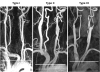
Figure 1
Types I, II, and III aortic arches on magnetic resonance angiograms of 3 patients with symptomatic significant left proximal internal carotid artery stenosis. The type of aortic arch is based on the vertical distance between the origin of the brachiocephalic artery (dotted line) and the top of the arch (solid line). In type I, this distance is less than 1 LCCA diameter. In type II, the distance is between 1 and 2 LCCA diameters, and in type III, the distance is greater than 2 LCCA diameters. Pre-procedural aortic arch evaluation using magnetic resonance angiograms not only helps to distinguish arch type, but also reduces the use of contrast agent by eliminating an aortogram from the procedure.
LCCA = left common carotid artery.
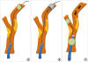
Most surgeons perform stenting via the femoral artery. However, the radial or brachial arteries can be used if there is obstruction of the access pathway or severe tortuosity of the aortic arch. Because approximately half of patients with carotid artery disease have significant co-morbid coronary artery disease, preoperative coronary angiography is mandatory prior to carotid engagement. It is safe to evaluate the degree of cerebral blood flow and collateral circulation by performing contralateral carotid artery angiography, vertebral artery angiography, or intra-cranial angiography before intubating the culprit carotid artery with the obstructive lesion. In cases of type I or II aortic arch, it is easy to enter the common carotid artery using a slightly curved catheter — such as a Judkins right (JR) or Headhunter catheter — which includes a 0.035-inch Terumo wire. During this procedure, it is advisable to contact the aortic arch as little as possible. In order to enter the left common carotid artery, it is often possible to insert the JR catheter into the innominate artery and then slowly pull the catheter with its tip facing toward the head. If this method does not work, insert the catheter by turning counterclockwise or entering the artery with clockwise rotation. For a bovine arch, it is advantageous to use Judkins left, Mammary, or Simmons catheters. Type III aortic arch with calcification is difficult to access; if access with a general catheter is not feasible, the surgeons can use Simmons or Sidewinder catheters. If the surgeon inserts a Simmons catheter into the opposite iliac artery and pushes it toward the abdominal aorta, it is possible to create an inverted U-shaped curve in the catheter (Figure 2A). After the surgeon enters the left subclavian artery, the Simmons catheter can be flexed, or it can be forced into curved flexion by pushing it with the support of the aorta (Figure 2B). The Simmons catheter can be introduced into either the innominate or left carotid arterial ostium by rotation and manipulation and, subsequently, can be deeply engaged with slow withdrawal of the catheter (Figure 2C).
Figure 2
Engagement methods with the Simmons catheter. (A) If the Simmons catheter is inserted into the opposite iliac artery and then pushed toward the abdominal aorta, an inverted U-shaped curve of the catheter can be made. (B) After entering the left subclavian artery, the Simmons catheter can be flexed, or the catheter can be forced into curved flexion by pushing it with the support of the aorta. (C) The Simmons catheter can be introduced into either the innominate or left carotid arterial ostium by rotating and manipulating and can subsequently be deeply engaged with slow withdrawal of the catheter.

Some interventionists use a telescoping technique that introduces a longer Headhunter or Vitek catheter into the Shuttle sheath, subsequently inserting the sheath directly into the common carotid artery.
Intubating the guiding catheter or sheath into the common carotid artery
If a surgeon is using a distal filter-type protection device, then a JR catheter of size 8 on the French scale (Fr) can be directly intubated into the common carotid artery (although, in most cases, the catheter can also be inserted following the insertion of a 6–7 Fr Shuttle sheath). With a protection device, the surgeon should enter the diagnostic catheter into the external carotid artery first. Moreover, the catheter should be inserted into the external carotid artery first in order to utilize a Mo.Ma Ultra device (Medtronic, Minneapolis, MN, USA), a balloon-shaped proximal protection device. A 0.035-inch soft Terumo guide wire is inserted into the external carotid artery following insertion of a 5 Fr JR catheter into the common carotid artery, and the JR catheter is fully inserted into the distal portion of the external carotid artery. At this point, if support from the Terumo induction wire is insufficient, a stiffer guide wire can be used. If the surgeon fully inserts the JR catheter into the distal portion of the external carotid artery, the Terumo guide wire should be removed, and a rigid 0.035-inch Amplatz wire should be inserted. Following insertion of the Amplatz wire, the JR catheter should be removed, and the surgeon should use a Shuttle sheath or Mo.Ma Ultra device to reach the desired portion of the common carotid artery.
CAS with a distal filter device
The surgeon begins CAS after inserting an 8 Fr JR catheter or 6–7 Fr Shuttle sheath into the common carotid artery to the proximal portion of the lesion. The surgeon can cautiously introduce a filter device through the lesion using road mapping or small contrast testing. In cases of severe stenotic lesion, the surgeon can insert a filter device after pre-dilation with a small 1.0–2.0-mm balloon. The filter device begins to expand after it is inserted into the internal carotid artery at the cranial level at least 3 cm above the lesion. In most cases, the lesion is pre-dilated with a 3.0-mm balloon to enable passage of the stent. Using road mapping, or placement of a carotid stent without contrast testing, with reference to the spine or chin line is beneficial for preventing air embolism. After the surgeon places the carotid stent, post-dilation is performed with a 5.0-mm balloon. For safety reasons, we prefer to expand the lesion with a balloon of this size (5.0 mm). It is advisable to avoid excessive post-dilation with balloons that are too large. Some interventionists do not perform post-dilation due to increased risk of carotid sinus reflex or embolism, but post-dilation gives the stent a larger minimum internal luminal diameter, thus reducing the incidence of re-stenosis or thrombosis within the stent. Post-dilation also facilitates removal of the filter device. Accordingly, we perform post-dilation in most cases. Following post-stent balloon expansion, the surgeon should remove the filter device using a prepared removal catheter for insertion to the point just before the filter. The surgeon should then pull out the filter device, insert the filter orifice properly in the catheter, and remove the catheter. If the filter device is pulled with too force, foreign materials trapped inside the filter can be squeezed out. Sometimes, access to the filter device can be difficult or the removal catheter can get caught at the edge or inside of the stent, particularly when the carotid artery is severely calcified or tortuous.30) In this situation, the removal catheter can usually be inserted by turning the patient's neck or manually compressing the stent insertion site to deform the flexion of the carotid artery. Replacing the retrieval catheter with an angulated 5 Fr catheter, such as a Headhunter, Davis, or multipurpose catheter, will allow the filter device to be extracted. This situation might require a guide wire extensor, and any 5 Fr catheter should be long, usually more than 125 cm. If it is difficult to insert the catheter after these attempts, an additional 0.014-inch guide wire can be inserted, after which the removal catheter should be inserted following ballooning inside the stent. Once the filter device has been removed, it is necessary to assess complications by checking for intracranial cerebral angiography as well as checking the site of the procedure.
CAS using a Mo.Ma Ultra device
Before inserting the Mo.Ma Ultra device, the surgeon attaches a Y-connector to the middle port and inserts a dilator. Occlusion balloons for the external carotid artery and common carotid artery should be deflated, air in the balloons should be removed using prepared valves and syringes, and the balloons should be prepared by connecting a 10-mL locking syringe filled with 3–4 mL of 1:3 diluted contrast medium. Then, the surgeon should insert a 9 Fr Mo.Ma Ultra device through a rigid Amplatz guide wire inserted into the internal carotid artery. At this time, it is advantageous to insert the 25-cm-long 9 Fr femoral sheath for the femoral artery, subsequently introducing the Mo.Ma Ultra device into the sheath. The sheath allows continuous monitoring of systemic pressure during the entire procedure (Figure 3) and facilitates entry of the Mo.Ma Ultra device because it alleviates iliac artery tortuosity and calcification to some degree. It is advantageous to use a 9 Fr Mo.Ma Ultra device to reduce air embolization and to more easily manipulate the device. When the end of the Mo.Ma Ultra device is inserted into the external carotid artery, the inner dilator with the remaining Amplatz guide wire should be removed. The occlusion site of the external carotid artery is about 0.5–1.0 cm distal of the carotid bifurcation site, and the superior thyroid artery should be blocked if possible. In some cases, the superior thyroid artery is located too close to the bifurcation, and the occlusion balloon can be placed near the carotid bifurcation to block the superior thyroid. If this artery originated from the distal common carotid artery or carotid bifurcation according to the unique anatomy of the patient, then it might not be possible to block it. If occlusion of the external carotid artery is deemed appropriate based on contrast testing, the Amplatz wire should be removed. It is advantageous to insert a 0.014-inch guide wire with a 3.0–4.0-mm balloon for pre-dilation just before the lesion. Because the use of contrast agent is not feasible during procedures with a Mo.Ma Ultra device, it is necessary to determine the precise location of balloon expansion and stent insertion using a landmark, such as the spine or the chin line, before continuing the procedure. If a small amount of contrast agent is injected immediately after the common carotid artery is blocked, the agent will remain at the lesion site for a while, which is helpful for the procedure. This contrast agent fades slowly when the Y-connector is opened during the procedure. After the lesion is pre-dilated, the surgeon should insert the carotid stent, immediately perform post-dilation, and then remove about 20 mL of blood 3 times using 3 prepared syringes of 30–50 mL. In this scenario, the patient's brain function can deteriorate, and there can be temporary impairment of cognitive function and involuntary movement, commonly the result of transient cerebral dysfunction. The surgeon should check all extracted blood through prepared mesh to find any debris. If there is no debris in the second syringe, the blood can be withdrawn from the third syringe. If debris continues to emerge, another 20 mL of blood should be removed. If there are no blood clots confirmed in the last syringe, another 20 mL of blood can be extracted. Following the procedure, the surgeon should normalize blood flow to the brain by releasing the balloons in the external carotid and common carotid arteries in turn. The surgeon can remove the Mo.Ma Ultra device after verifying the success of the procedure, including complications, with angiograms of the lesion site and intracranial artery.
Periprocedural patient care
Diffusion-weighted MRI is recommended for evaluating embolization associated with the CAS procedure. CAS is usually performed under local anesthesia. In the interest of continuous monitoring of a patient's cerebral function during the procedure, tranquilizers and narcotics should not be used. Dual antiplatelet agents, such as aspirin and clopidogrel, should be administered before the procedure. Active clotting time should be maintained for more than 250 seconds by administering 8,000–10,000 units of heparin or 120 units/kg of body weight. Because more than half of patients who undergo CAS have significant coronary artery disease, coronary angiography is advised before a surgeon engages a cerebral vessel. Blood pressure and electrocardiograms should be constantly monitored during the procedure. Because blood pressure and pulse rate frequently decrease due to carotid sinus reflex during procedural inflation of the balloon, it is good to expand the balloon following administration of 0.5–1.0 mg of atropine if the baseline systolic blood pressure (SBP) of the patient is less than 140 mmHg or if the patient's pulse is 60–70 beats/min. Sudden decreases in blood pressure or pulse due to carotid sinus reflex are usually transient; thus, there is no special treatment. In fact, simply encouraging the patient to cough will facilitate recovery if the pulse is too slow. However, hypotension can last for 12–24 hours following the procedure in some patients; in the absence of symptoms in such patients, no special treatments are necessary unless SBP drops below 80 mmHg. It is advantageous to maintain SBP above 90 mmHg in patients with severe stenosis of both carotid arteries, contralateral carotid occlusion, severe aortic stenosis, left ventricular systolic dysfunction, or significant coronary artery stenosis. To increase blood pressure, surgeons can administer a vasoconstrictor, such as dopamine. Although hypotension usually resolves within 24 hours, it is essential to assess the development of complications, such as intraperitoneal hemorrhage. In some patients, SBP remains above 140 mmHg following the procedure, which can cause cerebral hemorrhage or hyperperfusion. The appropriate SBP following CAS is 100–140 mmHg.
During the procedure, careful monitoring of the patient's cerebral function is necessary. One easy way to assess motor function is to have the patient hold in each hand a rubber doll that makes sounds and periodically clap the dolls together to make noise. If the patient's brain function deteriorates during the procedure, the operator should evaluate whether blood pressure or pulse is too low and whether there is a decreased blood supply to the lesion and should check for disturbance of blood flow or cerebral hemorrhage by reassessing the brain image. If an embolism develops in the brain, active clotting time should be restored immediately, and it is advisable to seek the help of a neuro-interventionist who can perform cerebrovascular intervention in the brain. If there is no specific cerebral lesion and no recovery of patient brain function, a CT scan should be performed immediately to confirm intracerebral hemorrhage.
Following the CAS procedure, it is most effective to have the neurologist, rather than the interventionist, monitor the patient's status by measuring blood pressure and cerebral function in the stroke care unit. On the day of the procedure or the next day, diffusion-weighted MRI can confirm whether a new brain lesion has occurred. Carotid ultrasound is often performed 6 months after the procedure and once every year thereafter.
SELECTING A STENT TYPE
Surgeons can use various types of stents for CAS. In cases with stenosis of the proximal part of the common carotid artery, it is advantageous to use a balloon-expandable stent for insertion in the correct position. In addition, a balloon-expandable stent is beneficial even in stenosis of the basal skull or petrous portion of the carotid artery. However, it is recommended to use self-expandable stents rather than balloon-expandable stents in the most common locations of carotid artery bifurcations or the proximal portion of the internal carotid artery. With balloon-expandable stents in these cases, there is a large possibility of embolism during the procedure that can damage or deform the stent due to physical stress from neck movement. A variety of nitinol stents have been developed, which can be tubular or tapered and can be categorized as open, closed, or hybrid according to cell design. Wallstent® (Boston Scientific, Natick, MA, USA) is a representative closed stent; it is not suitable for severely tortuous lesions, but it can be effective with relatively straight lesions with a small chance of embolization during the procedure. Moreover, it has a relatively small stent-mesh size to minimize embolization.
In contrast, open-type stents such as Precise (Cordis Endovascular, Miami, FL, USA), Acculink (Abbott Vascular, Santa Clara, CA, USA), and Protégé RX (ev3/Covidien, Plymouth, MN, USA) have the advantage of reliable insertion with good shape in tortuous lesions or with the irregular surfaces of atherosclerotic lesions. The Cristallo Ideale carotid stent (Medtronic) is a representative hybrid stent, but it was recently withdrawn from the market. The proximal and distal portions of the stent consist of open cells to fit the shape of the vascular bend, while the middle third is comprised of closed cells to prevent embolization. Recently, double-layer mesh stent technology, characterized by sequential aligned annular rings interconnected by bridges, was developed with a well-balanced mix of high flexibility and conformability to accommodate tortuous anatomy as well as proper plaque coverage and to prevent micro-embolization. Because one stent is not suitable for all lesions, it is advisable to choose an appropriate stent considering the lesion characteristics and the clinical status of the patient.
Recently, new-generation double-mesh stents have been made available in several countries. The Roadsaver® carotid artery stent system (Terumo Corp., Tokyo, Japan) is one type of new-generation carotid stent. It has a double-layer micromesh structure that reduces prolapse of atherosclerotic plaque and prevents distal embolization by improving plaque coverage. One recent study published favorable findings in which Roadsaver® stents reduced plaque prolapse and had good short-term clinical outcomes in 150 patients.31) The InspireMD C-Guard® stent (InspireMD, Israel) is another bare-metal stent covered with a micron-size mesh.32) However, large studies are needed to explain the long-term clinical outcomes of new-generation double-mesh stents in CAS.
SELECTING EPDs
The use of EPDs can reduce the incidence of periprocedural embolic events. A meta-analysis including 24 studies reported that EPD use was related to an about 41% decrease in embolic stroke occurrence (relative risk 0.59; p<0.001).33) A prospective registry showed the benefits of EPDs. The incidence of in-hospital death and stroke was 2.1% in patients with an EPD, whereas the rate was 4.9% in patients treated without EPD (p=0.004).34) According to recent guidelines, the use of EPDs should be considered in patients undergoing CAS (class IIa C indication).14) Three types of EPDs have been developed and introduced (Figure 3). The distal balloon occlusion device is rarely used, whereas distal filter and proximal balloon occlusion devices are currently used in clinical practice.
Distal filter devices with 100–150-µm holes prevent embolization with placement in the internal carotid artery. Commonly used instruments include the FilterWire EZ Embolic Protection System (Boston Scientific), Angioguard Guardwire (Cordis Endovascular), Emboshield NAV6 (Abbott Vascular), and the Spider FX (Covidien, Mansfield, MA, USA). The advantages of these devices include maintaining blood flow to the brain, which prevents deterioration of cerebral function, use of contrast testing to help properly position the stent, and the possibility of using a sheath with a relatively small lumen (6–7 Fr). However, distal filter devices must pass the lesion to activate protection. Because distal filter devices are relatively inflexible and bulky, it is difficult to pass the lesion with a guide wire smaller than 0.014 inch. Disadvantages of distal filter devices include limited protection in tortuous internal carotid arteries, failure to block microemboli, and difficulty in removing them following the procedure.
The Gore flow reversal system (WL Gore and Associates, Flagstaff, AZ, USA) and Mo.Ma Ultra (Medtronic) are 2 common proximal protection devices, but only Mo.Ma is available in Korea. The Gore device continuously draws carotid artery blood, filters foreign substances, and injects blood into the femoral vein. In contrast, the Mo.Ma device removes foreign materials by drawing 60–80 mL of blood after blocking the blood flow when stenting is completed. The major disadvantage of these proximal protection devices is complete blockage of the blood flow during the procedure. Thus, if there is poor collateral blood flow or total occlusion of the contralateral carotid artery, the blood supply to the brain can be blocked, and cerebral function in the patient can temporarily deteriorate during the procedure. In addition, the operator cannot perform contrast testing to determine adequate stent location and thus requires a catheter with a larger inner lumen more than 8 Fr. The use of these devices is not difficult with proper experience, and the patient's cerebral dysfunction is usually transient and typically improves after the procedure. The most important advantages include the use of a 0.014-inch guide wire for coronary intervention through the lesion following the onset of embolic protection, the irrelevance of any landing zone tortuosity, and lower incidence of embolization assessed by transcranial Doppler or diffusion-weighted MRI in comparison to distal filter devices.35)36) Meta-analysis data has reported few periprocedural events (2.25% of cases) following the use of Mo.Ma for protective purposes during carotid stenting.37) Table 2 summarizes a comparison of the 2 types of EPDs.
Table 2
Advantages and disadvantages of EPDs

A variety of anatomical structures in carotid bifurcation can interfere with proximal protection, in which cases, distal protection can be applied. Lesions with in-stent re-stenosis, significant stenosis of the common carotid artery, or ostium of the external carotid artery can be treated only with distal filter protection. External arterial occlusion can be treated with distal filter or mono Mo.Ma devices.
COMPLICATIONS ASSOCIATED WITH CAS
Complications associated with CAS include cerebral infarction, cerebral hemorrhage or hyperperfusion syndrome, complications associated with the site of stent insertion, protective device-related complications, systemic complications, and local complications at the puncture site.
Embolic stroke
When researchers evaluated all patients being treated with CAS with transthoracic Doppler, they observed microembolization in almost all procedures. Although diffusion-weighted MRI showed newly developed enhancements in 10–50% of cases, most patients did not develop symptoms. Researchers have reported that the use of proximal balloon-type EPDs has shown a lower incidence of embolic events than the use of distal protection devices.17)18)38) Based on experience and the advancement of instruments, especially the active use of EPDs, recent studies have shown that the incidence of cerebral events has decreased by 3%.37)38)
Hyperperfusion syndrome
The expansion of carotid artery stenosis by CAS rapidly resolves chronic pressure differences in patients, so that a large blood flow and high blood pressure are delivered to the brain parenchyma without adaptation. Most patients show cerebral vasoconstriction from auto-regulation of the brain, and elevated perfusion pressure is restored to a normal level within a few minutes due to this mechanism in CAS. However, in some patients, this auto-regulating ability is impaired due to long-term excessive lowering of cerebral blood flow, which can result in persistently elevated intracranial pressure (lasting from a few hours to days), thereby causing hyperperfusion syndrome.39) Although major manifestations of this complication include headache, vomiting, local seizures, and various degrees of unconsciousness due to increased intracranial pressure, the complication can lead to fatal cerebral hemorrhage. These findings are evidence of contralateral carotid occlusion, a poorly developed circle of Willis in the anatomy of patients, near total stenosis with excessive blood flow reduction, or simultaneous bilateral carotid intervention. Hyperperfusion usually develops over a few days following stent implantation; however, in some cases, symptoms develop immediately after the procedure. Because this complication can have very serious consequences, prevention efforts are important. It is better to avoid simultaneous expansion of the bilateral carotid arteries, and efforts should be made to maintain SBP below 140 mmHg during and after the procedure.
Cerebral hemorrhage
Cerebral hemorrhage is the most lethal complication of CAS; it occurs in about 0.7% of all cases and is often preceded by hyperperfusion.40) Because cerebral hemorrhage is usually associated with excessive anticoagulation, blood pressure control failure, immature manipulation of the guide wire, intracranial aneurysm, and reperfusion of a recently developed severe cerebral infarction, it is essential for surgeons to pay attention in these scenarios. Intracranial hemorrhage following CAS usually occurs within a few hours of the procedure and leads to catastrophic outcomes. If there is sudden loss of consciousness after complaints of severe headache, cerebral hemorrhage should be suspected. If this occurs, the operator should stop the procedure immediately and obtain intracranial cerebral angiograms to determine whether blood is leaking from the blood vessel or if there is local insufficient blood supply. In cerebral hemorrhage, anticoagulation should be attenuated using protamine sulfate, and brain CT scans should be obtained immediately. Stopping antiplatelet agents and anticoagulants for at least 3 days can decrease the risk of thrombosis at the stenting site.
Spasm and dissection of the carotid artery
Carotid artery spasm is commonly associated with tortuous carotid artery, placement of a filter device, and excessive wire manipulation. However, it usually improves when the wire is removed or nitroglycerin is administered into the spastic carotid artery.
Although carotid artery dissection is rare, it is a major complication that can occur due to severe tortuosity of the intracranial cerebral artery or excessive manipulation of instruments. Additional causes of dissection include excessive post-stenting ballooning of the distal portion of the stent and intensive manipulation of the guiding catheter. Because it is usually caused by surgeon carelessness, interventionists should pay attention to prevent it. Minor carotid artery dissection that does not disturb blood flow can be carefully observed in the course of treatment. However, most cases of carotid artery dissection require immediate additional stenting.
Stent thrombosis
Stent thrombosis is a very rare complication that can be prevented with the use of appropriate antiplatelet agents, the choice of appropriate stent size, and post-stenting balloon dilatation. Dual antiplatelet therapy with aspirin and clopidogrel should be used well ahead of the procedure. If the treatment duration is inappropriate, the initial loading dose should precede the procedure. Therapy should continue for more than 4 weeks following the procedure. Because late stent thrombosis is known to be more common in radiation-induced stenosis, a longer duration of antiplatelet therapy should be considered in this situation.
MEDICAL MANAGEMENT IN PATIENTS WITH CAROTID ARTERY STENOSIS
Although medical management is important to reduce the risk of stroke, a recent study claimed that the rate of optimal medical treatment is less than 20% in patients with carotid stenosis.41) Thus, clinics should strive to administer optimal medical management in patients with symptomatic severe carotid stenosis. Treatment should include antiplatelet agents, statins, and lifestyle modification.
Antiplatelet therapy is a cornerstone of recurrent stroke prevention. Standard treatment includes ongoing administration of one or more of aspirin, clopidogrel, and ticagrelor. In one trial comparing the effects of aspirin 325 mg and clopidogrel 75 mg, there were no significant changes in the clinical outcomes of patients including future stroke, myocardial infarction, and death within one year.42) Ticagrelor had better results in preventing early vascular events at 90 days following an initial event in a subgroup analysis.43) Recent European Stroke Conference (ESC) guidelines recommend single antiplatelet therapy in asymptomatic patients with carotid artery stenosis >50% (class IIa C indication).4)
Dual antiplatelet therapy with aspirin and clopidogrel can be beneficial for stroke prevention for a short duration. Short-term therapy has shown more favorable effects than aspirin alone in reducing microembolism and clinical ischemic events in patients with symptomatic carotid stenosis.44) However, long-term dual antiplatelet therapy (for a duration greater than 90 days) is not usually recommended for stroke prevention because of increased risk of bleeding.45) Recent guidelines recommend dual antiplatelet therapy with aspirin and clopidogrel for 4 weeks following CAS, followed by single antiplatelet therapy of aspirin or clopidogrel in CAS patients (class I A indication). In patients with carotid artery surgery, single antiplatelet therapy of aspirin or clopidogrel is recommended (class I A indication).10)
High-potency statin therapy can reduce the risk of future stroke in patients with symptomatic significant carotid stenosis. It is a standard treatment in all patients with stroke or TIA who do not have contraindications for statin therapy.46)47) In cases of insufficient lipid lowering, combinations of other agents with different mechanisms, including ezetimibe, can be an option. Serum low-density lipoprotein (LDL) cholesterol should be reduced to <70 mg/dL or decreased by ≥50% if the initial LDL cholesterol level is between 70 and 135 mg/dL in patients with carotid artery stenosis.48)
Antihypertensive medications should be included in hypertensive patients when blood pressure is higher than 140/90 mmHg, because patients with high blood pressure have increased stroke risk.46) According to current Korean Society of Hypertension and ESC/European Society of Hypertension guidelines, a target blood pressure <140/90 mmHg is recommended in general patients, and a diastolic blood pressure ≤85 mmHg is considered safe in diabetic patients.49)50)51)52) Other non-pharmacological treatments include dietary approaches to stop hypertension (DASH), regular exercise, and cessation of smoking. Because smoking cessation is essential for reducing cardiovascular events and mortality, it should be included in the category of lifestyle modification.14)
CONCLUSION
Symptomatic significant carotid artery stenosis is a major cause of ischemic stroke, and reducing risk requires carotid intervention. The decision to perform carotid revascularization should take into account the anatomical features of the access route, lesion type, patient's clinical characteristics and preferences, and the proficiency of the interventionists. Recently, CAS complications incidence has decreased due to improved procedure techniques and new instruments, including proximal EPDs. Although CAS should be performed sequentially in a standard sequence, it is important to increase success rates through familiarity with various ways to solve procedure-related problems, continuous assessment of the patient's clinical status, and appropriate responses to in-procedure developments.
 XML Download