Journal List > J Gynecol Oncol > v.22(3) > 1093923

Aktürk, Karaca, Alanbay, Dede, Karaşahin, Yenen, and Başer: Comparison of four malignancy risk indices in the detection of malignant ovarian masses

Abstract

Objective

The aim of this study was to evaluate the ability of four risk of malignancy indices (RMI) to detect malignant ovarian tumors.

Methods

This is a prospective study of 100 women admitted to the Department of Obstetrics and Gynecology of Gulhane Military Medicine Academy for surgical exploration of pelvic masses. To diagnose malignant ovarian tumors, the sensitivity, specificity, negative and positive predictive values and diagnostic accuracy of four RMIs (RMI 1, RMI 2, RMI 3, and RMI 4) were obtained.

Results

In our study we found that there is no statistically significant difference in the performance of four different RMIs in discriminating malignancy. We think that malignancy risk indices is more reliable than the menopausal status, serum CA-125 levels, ultrasound features and tumor size separately in detecting malignancy.

Conclusion

We concluded that any of the four malignancy risk indices described can be used for selection of cases for optimal therapy. These methods are simple techniques that can be used even in less-specialized gynecology clinics to facilitate the selection of cases for referral to an oncological unit.

INTRODUCTION

A pelvic mass is one of the most frequent indications for referral to specialist gynecologists. Often, these pelvic masses are malignant and require surgical treatment. Up to 24% of ovarian tumors in premenopausal women are malignant and up to 60% are malignant in postmenopausal women [1-3]. The preoperative diagnosis of whether a mass is malignant cannot always be made with current diagnostic modalities. Surgery can be optimally planned if an ovarian neoplasm is known to be benign or malignant in advance. The type of surgical procedure and the experience of the surgeon are important factors for the prognosis of ovarian cancer. An improved method for preoperative discrimination of a pelvic mass would result in more women receiving first-line therapy from appropriately trained and experienced personnel [4,5]. For such referrals to be efficient, improved specific and sensitive methods for diagnosing ovarian cancers are needed.
Many investigators have employed a variety of sonographic variables in an attempt to predict a malignancy, including Doppler analysis [6-12]. A number of articles have discussed ovarian tumors and the panel of different tumor markers [13-16]. Various combined methods for evaluating the risk of ovarian cancer in women have been proposed [17,18]. The risk of malignancy index (RMI) is a simple scoring system based on menopausal status, ultrasound, and serum concentrations of CA-125. This has given much better results than a single parameter [18-23]. The RMI can be applied in less specialized centers. The risk of malignancy index is the product of the ultrasound scores (U), the menopausal score (M), and the absolute value of serum CA-125 levels: RMI = U × M × CA-125.
In the 1990s, Jacobs et al. [18] originally developed the RMI, which is now termed RMI 1. Tingulstad et al. [19] developed their version of the RMI in 1996 and it is known as RMI 2. In 1999, Tingulstad et al. [20] modified the RMI, which is termed RMI 3. Yamamoto et al. [24] created their own model of a malignancy risk index. They added the parameter of the tumor size (S) to the RMI and have termed it the RMI 4.
The purpose of this study was to evaluate the ability of the four malignancy risk indices to discriminate a benign from a malignant pelvic mass and to evaluate the performances of the four malignancy risk indices.

MATERIALS AND METHODS

The clinical data of 100 women with pelvic masses appointed for laparotomy or laparoscopy between October 1, 2008, and February 3, 2010, to our hospital were obtained. We began our trial to compare RMI 1, RMI 2, and RMI 3 with each other. During the data collection period, in 2009, Yamomoto et al. [24] published their study about RMI 4. So we calculated RMI 4 scores of masses retrospectively before 2009 and prospectively after that year. Preoperative serum CA-125 levels, ultrasound findings, and menopausal status were noted. The ultrasound was performed transvaginally by a 7.5-MHz transducer (Siemens, Antares Sonoline, CA, USA). A transabdominal repeat examination with a full bladder was obtained if a mass was found to be too large to be observed completely transvaginally. A score was assigned for the following ultrasound features suggestive of malignancy: the presence of a multilocular cystic lesion, solid areas, bilateral lesions, ascites, and intraabdominal metastases, scored as one point for each. A total ultrasound score (U) was thus calculated for each patient. Tumor size (S) was measured by ultrasound for each patient. Postmenopausal status was defined as more than 1 year of amenorrhea or age older than 50 years in women who had undergone hysterectomy. All other women were considered premenopausal. Serum samples were collected preoperatively and serum CA-125 levels were measured using Electrochemiluminescence Immunoassaay (ECLIA) (Roche Elecsys E 170-1) in accordance with the manufacturer's instructions. Based on the data obtained, RMI 1, RMI 2, RMI 3, and RMI 4 were calculated for all patients together with the sensitivity, specificity, diagnostic accuracy and positive and negative predictive values of the four methods as follows:
  1. RMI 1 (Jacobs et al. 1990) = U × M × CA-125, where a total ultrasound score of 0 made U=0, a score of 1 made U=1, and a score of ≥2 made U=3; premenopausal status made M=1 and postmenopausal M=3. The serum level of CA-125 was applied directly to the calculation [18].

  2. RMI 2 (Tingulstad et al. 1996) = U × M × CA-125, where a total ultrasound score of 0 or 1 made U=1, and a score of ≥2 made U=4; premenopausal status made M=1 and postmenopausal M=4. The serum level of CA-125 was applied directly to the calculation [19].

  3. RMI 3 (Tingulstad et al. 1999) = U × M × CA-125, where a total ultrasound score of 0 or 1 made U=1, and a score of ≥2 made U=3; premenopausal status made M=1 and postmenopausal M=3. The serum level of CA-125 was applied directly to the calculation [20].

  4. RMI 4 (Yamamoto et al. 2009) = U × M × S (size in centimeters) × CA-125, where a total ultrasound score of 0 or 1 made U=1, and a score of ≥2 made U=4. Premenopausal status made M=1 and postmenopausal status made M=4. A tumor size (single greatest diameter) of <7 cm made S=1, and ≥7 cm made S=2. The serum level of CA-125 was applied directly to the calculation [24].

The histopathological diagnosis was considered as the gold standard for definite outcome. We did not include borderline tumors in our study. When a gynecological cancer was found, it was staged according to the International Federation of Gynecology and Obstetrics classification [21]. All statistical analyses were performed using the SPSS ver. 15.0 (SPSS Inc., Chicago, IL, USA). The x2 test was used to test differences in distribution of age, menopausal status, and ultrasound score. The Mann-Whitney U-test was applied when testing differences in distribution of CA-125 among women with benign and malignant pelvic masses. The McNemar's test was used when testing differences in performances between RMI 1, RMI 2, RMI 3, and RMI 4. The sensitivity was defined as the percentage of patients with malignant disease having a positive test result. The specificity was defined as the percentage with benign disease having a negative test result. The positive predictive value was defined as the percentage of patients with a positive test result having malignant disease and the negative predictive value was defined as the percentage of patients with a negative test result having benign disease.

RESULTS

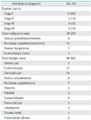
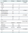
As a result of the histological examination of the surgical specimens of 100 patients, 80 (80%) had benign and 20 (20%) had malignant disease (Table 1). The histopathological classification of all cases and the stage distribution of malignant ones are given in Table 1. The distribution of benign and malignant cases by age, menopausal status, tumor size and ultrasound score is described in Table 2. In univariate analysis a significant linear trend for malignancy was found by increasing ultrasound score and the occurrence of malignancy in the pre- and postmenopausal patients. Although the risk of malignancy was increasing by age, it did not reach the statistical significance (p=0.051).
The mean serum level of CA-125 was significantly higher among women with malignancy when compared with women with benign tumors (329.2310 U/mL vs. 28.0374 U/mL). The sensitivity, specificity, and positive and negative predictive values and diagnostic accuracy of serum CA-125 level of 35 U/mL, the ultrasound score of 2, postmenopausal status and the size of 7 centimeters are reported in Table 3. When individual parameters were compared in Table 3, CA-125 had better sensitivity than the ultrasound score, size and menopausal status, even though the others had higher specificity than CA-125, but with considerable loss of sensitivity which is important in suspecting a malignancy. The performance of RMI 1, RMI 2, RMI 3, and RMI 4 at different cutoff values is shown in Table 4. The overall best performance in RMI 1, RMI 2, and RMI 3 was obtained at a cutoff level of 200 and at 500 for RMI 4. Although, at a cutoff level of 200 for RMI 1, RMI 2, RMI 3, and at the level of 450 for RMI 4, RMI 4 looked like better than the others with its sensitivity, specificity and the diagnostic accuracy rates, direct comparison of the four indices revealed that there was no statistically significant difference in performance of the four methods (McNemar test, p=0.063). Receiver operating characteristic (ROC) analysis of the RMI 1, RMI 2, RMI 3, and RMI 4 showed that the values of area under the curve were significantly high with a value of 0.825, 0.806, 0.825, 0.856, respectively (p<0.001). Area under the curves values of menopausal status, serum CA-125, US features, and tumor size are 0.675, 0.750, 0.706, and 0.750, respectively. We think that risk of malignancy indices were more reliable in detecting malignancy in terms of area under the curves. The diagnostic performance of ultrasound score, CA-125, menopausal status, tumor size, RMI 1, RMI 2, RMI 3, and RMI 4 is shown in the receiver-operating characteristic curves (Fig. 1).

DISCUSSION

This study has revealed the usefulness of RMI to correctly discriminate benign from malignant pelvic masses. The RMI was originally developed by Jacobs et al. [18], and subsequently the same group reproduced the results in a second patient group, establishing the superiority of RMI over the individual parameters [22].
In our patient group we found that there is no statistically significant difference in the performance of these four different malignancy risk indices (RMI 1, RMI 2, RMI 3, and RMI 4) in discriminating malignancy. But RMI 2 was found to be more reliable in discriminating benign and malignant disease by other investigators [19,23]. Tingulstad et al. [19] developed their RMI in 1996 and called it RMI 2. They compared it with that established by Jacobs et al. [18] and found that at a cutoff level of 200, a direct comparison of the two indices showed that RMI 2 was significantly better than RMI 1 (McNemar test, p=0.001). Morgante et al. [23] also found a similar result that for all cutoff values between 80 and 250, RMI 2 performed better than RMI 1 (p=0.0001). Tingulstad et al. [20] modified the RMI and defined it RMI 3, and they observed that at a cut-off level of 200 the sensitivity and specificity were 71 and 92%, respectively.
In 2001 Manjunath et al. [25] compared RMI 1, RMI 2, and RMI 3 with each other and also confirmed that there was no statistical difference between these three indices in benign - malignancy discrimination. In their study, Clarke et al. [26], using a cut-off of 120, found that RMI 1 had a sensitivity of 72% and a specificity of 87%; RMI 2 had a sensitivity of 76% and a specificity of 81%; RMI 3 had a sensitivity of 74% and a specificity of 84%. In 2009 Yamamoto et al. [24] developed their own RMI by using tumor size and called it RMI 4. Their study confirms that, at a cutoff level of 450, the accuracy of the RMI 4 was better than RMI 1 (p=0.0013), RMI 2 (p=0.0009) and RMI 3 (p=0.0013) with a cutoff level of 200. They observed that at a cutoff level of 450 the sensitivity, specificity, positive predictive value, negative predictive value and accuracy were respectively, 86.8%, 91.0%, 63.5%, 97.5%, and 90.4% [24]. In our study we found a sensitivity, specificity, positive predictive value, negative predictive value and accuracy of 85%, 87%, 60%, 95%, and 86%, respectively, which is comparable with the results of Yamamoto et al. [24]. But we also found that the other three indices' diagnostic performances were reliable, being different than Yamomoto et al's results.
The risk of malignancy index is a simple scoring system, appears to be very accurate, is useful in clinical practice, and should therefore be the test of choice in the preoperative evaluation of the adnexal mass. Any of the four malignancy risk indices (RMI 1, RMI 2, RMI 3, RMI 4) described can be used for selection of cases for optimal therapy. Since the specificity of risk of malignancy index is high, there is a potential role for this index in the selection of cases for conservative management or minimal invasive surgery of benign cases, like ultrasound guided aspiration or laparoscopic excision of other cysts.

Figures and Tables

Fig. 1
Receiver operator characteristic curve showing the performance of RMI 1, RMI 2, RMI 3, RMI 4, CA-125, tumor size, ultrasound score, and menopausal status. RMI, risk of malignancy index.
jgo-22-177-g001
Table 1
Histopathological clasification and stage distribution of cases
jgo-22-177-i001
Table 2
The distribution of benign and malignant cases by age, menopausal status, serum CA-125, tumor size and ultrasound score
jgo-22-177-i002
Table 3
The sensitivity, specificity, and positive (PPV) and negative (NPV) predictive values and diagnostic accuracy (DA) of serum CA-125, ultrasound score, postmenopausal status and tumor size
jgo-22-177-i003
Table 4
Sensitivity, specificity, positive (PPV) and negative (NPV) predictive values, and diagnostic accuracy (DA) for detecting malignancy at different cutoff levels of RMI 1, RMI 2, RMI 3, and RMI 4
jgo-22-177-i004

RMI, risk of malignancy index.

Notes

No potential conflict of interest relevant to this article was reported.

References

1. Gillis CR, Hole DJ, Still RM, Davis J, Kaye SB. Medical audit, cancer registration, and survival in ovarian cancer. Lancet. 1991. 337:611–612.
2. Finkler NJ, Benacerraf B, Lavin PT, Wojciechowski C, Knapp RC. Comparison of serum CA-125, clinical impression, and ultrasound in the preoperative evaluation of ovarian masses. Obstet Gynecol. 1988. 72:659–664.
3. Vasilev SA, Schlaerth JB, Campeau J, Morrow CP. Serum CA-125 levels in preoperative evaluation of pelvic masses. Obstet Gynecol. 1988. 71:751–756.
4. Eisenkop SM, Spirtos NM, Montag TW, Nalick RH, Wang HJ. The impact of subspecialty training on the management of advanced ovarian cancer. Gynecol Oncol. 1992. 47:203–209.
5. Kehoe S, Powell J, Wilson S, Woodman C. The influence of the operating surgeon's specialisation on patient survival in ovarian carcinoma. Br J Cancer. 1994. 70:1014–1017.
6. Manjunath AP, Desai MR, Desai PD, Modi DA. Value of sonography in evaluation of gynecologic pelvic masses. J Obstet Gynecol India. 1999. 49:60–64.
7. Sassone AM, Timor-Tritsch IE, Artner A, Westhoff C, Warren WB. Transvaginal sonographic characterization of ovarian disease: evaluation of a new scoring system to predict ovarian malignancy. Obstet Gynecol. 1991. 78:70–76.
8. Wu CC, Lee CN, Chen TM, Lai JI, Hsieh CY, Hsieh FJ. Factors contributing to the accuracy in diagnosing ovarian malignancy by color Doppler ultrasound. Obstet Gynecol. 1994. 84:605–608.
9. Predanic M, Vlahos N, Pennisi JA, Moukhtar M, Aleem FA. Color and pulsed Doppler sonography, gray-scale imaging, and serum CA-125 in the assessment of adnexal disease. Obstet Gynecol. 1996. 88:283–288.
10. Weiner Z, Thaler I, Beck D, Rottem S, Deutsch M, Brandes JM. Differentiating malignant from benign ovarian tumors with transvaginal color flow imaging. Obstet Gynecol. 1992. 79:159–162.
11. Anandakumar C, Chew S, Wong YC, Chia D, Ratnam SS. Role of transvaginal ultrasound color flow imaging and Doppler waveform analysis in differentiating between benign and malignant ovarian tumors. Ultrasound Obstet Gynecol. 1996. 7:280–284.
12. Osmers R. Sonographic evaluation of ovarian masses and its therapeutical implications. Ultrasound Obstet Gynecol. 1996. 8:217–222.
13. Einhorn N, Bast RC Jr, Knapp RC, Tjernberg B, Zurawski VR Jr. Preoperative evaluation of serum CA-125 levels in patients with primary epithelial ovarian cancer. Obstet Gynecol. 1986. 67:414–416.
14. Chen DX, Schwartz PE, Li XG, Yang Z. Evaluation of CA-125 levels in differentiating malignant from benign tumors in patients with pelvic masses. Obstet Gynecol. 1988. 72:23–27.
15. Vasilev SA, Schlaerth JB, Campeau J, Morrow CP. Serum CA-125 levels in preoperative evaluation of pelvic masses. Obstet Gynecol. 1988. 71:751–756.
16. Jacobs IJ, Rivera H, Oram DH, Bast RC Jr. Differential diagnosis of ovarian cancer with tumour markers CA-125, CA 15-3 and TAG 72.3. Br J Obstet Gynaecol. 1993. 100:1120–1124.
17. Twickler DM, Forte TB, Santos-Ramos R, McIntire D, Harris P, Scott D. The Ovarian Tumor Index predicts risk for malignancy. Cancer. 1999. 86:2280–2290.
18. Jacobs I, Oram D, Fairbanks J, Turner J, Frost C, Grudzinskas JG. A risk of malignancy index incorporating CA-125, ultrasound and menopausal status for the accurate preoperative diagnosis of ovarian cancer. Br J Obstet Gynaecol. 1990. 97:922–929.
19. Tingulstad S, Hagen B, Skjeldestad FE, Onsrud M, Kiserud T, Halvorsen T, et al. Evaluation of a risk of malignancy index based on serum CA-125, ultrasound findings and menopausal status in the pre-operative diagnosis of pelvic masses. Br J Obstet Gynaecol. 1996. 103:826–831.
20. Tingulstad S, Hagen B, Skjeldestad FE, Halvorsen T, Nustad K, Onsrud M. The risk-of-malignancy index to evaluate potential ovarian cancers in local hospitals. Obstet Gynecol. 1999. 93:448–452.
21. Staging announcement: FIGO cancer committee. Gynecol Oncol. 1986. 25:383–385.
22. Davies AP, Jacobs I, Woolas R, Fish A, Oram D. The adnexal mass: benign or malignant? Evaluation of a risk of malignancy index. Br J Obstet Gynaecol. 1993. 100:927–931.
23. Morgante G, la Marca A, Ditto A, De Leo V. Comparison of two malignancy risk indices based on serum CA-125, ultrasound score and menopausal status in the diagnosis of ovarian masses. Br J Obstet Gynaecol. 1999. 106:524–527.
24. Yamamoto Y, Yamada R, Oguri H, Maeda N, Fukaya T. Comparison of four malignancy risk indices in the preoperative evaluation of patients with pelvic masses. Eur J Obstet Gynecol Reprod Biol. 2009. 144:163–167.
25. Manjunath AP, Pratapkumar , Sujatha K, Vani R. Comparison of three risk of malignancy indices in evaluation of pelvic masses. Gynecol Oncol. 2001. 81:225–229.
26. Clarke SE, Grimshaw R, Rittenberg P, Kieser K, Bentley J. Risk of malignancy index in the evaluation of patients with adnexal masses. J Obstet Gynaecol Can. 2009. 31:440–445.
TOOLS
Similar articles