

 PDF
PDF Citation
 Print
Print


INTRODUCTION
The number of cases of ovarian cancer has increased in Japan and has now reached 10,000 per year. Similarly, the number of deaths from ovarian cancer has also increased, with about 4,800 deaths annually [1]. This tendency is also seen worldwide. More than half of cases of epithelial ovarian cancer are diagnosed as advanced cancer including stage III disease, accompanied by extrapelvic peritoneal dissemination (P) and retroperitoneal lymph node metastasis, or stage IV disease with distant metastasis; the prognosis in these advanced cases is poor.
In contrast, stage I epithelial ovarian cancer accounts for about 40% of all cases and is localized to the ovary in Japan [2]. This allows complete surgical excision and prognosis is favorable, with a 5-year survival rate of about 90% [345]. Surgery for stage I ovarian cancer includes adnexectomy on the side of the tumor, resection of the contralateral adnexa, hysterectomy, and resection of the greater omentum, retroperitoneal lymph node dissection, and peritoneal biopsy at several sites as part of staging surgery to detect metastasis or dissemination. Prevention of recurrence is important because the outcome after recurrence is poor, even in ovarian cancer diagnosed as International Federation of Gynecologists and Obstetricians (FIGO) stage I. Therefore, careful evaluation of histologic type, FIGO stage subclassification, and degree of histological differentiation, and postoperative adjuvant chemotherapy are recommended in most cases.
Several studies have reported on the recurrence of stage I epithelial ovarian cancer, but the majority of these studies have included only a small number of patients, while there have been only a few studies on detailed recurrence sites or investigations of the first surgery, initial adjuvant therapy, or histologic type [678910]. Details of the recurrence site and patient background in recurrence cases of stage I epithelial ovarian cancer are important to improve the operative procedures in the first surgery and to select appropriate adjuvant therapy for stage I ovarian cancer. Therefore, in this study, we performed a retrospective investigation on a large series of cases of stage I epithelial ovarian cancer treated at multiple affiliated institutions.
MATERIALS AND METHODS
1. Subjects
Medical records of 602 patients diagnosed with stage I epithelial ovarian cancer between 2000 and 2013 at 4 hospitals related to The Jikei University School of Medicine were evaluated retrospectively. Age, surgical procedure, substage, histologic type, chemotherapy, recurrence, initial recurrence site, and survival outcomes were investigated. Patients for whom medical records were discarded were excluded as data were insufficient. Cases with non-epithelial malignant ovarian tumors classified as sex cord-stromal tumor, malignant germ cell tumor, and borderline malignant tumor were also excluded.
2. Histologic type classification
Tumors were classified into histologic classifications of serous carcinoma, endometrioid carcinoma, clear cell carcinoma, mucinous carcinoma, mixed type, and others.
3. Stage classification
Stages were evaluated using the FIGO classification (published in January 2014) [11]. In addition to findings on laparotomy, the substage was determined by examining the peritoneal cell diagnosis (washing cytology) and the presence or absence of tumor capsule rupture.
4. Classification by surgical procedure
Patients who received hysterectomy, bilateral salpingo-oophorectomy, and resection of the great omentum were defined as the Basic Lapa (BL) group, and those who additionally received systematic lymph node dissection including both pelvic and para-aortic lymph nodes or only pelvic lymph node dissection were designated as the BL + pelvic lymphadenectomy (PLNx) + para-aortic lymphadenectomy (PANx) and BL + PLNx groups, respectively. The number of excised lymph nodes was not included in evaluation of completeness of dissection. Patients diagnosed as stage I by a surgical procedure less extensive than BL, such as fertility-sparing surgery and reduction surgery, were designated as the Limited Ope (LO) group.
5. Initial site of recurrence
Cases were divided into 4 categories based on the initial recurrence site: peritoneal dissemination (P), based on a lesion evaluable by imaging in the intra- and extrapelvic peritoneum (these included cases with ascites retention without an evaluable lesion, but with a cancer antigen 125 [CA125] level meeting Gynecological Cancer Intergroup [GCIG] criteria for recurrence) [12]; hematogenous (H), recurrence in the liver and spleen parenchyma, lung, bone, and brain; lymphogenous (L), recurrence in retroperitoneal lymph nodes, such as pelvic and para-aortic lymph nodes, and in inguinal, mediastinal, and supraclavicular lymph nodes; and others (O), including pleural recurrence in which only pleural effusion was observed, recurrence diagnosed based on a positive pleural cytology, and cases with elevation of tumor marker CA125 and meeting GCIG criteria. Cases with overlapping recurrence sites were included in each category. For example, a case with an evaluable intrapelvic peritoneal recurrent lesion and pelvic lymph node recurrence was included in both the P and L categories.
6. Statistical analysis
Logistic regression analysis was performed to identify risk factors for recurrence. A χ2 test was performed to examine the relationship between type of surgery and lymphogenous recurrence. A p-value of <0.05 was regarded as significant. Statistical analysis was performed using R software ver. 3.3.1 (R Foundation, Vienna, Austria).
RESULTS
1. Patient characteristics
The median age of the 602 subjects was 54 years (range: 14–99 years) and the median follow-up period was 60 months (range: 0–182 months) (Table 1). Since the treatment strategies for staging surgery and adjuvant therapy differed among institutions and over time, the patient background was diverse. Treatment strategies for lymph node dissection and adjuvant chemotherapy were selected for each patient based on complications and the patient's consent.
Table 1
Characteristics of all patients
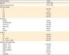
Values are presented as median (range) or number (%).
BL, basic lapa; CBDCA, carboplatin; CDDP, cisplatin; CPT-11, irinotecan; DTX, docetaxel; FIGO, International Federation of Gynecologists and Obstetricians; LO, limited ope; PANx, para-aortic lymphadenectomy; PLNx, pelvic lymphadenectomy; PTX, paclitaxel.
The most histologic type was clear cell carcinoma in 277 patients (46%), which is the most frequent type in Japan and consistent with previously reported rates. Tumors of stage IC1 were most frequent, in 247 patients (41%). Systematic lymph node dissection was performed in 224 patients (37%). One or more cycles of chemotherapy were administered in 412 patients (68%), whereas 32% received no chemotherapy, including patients judged not to require postoperative adjuvant chemotherapy based on the disease stage and histologic type and those for which chemotherapy was not used due to age, complications, and the patient's wishes.
Of the 602 patients with stage I epithelial ovarian cancer, recurrence occurred in 70 cases (11.6%) (Table 2). The median age of recurrence cases was 52 years (range: 26–84 years) and the median follow-up period was 41 months (range: 7–178 months) (Table 2). The median time to recurrence was 18 months (range: 3–126 months) and recurrence occurred within one year in 29 patients (41%), at 1–2 years in 21 patients (30%), at 2–3 years in 3 patients (4%), and at 3–5 years in 8 patients (11%), and after 5 years in 9 patients (13%). The most frequent histologic type was clear cell carcinoma in 39 patients (56%). The most frequent stage was IC1 in 24 cases (34%), followed by IC3 in 23 (33%). Of the recurrent cases, 22 (31%) had received systematic lymph node dissection, and 45 (64%) had received one or more cycles of chemotherapy.
Table 2
Characteristics of patients with recurrence
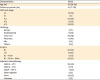
Values are presented as median (range) or number (%).
BL, basic lapa; CBDCA, carboplatin; CDDP, cisplatin; CPT-11, irinotecan; DTX, docetaxel; FIGO, International Federation of Gynecologists and Obstetricians; LO, limited ope; PANx, para-aortic lymphadenectomy; PLNx, pelvic lymphadenectomy; PTX, paclitaxel.
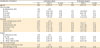
Associations of age, stage, histologic type, surgical procedure, and postoperative adjuvant therapy with recurrence were investigated by univariate and multivariate logistic regression analyses (Table 3). In multivariate analysis, the recurrence rate was significantly higher in stages IC2 and IC3 than IA (odds ratio [OR]=3.22 and 5.32, respectively), and significantly lower in endometrioid carcinoma compared to clear cell carcinoma (OR=0.40) and in the BL + PLNx + PANx group compared to the BL group (OR=0.47).
Table 3
OR of recurrence based on patient characteristics
2. Recurrence site
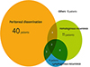
Recurrence sites are shown in Table 4 and Fig. 1. Recurrence was classified as peritoneal (P), hematogenous (H), lymphogenous (L) and other (O) in 49 (70%), 18 (26%), 9 (13%), and 6 (9%) cases, including overlapping classifications in 9 cases: P and H in 4 cases, P and L in 2 cases, and P, H, and L in 3 cases. Diagnosis of recurrence by pathological examination of tissue biopsy specimens was made in 16 cases (23%). Of the 49 patients (70%) with peritoneal recurrence, intrapelvic peritoneal recurrence occurred in 43 patients (61%), accounting for more than half of all recurrences.
Table 4
Classification of sites of initial recurrence
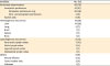
Fig. 1
Summary of numbers of recurrent patients and degree of overlap between the initial sites of recurrence.
In the 39 patients with recurrent clear cell carcinoma, 25 patients (64%) had peritoneal recurrence, including 19 patients (49%) with intrapelvic recurrence; and 12 patients (31%) and 6 patients (15%) had hematogenous and lymphogenous recurrence, respectively (Supplementary Table 1). There was no specific recurrence pattern in clear cell carcinoma compared with those in other histologic types. In contrast, all 5 cases of recurrent serous carcinoma had initial peritoneal recurrence.
Among 226 patients who received systematic lymph node dissection including 2 patients of fertility-sparing surgery in LO group, only 2 (0.9%) had lymphogenous recurrence: one patient each demonstrated recurrence to para-aortic and to cardiophrenic lymph nodes. In 376 patients who did not receive systematic lymph node dissection including the para-aortic lymph node, lymphogenous recurrence occurred in 7 patients (1.9%). The difference between these groups was not significant (p=0.339 by χ2 test).
DISCUSSION
Several articles have examined the sites of recurrence of stage I ovarian cancer [678]. However, details of recurrences sites of stage I epithelial ovarian cancer have only been reported in 2 studies from multiple institutions in Italy in 1997 and 2013, including overlapping cases. The first study was a retrospective investigation of 224 FIGO stage I patients, in which recurrence occurred in 39 (17%), including intrapelvic peritoneal recurrence in 54% of recurrence cases [9]. In the second study, recurrence occurred in 87 (19%) of 467 FIGO stage IA–IIA patients, including intrapelvic peritoneal recurrence in 44% of recurrence cases [10]. In our study in 602 patients with stage I epithelial ovarian cancer, the recurrence rate was 11.6%, with peritoneal and intrapelvic peritoneal recurrence accounting for 70% and 61% of recurrence cases, respectively. These findings clarify that peritoneal dissemination, especially in the intrapelvic peritoneum, is the most frequent site for recurrence of stage I ovarian cancer.
The high recurrence rate in the peritoneum may partly be due to insufficient investigation of peritoneal lesions at initial surgery. In a study of peritoneal biopsy (6 sites in the pelvis) in the treatment of early-stage ovarian cancer, microscopic disseminated lesions were detected by intrapelvic peritoneal biopsy in 5 (4.1%) of 122 patients, and these cases were later upstaged [12]. In another study in which the peritoneum (including the subphrenic peritoneum) was randomly biopsied in 129 patients, no phrenic peritoneal dissemination was found, but microscopic dissemination was present in 7% of cases as lesions of occult dissemination in the intrapelvic peritoneum, and these cases were similarly upstaged [13]. In the National Comprehensive Cancer Network (NCCN) guidelines [14], random peritoneal biopsies should be taken from the pelvis, paracolic gutters, and undersurfaces of the diaphragm (diaphragm scraping for Papanicolaou stain is an acceptable alternative). Furthermore, the Gynecologic Oncology Group (GOG) surgical manual recommends peritoneal biopsies from cul-de-sac, vesical peritoneum, right and left pelvic sidewalls, right and left paracolic gutters and right diaphragm for clinical stage I ovarian cancer. These guidelines strongly suggest that patients with occult dissemination in the intrapelvic peritoneum could be included in the cases of stage I epithelial ovarian cancer, and that routine peritoneal biopsy in the first surgery is necessary. In a basic study of peritoneal dissemination, Mitsui et al. [15] found that ovarian cancer stem cells and cancer stem cell niches in the pelvic peritoneum are important treatment targets, and that the pelvic peritoneum may have a pre-metastatic niche as a microenvironment for metastasis of tumor cells [1617]. Therefore, determination of the frequency of intrapelvic occult dissemination lesions in pelvic peritoneal biopsy samples and elucidation of the mechanism of intrapelvic peritoneal dissemination are required.
Clear cell carcinoma occurs at high frequency in Japan compared with that in western countries, and accounts for about 40% of cases of stage I ovarian cancer [1819202122]. In the present study, 46% of all stage I epithelial ovarian cancer cases and more than half (56%) of recurrent cases were clear cell carcinoma. Of 277 cases of clear cell carcinoma, 39 cases (14%) developed recurrence, and the recurrence rate of clear cell carcinoma was significantly higher than that of endometrioid carcinoma. However, no specific pattern of recurrence site was found in clear cell carcinoma.
Clear cell carcinoma has been shown to be less sensitive to initial chemotherapy than serous carcinoma and endometrioid carcinoma [23]. Therefore, new treatments limited to clear cell carcinoma, such as regimens other than platinum/taxane therapy and adjuvant therapy other than chemotherapy, have been developed. A recent international randomized controlled study (the GCIG/Japanese Gynecologic Oncology Group [JGOG] 3017 trial) centered in Japan, in which two-thirds of the cases were stage I, failed to show superiority of combination with irinotecan and cisplatin (CPT-P) therapy over combination with paclitaxel and carboplatin (TC) therapy [24]. Clinical studies of the effects of molecular targeted drugs on clear cell carcinoma have also been performed, but there is currently no specific adjuvant chemotherapy with a greater effect than that of paclitaxel + carboplatin for stage I clear cell carcinoma.
Radiotherapy to the entire pelvis has also been investigated as postoperative adjuvant therapy for stage I ovarian clear cell carcinoma. Nagai et al. [25] found that whole pelvis irradiation given postoperatively for stage I–III ovarian clear cell carcinoma was superior for control of localized lesions, compared to postoperative chemotherapy. In a comparison of outcomes of postoperative chemotherapy and radiotherapy for stage I and II clear cell carcinoma, Hoskins et al. [26] reported that PFS was improved by 20% by radiotherapy in the high-risk group (stage IC positive cytology, surface involvement, and stage II). In contrast, in a similar comparison for stage I–II clear cell carcinoma, Hogen et al. [27] observed no survival benefit in the high-risk group. Therefore, evidence for the efficacy of radiotherapy for control of intrapelvic peritoneal recurrence of stage I ovarian clear cell carcinoma has not been established, and development of treatment with consideration of the recurrence site is awaited.
And our study showed the tendency that patients who received systematic lymph node dissection occurred fewer lymphogenous recurrences. It remains controversial whether systematic lymph node dissection leads to decreased recurrence. Chan et al. [28] found a significantly better outcome in patients who underwent systematic lymph node dissection in massive US Surveillance, Epidemiology, and End Results (SEER) data. This result was due to elimination of cases with lymph node metastasis, leading to extraction of stage I cases with a favorable prognosis. Several studies have shown that systematic lymph node dissection reduces the incidence of retroperitoneal recurrence: in a prospective study, Maggioni et al. [29] found a significantly lower recurrence rate in the retroperitoneal cavity in patients with retroperitoneal lymph node dissection; Sakuragi et al. [30] found no recurrence in retroperitoneal lymph nodes in 94 patients treated with retroperitoneal lymph node dissection; and Petru et al. [31] reported a higher rate of retroperitoneal recurrence in patients without retroperitoneal lymph node dissection. In our study, lymph node recurrence was found in only 2 (1%) of 224 patients with systematic lymph node dissection, and recurrence in the dissected lymph node region was found in only one case of para-aortic lymph node metastasis. These findings are consistent with the above reports. The rate of retroperitoneal lymph node metastasis has been reported to be higher than 10% in stage I epithelial ovarian cancer [32], whereas in our study only 9 (1%) of 602 patients and 9 (13%) of 70 recurrent cases had lymphogenous recurrence. The NCCN treatment guidelines recommend investigation of retroperitoneal lymph nodes in early-stage ovarian cancer [14], and our findings in this study support use of retroperitoneal lymph node dissection in stage I ovarian cancer.
This study showed a high frequency of recurrence of stage I epithelial ovarian cancer by peritoneal dissemination, especially intrapelvic peritoneal recurrence. The report also provides the first description of the histologic type and subtype and detailed recurrence patterns with regard to the surgical procedure. Further clinical and basic studies are needed to improve diagnostic methods and surgical procedures, and adjuvant therapy may be necessary for patients with stage I epithelial ovarian cancer.
 XML Download