Journal List > J Gynecol Oncol > v.28(1) > 1093846

Jeon, Lee, Lim, Song, Bae, Kim, Lee, Kim, Chang, and Lee: Surgical manual of the Korean Gynecologic Oncology Group: ovarian, tubal, and peritoneal cancers

Abstract

The Surgery Treatment Modality Committee of the Korean Gynecologic Oncology Group has determined to develop a surgical manual to facilitate clinical trials and to improve communication between investigators by standardizing and precisely describing operating procedures. The literature on anatomic terminology, identification of surgical components, and surgical techniques were reviewed and discussed in depth to develop a surgical manual for gynecologic oncology. The surgical procedures provided here represent the minimum requirements for participating in a clinical trial. These procedures should be described in the operation record form, and the pathologic findings obtained from the procedures should be recorded in the pathologic report form. Here, we describe surgical procedure for ovarian, fallopian tubal, and peritoneal cancers.

INTRODUCTION

This surgical manual is for all cases of suspicious ovarian, tubal, and peritoneal cancers. It is organized into five sections including surgical procedures in ovarian, tubal, and peritoneal cancers, perioperative preparation, operation record form (ORF), pathologic report form (PRF), and tumor burden index (TBI).
We emphasize that surgical procedures in this manual represent the minimum requirements for clinical trials. This manual is the first version and will be updated to accommodate various clinical trials.

SURGICAL PROCEDURES IN OVARIAN, TUBAL, AND PERITONEAL CANCERS

In cases of suspected early stage diseases, the primary objective of surgical staging of ovarian, tubal, and peritoneal cancers is to establish adjuvant treatment strategies and in cases of suspected advanced stage diseases, optimal debulking surgery of ovarian, tubal, and peritoneal cancers should be achieved with acceptable morbidity.

1. Contents of surgical procedure

Midline vertical abdominal incision from the pubic symphysis to the xiphoid process is recommended for adequate exposure and evaluation of the whole abdomen. Minimally invasive surgical techniques (laparoscopy or robotic surgery) may be performed to accomplish surgical staging for selected patients based on preoperative imaging, such as computed tomography (CT), magnetic resonance imaging, or positron emission tomography/CT [1234567].
Prior to systemic exploration, free peritoneal fluid should be aspirated for cytology. Washing cytology with at least 20 to 50 mL of saline should be obtained in case of no free fluid in abdominal cavity. Patients with stage III or IV disease do not require cytologic assessment [168].
A systematic exploration is recommended to check the tumor involvement in the pelvic and abdominal organs, and peritoneal surface; clockwise or counterclockwise examination is usually performed from the cecum cephalad along the right paracolic gutter. The followings are investigated sequentially: ascending colon, liver, right diaphragm, stomach, lesser sac, porta hepatis, transverse colon, left diaphragm, spleen, distal pancreas, descending colon, left paracolic gutter, rectosigmoid colon, uterus, ovary, and bladder [16].
Biopsy should be performed at any suspicious site with tumor involvement if the suspected disease affects the surgical staging or adjuvant treatment. Multiple intraperitoneal biopsies from the cul-de-sac, vesical peritoneum, both pelvic sidewalls, and both paracolic gutters should be conducted in case of no evidence of disease [16].
Ovarian tumor should be removed intact, and frozen biopsy is strongly recommended during operation, if possible. Hysterectomy with bilateral salpingo-oophorectomy is recommended. Tumors throughout the abdomen should be removed as much as possible. Omentectomy should be fulfilled during surgical staging [9].
All visible and palpable tumor volume should be minimized as much as possible with debulking operations, such as visceral and parietal peritonectomy: peritoneal stripping, diaphragmatic resection, cholecystectomy, hepatic resection, splenectomy, distal pancreatectomy, appendectomy, bowel resection, urinary tract resection, partial cystectomy, and lymph node dissection [7101112131415].
Retroperitoneal inspection should be carried out to check for metastasis to pelvic and para-aortic lymph nodes. Pelvic and para-aortic lymph node should be systematically evaluated in case of stage I or II, and the extent of retroperitoneal lymph node dissection could be modified based on the degree of the intraperitoneal residual tumor and the status of the lymph node on the preoperative image (see the description of lymphadenectomy in ORF) [161718]. Unilateral salpingo-oophorectomy with preservation of the uterus may be considered to preserve fertility for selected patients [1920].
Before the neoadjuvant chemotherapy (NAC), the methods for pathologic diagnosis of ovarian, tubal, and peritoneal cancers are recommended as follows: laparoscopic biopsy, image-guided gun biopsy or aspiration, or cell block from the aspiration of ascites. In case of interval debulking surgery, the traced lesion after NAC should be evaluated carefully and its management should be recorded clearly [2122].
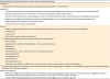
Medical record of surgery is recommended to describe the extent of initial tumors before surgery at pelvis, mid-abdomen, or upper abdomen. Demonstration of the status of residual tumors after surgery, complete or incomplete, is recommended to identify the size and number of remaining lesions. Photograph or video recording is one of the methods used to describe the preoperative and postoperative tumor, and surgical procedures. We provide schematic overview of this surgical manual (Table 1).
Table 1

Schematic overview of surgical procedure in ovarian, tubal, and peritoneal cancers

jgo-28-e6-i001
Recommendation for surgical technique in ovarian, tubal, and peritoneal cancer
Preparation
Preoperative intravenous antibiotics injection with adequate bowel preparation is recommended
Assessment
A midline vertical incision is recommended. Minimally invasive techniques (laparoscopy, robotic) may be performed for selected patients
Systematic exploration for tumor involvement on the pelvic and abdomen organs, and peritoneum
Aspiration of peritoneal fluid or washing cytology in case of no free peritoneal fluid (pelvis, paracolic gutters and infradiaphragmatic area)
Inspection and palpation of all peritoneal surfaces including diaphragms, serosa, and mesentery of the entire gastrointestinal tract
Random biopsies in the absence of any suspicious area
Intraoperative frozen biopsy (recommended)
Surgery
Bilateral salpingo-oophorectomy, but unilateral salpingo-oophorectomy may be considered in case of preserving fertility
Hysterectomy
Omentectomy
Pelvic and para-aortic lymph node dissection
The following procedures can be considered for the optimal cytoreduction
Bowel resection
Stripping and/or resection of the diaphragm or other peritoneal surfaces
Splenectomy
Appendectomy
Partial cystectomy, uretero-neoureterostomy, or ureteroneocystotomy
Partial hepatectomy
Partial gastrectomy
Cholecystectomy
Distal pancreatectomy
Suprarenal, porta hepatis, cardiophrenic, internal mammary, axillary, or supraclavicular lymph node dissection
*All visible and palpable tumor should be tried to be resected by experienced gynecologic oncologists or multidisciplinary surgical team if surgical procedure is feasible with acceptable morbidity
Special circumstances
Before the neoadjuvant chemotherapy (NAC), the method for microscopic diagnosis of ovarian, tubal, and peritoneal cancer is suggested as follows: laparoscopic biopsy, image-guided gun biopsy or aspiration, and cell block from the aspiration of ascites
In case of interval debulking surgery, the traced lesion after NAC is suggested to be explored surgically

PERIOPERATIVE PREPARATION

We provide perioperative preparation that includes antibiotic prophylaxis, prevention of thromboembolic disease, and patient’s position.

1. Antibiotic prophylaxis

The use of prophylactic antibiotics before surgery is suggested for the prevention of postoperative gynecological infections. Antibiotics are recommended to be given immediately before skin incision. Antibiotic regimen can be selected according to the types of surgery or surgeon’s preference. Additional use of prophylactic antibiotics is recommended to maintain effective levels of intravascular antibiotics in certain clinical situations, like massive bleeding or prolonged operative time [2324].

2. Prevention of thromboembolic disease

Prophylaxis with anti-coagulants can be selectively suggested to cancer patients with high risk of deep-vein thrombosis and thromboembolic disease (Table 2) [2526272829].
Table 2

The methods for the prevention of thromboembolic events [2526272829]

jgo-28-e6-i002
Class Example
Pharmacologic Unfractionated heparin, low-molecular weight heparin, fondaparinux, warfarin, dextran
Mechanical External pneumatic compression, elastic stocking
Behavioral Short preoperative hospitalization, early postoperative mobilization, feet elevation above heart level

3. Patient position

If concomitant bowel resection is expected during operation, lithotomy position is recommended for patients who undergo laparotomy, and gel pads can be used for prevention of pressure sores [12].

OPERATION RECORD FORM

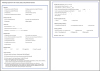
In the debulking surgery for the advanced stage disease, multidisciplinary surgical teams including gynecologic oncologic surgeons, colorectal surgeons, hepatobiliary surgeons, and even thoracic surgeons usually perform a lot of surgical procedures to minimize residual lesion and these surgical procedures should be described systematically and properly in the operation record. ORF for ovarian tubal and peritoneal cancers has been established on the basis of the Synoptic Operative Template for Ovarian Cancer of National Cancer Center of Korea. Standardized ORF may encourage to record all required information and surgical procedures and can save time. In the clinical trial setting, by looking at ORF, investigators can identify all procedures. ORF includes the following information (Fig. 1, Supplementary Fig. 1).
Fig. 1
Operation record form for ovarian, tubal, and peritoneal cancers. CA-125, cancer antigen 125; CA-19-9, cancer antigen 19-9; CEA, carcinoembryonic antigen; FFP, fresh frozen plasma; FIGO, International Federation of Gynecology and Obstetrics; HE-4, human epididymis protein 4; KGOG, Korean Gynecologic Oncology Group; LN, lymph node; LND, lymph node dissection; LNS, lymph node sampling; LLQ, left lower quadrant; Lt, left; LUQ, left upper quadrant; Plt conc, platelet concentration; p-RBC, packed red blood cells; RLQ, right lower quadrant; Rt, right; RUQ, right upper quadrant; WB, whole blood.
jgo-28-e6-g001

TUMOR BURDEN INDEX

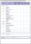
To estimate perioperative tumor burden, Korean Gynecologic Oncology Group (KGOG) developed TBI by modifying the peritoneal carcinomatosis index of Korean National Cancer Center. The peritoneal cavity is divided into nine well defined regions (Fig. 2, Supplementary Fig. 2). Investigators should describe pre- and post-operative largest tumor diameter, operative finding, operation name in each region, and the largest residual tumor at the end of the operation.
Fig. 2
Korean Gynecologic Oncology Group tumor burden index (TBI) for ovarian, tubal, and peritoneal cancers. IMA, inferior mesenteric artery; LN, lymph node; LUQ, left upper quadrant; PALN, paraaortic lymph node; RUQ, right upper quadrant.
jgo-28-e6-g002

PATHOLOGIC REPORT FORM

Surgery Treatment Modality Committee of KGOG collected and analyzed several ovarian cancer PRFs from committee members’ hospitals and decided that PRF should be made with Gynecologic Pathology Study Group. There were in-depth discussions with the Gynecologic Pathology Study Group about how to develop the PRF for ovarian, tubal and peritoneal cancer. PRF includes the following information (Fig. 3, Supplementary Fig. 3).
Fig. 3
Pathologic report form for ovarian, tubal, and peritoneal cancers. pTNM, pathological tumor node metastasis.
jgo-28-e6-g003

Notes

Funding This work was supported in part by the grant (NCC1310311 and NCC1610070) of the National Cancer Center, Korea.

Conflict of Interest No potential conflict of interest relevant to this article was reported.

References

1. Nezhat FR, Pejovic T, Finger TN, Khalil SS. Role of minimally invasive surgery in ovarian cancer. J Minim Invasive Gynecol. 2013; 20:754–765.
2. Stier EA, Barakat RR, Curtin JP, Brown CL, Jones WB, Hoskins WJ. Laparotomy to complete staging of presumed early ovarian cancer. Obstet Gynecol. 1996; 87((5 Pt 1)):737–740.
3. Rutten MJ, Leeflang MM, Kenter GG, Mol BW, Buist M. Laparoscopy for diagnosing resectability of disease in patients with advanced ovarian cancer. Cochrane Database Syst Rev. 2014; (2):CD009786.
4. Ditto A, Martinelli F, Lorusso D, Haeusler E, Carcangiu M, Raspagliesi F. Fertility sparing surgery in early stage epithelial ovarian cancer. J Gynecol Oncol. 2014; 25:320–327.
5. Muñoz KA, Harlan LC, Trimble EL. Patterns of care for women with ovarian cancer in the United States. J Clin Oncol. 1997; 15:3408–3415.
6. Colombo PE, Mourregot A, Fabbro M, Gutowski M, Saint-Aubert B, Quenet F, et al. Aggressive surgical strategies in advanced ovarian cancer: a monocentric study of 203 stage IIIC and IV patients. Eur J Surg Oncol. 2009; 35:135–143.
7. Chi DS, Eisenhauer EL, Zivanovic O, Sonoda Y, Abu-Rustum NR, Levine DA, et al. Improved progression-free and overall survival in advanced ovarian cancer as a result of a change in surgical paradigm. Gynecol Oncol. 2009; 114:26–31.
8. Young RC, Decker DG, Wharton JT, Piver MS, Sindelar WF, Edwards BK, et al. Staging laparotomy in early ovarian cancer. JAMA. 1983; 250:3072–3076.
9. Nezhat FR, Ezzati M, Chuang L, Shamshirsaz AA, Rahaman J, Gretz H. Laparoscopic management of early ovarian and fallopian tube cancers: surgical and survival outcome. Am J Obstet Gynecol. 2009; 200:83.e1–83.e6.
10. Eisenhauer EL, Abu-Rustum NR, Sonoda Y, Aghajanian C, Barakat RR, Chi DS. The effect of maximal surgical cytoreduction on sensitivity to platinum-taxane chemotherapy and subsequent survival in patients with advanced ovarian cancer. Gynecol Oncol. 2008; 108:276–281.
11. Elattar A, Bryant A, Winter-Roach BA, Hatem M, Naik R. Optimal primary surgical treatment for advanced epithelial ovarian cancer. Cochrane Database Syst Rev. 2011; (8):CD007565.
12. Estes JM, Leath CA 3rd, Straughn JM Jr, Rocconi RP, Kirby TO, Huh WK, et al. Bowel resection at the time of primary debulking for epithelial ovarian carcinoma: outcomes in patients treated with platinum and taxane-based chemotherapy. J Am Coll Surg. 2006; 203:527–532.
13. Winter WE 3rd, Maxwell GL, Tian C, Sundborg MJ, Rose GS, Rose PG, Gynecologic Oncology Group, et al. Tumor residual after surgical cytoreduction in prediction of clinical outcome in stage IV epithelial ovarian cancer: a Gynecologic Oncology Group Study. J Clin Oncol. 2008; 26:83–89.
14. Zivanovic O, Eisenhauer EL, Zhou Q, Iasonos A, Sabbatini P, Sonoda Y, et al. The impact of bulky upper abdominal disease cephalad to the greater omentum on surgical outcome for stage IIIC epithelial ovarian, fallopian tube, and primary peritoneal cancer. Gynecol Oncol. 2008; 108:287–292.
15. Narasimhulu DM, Khoury-Collado F, Chi DS. Radical surgery in ovarian cancer. Curr Oncol Rep. 2015; 17:16.
16. Schorge JO, Clark RM, Lee SI, Penson RT. Primary debulking surgery for advanced ovarian cancer: are you a believer or a dissenter? Gynecol Oncol. 2014; 135:595–605.
17. Mikami M. Role of lymphadenectomy for ovarian cancer. J Gynecol Oncol. 2014; 25:279–281.
18. Burghardt E, Pickel H, Lahousen M, Stettner H. Pelvic lymphadenectomy in operative treatment of ovarian cancer. Am J Obstet Gynecol. 1986; 155:315–319.
19. Bentivegna E, Morice P, Uzan C, Gouy S. Fertility-sparing surgery in epithelial ovarian cancer. Future Oncol. 2016; 12:389–398.
20. Eskander RN, Randall LM, Berman ML, Tewari KS, Disaia PJ, Bristow RE. Fertility preserving options in patients with gynecologic malignancies. Am J Obstet Gynecol. 2011; 205:103–110.
21. Fujiwara K, Kurosaki A, Hasegawa K. Clinical trials of neoadjuvant chemotherapy for ovarian cancer: what do we gain after an EORTC trial and after two additional ongoing trials are completed? Curr Oncol Rep. 2013; 15:197–200.
22. Bristow RE, Eisenhauer EL, Santillan A, Chi DS. Delaying the primary surgical effort for advanced ovarian cancer: a systematic review of neoadjuvant chemotherapy and interval cytoreduction. Gynecol Oncol. 2007; 104:480–490.
23. Stumpf PG. Practical solutions to improve safety in the obstetrics/gynecology office setting and in the operating room. Obstet Gynecol Clin North Am. 2008; 35:19–35.
24. Gadducci A, Cosio S, Spirito N, Genazzani AR. The perioperative management of patients with gynaecological cancer undergoing major surgery: A debated clinical challenge. Crit Rev Oncol Hematol. 2010; 73:126–140.
25. Wille-Jørgensen P, Rasmussen MS, Andersen BR, Borly L. Heparins and mechanical methods for thromboprophylaxis in colorectal surgery. Cochrane Database Syst Rev. 2003; (4):CD001217.
26. Heparin HJ. Heparin. N Engl J Med. 1991; 324:1565–1574.
27. Samama MM, Gerotziafas GT. Evaluation of the pharmacological properties and clinical results of the synthetic pentasaccharide (fondaparinux). Thromb Res. 2003; 109:1–11.
28. Dinwoodey DL, Ansell JE. Heparins, low-molecular-weight heparins, and pentasaccharides: use in the older patient. Cardiol Clin. 2008; 26:145–155.
29. Bauer KA. New anticoagulants. Curr Opin Hematol. 2008; 15:509–515.

Supplementary Materials

Supplementary Fig. 1

Operation record form for ovarian, tubal, and peritoneal cancers

Supplementary Fig. 2

Tumor burden index (TBI) for ovarian, tubal, and peritoneal cancers

Supplementary Fig. 3

Pathologic report form for ovarian, tubal, and peritoneal cancers
TOOLS
ORCID iDs

Seob Jeon
https://orcid.org/http://orcid.org/0000-0001-5230-1997

Sung Jong Lee
https://orcid.org/http://orcid.org/0000-0002-6077-2649

Myong Cheol Lim
https://orcid.org/http://orcid.org/0000-0001-8964-7158

Taejong Song
https://orcid.org/http://orcid.org/0000-0003-0288-1254

Jaeman Bae
https://orcid.org/http://orcid.org/0000-0001-7453-1443

Kidong Kim
https://orcid.org/http://orcid.org/0000-0001-9254-6024

Jung-Yun Lee
https://orcid.org/http://orcid.org/0000-0001-7948-1350

Sang Wun Kim
https://orcid.org/http://orcid.org/0000-0002-8342-8701

Suk-Joon Chang
https://orcid.org/http://orcid.org/0000-0002-0558-0038

Jong-Min Lee
https://orcid.org/http://orcid.org/0000-0002-0562-5443

Similar articles