

 PDF
PDF Citation
 Print
Print


INTRODUCTION
To date, no surgical manual or standardized anatomical description of gynecologic oncology has been developed by the Korean Gynecologic Oncology Group (KGOG). The members of the Surgery Treatment Modality Committee (Surgery TMC) of the KGOG identified a need for a surgical manual to facilitate clinical trials and to improve communication between investigators by standardizing and precisely describing operating procedures.
We reviewed the current literature on anatomic terminology, identification of surgical components, and surgical techniques, and discussed them in depth in order to create a surgical manual for gynecologic oncology. Here, we focused on radical hysterectomy and lymphadenectomy, and we developed a KGOG classification for those conditions.
ANATOMICAL NOMENCLATURE
To simplify the anatomical terms for classification, we will consistently use the terminology ‘paracervix,’ ‘vesicouterine ligament,’ and ‘uterosacral ligament’ for the structures usually referred to by surgeons as the cardinal ligament (or lateral parametrium), anterior parametrium, and posterior parametrium, respectively [1].
The term ‘paracervix’ (cardinal ligament, Mackenrodt’s ligament, or parametrium) refers to the dorsolateral attachment of the cervix, tissues that surround the uterine artery between the uterine corpus and pelvic sidewall cranial to the ureter, connective tissue, and lymph channels.
The term ‘vesicouterine ligament’ (ventral parametrium) includes the anterior and posterior leaflets. The connective tissue of the anterior leaflet of the vesicouterine ligament; that is, the anterior portion of the so-called ureteral tunnel, can be identified following the complete separation of the uterine artery and superficial uterine vein from the ureter. The posterior leaflet of the vesicouterine ligament is the tissue that resides under the ureter and connects the posterior wall of the bladder and the lateral cervix/upper lateral vagina [2].
The term ‘uterosacral ligament’ (dorsal parametrium) refers to the fibrous tissue and non-striated muscular fibres that are attached to the front of the sacrum and travel from the uterus to the anterior aspect of the sacrum.
Throughout this classification, the term ‘nerve preservation’ refers to identify the hypogastric nerves, the inferior hypogastric nerve plexus (pelvic plexus), and its bladder branches, and indicates allowing the resection of oncologically relevant pericervical structures while preserving the sympathetic and parasympathetic innervations of the pelvic organs [3].
KGOG CLASSIFICATION OF HYSTERECTOMY
The Piver-Rutlege-Smith classification published in 1974 has achieved popularity [45]. However, several limitations, including inconsistencies in terminology, drawbacks associated with a uterocentric concept, exclusion of nerve-sparing and fertility-preserving techniques, and difficulties adapting the guidelines to advances in vaginal, laparoscopic, or robotic surgery, makes continued use of the classification challenging. To overcome these limitations, Querleu and Morrow [1] proposed a new classification consisting of four types of radical hysterectomy, the Kyoto classification. We decided to create a KGOG classification of hysterectomy and lymphadenectomy based on the Kyoto classification, because it is considered contemporary and adequate for worldwide communication.
The classification has been modified and adapted to Korean circumstances by the Surgery TMC of the KGOG. There are two major changes. One is the deletion of the type B subclassification (B1 and B2), and the other is the inclusion of a fertility-preserving procedure, trachelectomy (cervicectomy), as a subclassification of types C and D.
The reasons for the elimination of the type B subclassification are as follows: (1) It is not easy and is sometimes impossible to differentiate the paracervical lymph nodes from the pelvic lymph nodes. (2) The basic reason for developing this classification is to provide separate descriptions of hysterectomy and lymphadenectomy. (3) Separate clinical indications for each operation have not been identified. (4) The clinical significance of the differentiation within type B is considered minimal. Table 1 summarises the four types of KGOG classification of hysterectomy.
Table 1
Korean Gynecologic Oncologic Group classification of hysterectomy*
*Modification of the new classification of radical hysterectomy by Querleu et al. [1]. †It is similar to type I of the “Piver-Rutledge-Smith (PRS) classification” [45]. ‡(T) means trachelectomy (cervicectomy). §This is similar to type II under the PRS classification. ǁThis is similar to type III under the PRS classification.
![]()
1. Type A: minimum resection of the paracervix
This is an extrafascial hysterectomy. The paracervix is transected medial to the ureter and lateral to the cervix. The ureter does not need to be unroofed. The uterosacral and vesicouterine ligaments are transected close to the uterus. The length of the vaginal resection is generally less than 10 mm, and the vaginal part of the paracervix is not removed.
2. Type B: transection of the paracervix at the ureter
Partial resection of the uterosacral and vesicouterine ligaments is the key element of this category. The ureter is unroofed and rolled laterally, permitting transection of the paracervix at the level of the ureteral tunnel. The neural component of the paracervix caudal to the deep uterine vein is not resected. At least 10 mm of the vagina from the cervix or tumor is resected.
3. Type C: transection of the paracervix at the junction with the internal iliac vascular system
Following complete mobilization of the ureter, transection of the uterosacral ligament at the rectum and transection of the vesicouterine ligament at the bladder are characteristics of type C. In addition, 15 to 20 mm of the vagina from the tumor or cervix and the corresponding paracolpos is resected, depending on the extent of vaginal and paracervical involvement and the surgeon’s preference. Type C is divided into two types: C1, with autonomic nerve preservation; C2, without autonomic nerve preservation.
In type C1, the uterosacral ligament is transected after separation of the hypogastric nerves. The bladder branches of the pelvic plexus are preserved in the lateral ligament of the bladder (i.e., the lateral part of the bladder pillar). If the caudal part of the paracervix is transected, careful identification of bladder nerves is subsequently required. In type C2, the paracervix is transected completely, including the part caudal to the deep uterine vein.
4. Type D: entire resection of paracervix with vessels
This rare type of surgery is characterised by additional ultraradical procedures, primarily indicated at the time of pelvic exenteration. In this type of surgery, the entire paracervix is resected. Type D is divided into two types: D1, resection of the entire paracervix along with the internal iliac vessels; D2, resection of the entire paracervix, with the internal iliac vessels and adjacent fascial or muscular structures.
Type D1 is a resection of the entire paracervix at the pelvic sidewall along with the internal iliac vessels, exposing the roots of the sciatic nerve. The procedure involves a total resection of the vessels of the lateral part of the paracervix. These vessels (i.e., inferior gluteal, internal pudendal, and obturator vessels) arise from the internal iliac vessel system. Type D2 is the same as D1 plus resection of the entire paracervix with the internal iliac vessels and adjacent fascial or muscular structures (i.e., pubococcygeus, iliococcygeus, coccygeus, and obturator muscles).
LYMPHADENECTOMY
We classified lymphadenectomy by its level and radicality. Anatomically, arteries are the most stable landmarks for lymphadenectomy. Four areas or levels are defined according to the corresponding arterial anatomy: (1) level 1, external and internal iliac (including obturator), (2) level 2, common iliac (including presacral), (3) level 3, para-aortic infra-inferior mesenteric artery (IMA), and (4) level 4, para-aortic infrarenal. If other lymph nodes are resected, specification of the procedure is necessary.
Although lymph nodes can cross borders, the limits between levels 1 and 2, levels 2 and 3, and levels 3 and 4 are the bifurcation of the common iliac artery, the bifurcation of the aorta, and the IMA, respectively.
We also defined types of lymphadenectomy by radicality, lymph node sampling (LNS), systematic lymph node dissection (LND), and debulking. LNS is defined as sampling of a sentinel node, suspicious nodes, or random sampling [6]. In a systematic pelvic LND (PLND), all lymph nodes and fatty tissues between the external and internal iliac arteries, from the bifurcation of the common iliac artery up to the circumflex vein and above the obturator nerve, are removed. A systematic para-aortic LND includes resection of all lymph nodes and fatty tissue surrounding the aorta, inferior vena cava, and renal vessels from the renal vein cranially to the midpoint of the common iliac vessels caudally, and extending laterally to the edge of the psoas major muscle. The range of the minimum number of lymph nodes for an adequate systematic PLND has been previously found to be 10 to 25 [67891011]. The number of LNs required can be modified according to the characteristics of a clinical trial. Debulking of lymph nodes is defined as resection of bulky nodes [912].
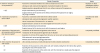
OPERATION RECORD FORM
An operation record form (ORF) for cervical cancer has been established on the basis of the Synoptic Operative Template for Ovarian Cancer of the National Cancer Center of Korea. A standardized ORF will encourage keeping of full and accurate records with all required information and surgical procedures for clinical trials on cervical cancer. The ORF for cervical cancer includes the following information (Fig. 1, Supplementary Fig. 1).
 | Fig. 1Operation record form for cervical cancer. CA-125, cancer antigen 125; CEA, carcinoembryonic antigen; CIN, cervical intraepithelial neoplasia; CIS, cervical carcinoma in situ; FIGO, International Federation of Gynecology and Obstetrics; FFP, fresh frozen plasma; IMA, inferior mesenteric artery; KGOG, Korean Gynecologic Oncology Group; LEEP, loop electrosurgical excision procedure; LLETZ, large loop excision of the transformation zone; LLQ, left lower quadrant; LN, lymph node; LND, lymph node dissection; LNS, lymph node sampling; Lt, left; LUQ, left upper quadrant; Plt conc, platelet concentration; p-RBC, packed-red blood cell; RLQ, right lower quadrant; Rt, right; RUQ, right upper quadrant; SCC-Ag, squamous cell carcinoma antigen; WB, whole blood.
|
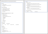
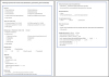
PATHOLOGIC REPORT FORM
The surgery TMC of the KGOG, in conjunction with the Gynecological Pathology Study Group, reviewed and analyzed several pathologic report forms (PRFs) for cervical cancer from domestic and international institutes. Finally, two kinds of PRF for cervical cancer were developed; one is for excision, and the other is for trachelectomy, hysterectomy, and pelvic exenteration. PRFs for cervical cancer include the following information (Figs. 2, 3, Supplementary Figs. 2, 3).
CONCLUSIONS
The purpose of this surgical manual is to facilitate future clinical trials and to improve communication between investigators by standardizing and describing operative procedures. The surgical procedures provided here represent the minimum requirements for participating in a clinical trial. These procedures should be systematically and properly described in the ORF, and the pathologic findings obtained from the procedures should be recorded in the PRF. This manual will be updated as necessary to include various clinical trials and to reflect the latest trends.

 XML Download