

 PDF
PDF Citation
 Print
Print


INTRODUCTION
Gynecologic malignant tumors, particularly cervical, endometrial, and ovarian cancer have both high morbidity and mortality. It is important for the diagnosis and treatment of these cancers to know their characteristics, such as prevalence, stages, treatment methods, survival rates, etc.
There are 3 cancer statistical registries in Japan. First, there is a population-based cancer registry that was legislated in 2016 as a national cancer registry [1]. A total of 26 parameters related to each cancer are included for each patient, and these parameters are common to all malignant tumors. Medical institutions that have diagnosed malignant tumors are required to register the information on the cancer, and patients have no right to refuse this registration. The National Cancer Center Japan collects and analyzes the data in this registry. Prognostic information collected from medical institutions and obituary notices that were provided to local governments are also included.
The second cancer registry is a hospital-based cancer registry (HCR). This is a registry project involving the leading hospitals in all prefectures and includes cancer cooperation hospitals. This registry is in accordance with the guidelines for hospital cancer registries, and 633 institutions participated in this registry in 2012. Approximately 80% of all cancer patients in Japan are estimated to be registered with HCR. The information collected includes a total of 100 parameters that are common to all malignant tumors. Cancer cooperation hospitals are subject to registration, and trained staff are in charge of this registry.
The third registry is an organ-based cancer registry. This registry is organized by institutions participating in the societies and scientific meetings of the relevant diseases. Unlike the other 2 registries, the organ-based cancer registry records parameters according to cancer type and characteristics. Thus, this registry has flexibility over time with the type of data collected, and it also can register rare cancers. The organ-based cancer registry for gynecologic malignancy is a gynecologic cancer registry that is maintained by the gynecologic tumor committee, Japan Society of Obstetrics and Gynecology.
The present study was conducted using both the population-based cancer registry and the gynecologic cancer registry to elucidate the characteristics of gynecologic malignant tumors in Japan.
1. Population-based cancer registry
The Cancer Information Service in National Cancer Center Japan analyzes the data provided by local governments in Japan, determines the prevalence of cancer, estimates nationwide morbidity of cancer, and publishes the “Cancer Registry and Statistics. Cancer Information Service, National Cancer Center, Japan” on their website [2]. Based on the nationwide estimates from the population-based cancer registry from 1975 to 2012, the morbidities of cervical cancer (without CIN3), endometrial cancer, and ovarian cancer were obtained and used for analysis.
The data on Vital Statistics in Japan (Ministry of Health, Labour and Welfare, Tokyo, Japan) are published as the “Cancer Registry and Statistics. Cancer Information Service, National Cancer Center, Japan” on their website [2]. The age-adjusted mortality due to cervical cancer (without CIN3), endometrial cancer, and ovarian cancer from 1975 to 2012 based on the world population were obtained and used for analysis.
2. Gynecologic cancer registry
The Gynecologic Tumor Committee of the Japan Society of Obstetrics and Gynecology has published the annual patient reports on patients who started treatment in 2005, 2010, and 2014 [345]. Clinicopathologic factors for cervical cancer (without CIN3), endometrial cancer, and ovarian cancer, including age, clinical stage, postsurgical stage, histological type, and therapeutic strategy were retrieved from the gynecologic cancer registry and used for analysis. The clinical and postsurgical staging for cervical and endometrial cancer patients in 2005 and 2010 was consistent with International Federation of Gynecology and Obstetrics (FIGO) 1988 staging system and that in 2014 with FIGO 2008 staging system. The clinical and postsurgical staging for ovarian cancer patients was consistent with FIGO 1988 staging system. Furthermore, the clinical stage and postsurgical stage were classified and calculated by dividing patients into 2 groups: those who were 39 years or younger and those who were 40 years or older. The histological type of cervical, endometrial and ovarian cancer was consistent with World Health Organization (WHO) 2003 classification.
With regard to prognostic information, the 5-year overall survival rates of cervical, endometrial, and ovarian cancer, which were estimated by the Kaplan-Meier method, were retrieved from the annual treatment reports on patients who started treatment in 2003, 2006, and 2009 [678]. The clinical and postsurgical staging for cervical, endometrial, and ovarian cancer patients in 2003, 2006, and 2009 was consistent with FIGO 1988 staging system.
INCIDENCE AND MORTALITY OF GYNECOLOGIC MALIGNANT TUMORS
The changes in the nationwide estimated morbidity of cervical, endometrial, and ovarian cancer in the population-based cancer registry in Japan are shown in Fig. 1A. The morbidity of cervical cancer ranged from 9,000 to 11,000 women in the 1970s and decreased to 7,000–8,000 women in the 1990s. However, the morbidity increased to 10,908 women in 2012. The morbidity of endometrial cancer was approximately 1,000 women in 1970s and increased each year, exceeding 5,000 women in 1999. In 2007, endometrial cancer morbidity exceeded that of cervical cancer and rose to 13,606 women in 2012, which was the largest number among the gynecologic malignant tumors in Japan. The morbidity of ovarian cancer was 2,000 to 3,000 women in the 1970s, and increased each year, exceeding 5,000 women in 1988. In 2012, the morbidity of ovarian cancer was 9,384 women.
Fig. 1
Incidence and mortality of gynecologic malignancies in Japan. (A) Incidence of gynecologic malignancies (cervical, endometrial, and ovarian cancers). (B) Age-adjusted mortality of gynecologic malignancies (cervical, endometrial, and ovarian cancers).
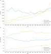
The age-adjusted mortality of cervical, endometrial, and ovarian cancer based on the world population is shown in Fig. 1B. The mortality of cervical cancer remained flat, with approximately 2.0 per 100,000 women, and was 2.1 per 100,000 women in 2012. The mortality of endometrial cancer gradually increased with a rapid increase in the morbidity, and was 1.3 per 100,000 women in 2012, which was lower than that of cervical and ovarian cancer. The mortality of ovarian cancer was the highest and exceeded 3.5 per 100,000 women in the late 1990s but decreased to 3.2 per 100,000 in 2012.
CERVICAL CANCER
1. Clinical stages
Changes in the clinical stages of cervical cancer are shown in Fig. 2A. In 2014, 12.8% of the cervical cancer patients were classified as stage IA1, 2.1% as stage IA2, 29.8% as stage IB1, 9.5% as stage IB2, 3.0% as stage IIA1, 2.5% as stage IIA2, 17.0% as stage IIB, 1.1% as stage IIIA, 9.1% as stage IIIB, 2.8% as stage IVA, 8.4% as stage IVB, and 1.9% as unknown stage.
Fig. 2
Characteristics of cervical cancer. (A) Distribution of clinical stages in cervical cancer. (B) Distribution of clinical stages in younger or elder patients with cervical cancer. (C) Distribution of histological types in cervical cancer. (D) Distribution of treatment methods in cervical cancer.
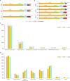
Changes in the clinical stages by age are shown in Fig. 2B. In young patients aged 39 years or less, 73%–79% of the patients were classified as stage I and 11%–14% were classified as stage II, suggesting that most of these patients had early cancer. On the other hand, older patients aged 40 years or more, 41%–48% were classified as stage I and 24%–27% were classified as stage II, indicating that the percent with early cancer was significantly lower (both stages I and II, p<0.001).
2. Histological types
The histological classification of cervical cancer is shown in Fig. 2C. In 2005, 2010, and 2014, patients with squamous cell carcinoma or adenocarcinoma accounted for 93%–94% while there are fewer patients with other histological types. Patients with squamous cell carcinoma comprised 75.5% of the cervical cancer patients in 2005 but the percent significantly decreased to 72.8% in 2014 (p=0.001). In contrast, patients with adenocarcinoma accounted for 16.9% of the cervical cancer patients in 2005 but significantly increased to 20.1% in 2014 (p<0.001).
3. Treatments
The changes in treatment methods for cervical cancer are shown in Fig. 2D. Patients who underwent surgery as a main treatment accounted for 63%–65% of the patients and were significantly more than that who underwent radiotherapy as the main treatment (30%–33%; p<0.001). However, patients who underwent radiotherapy as a main treatment in 2014 were significantly more than those in 2005 (p<0.001), and in particular concurrent chemoradiation therapy (CCRT) significantly increased (p<0.001).
Patients who underwent radiotherapy as adjuvant therapy have been decreasing, while those who underwent chemotherapy have been increasing (p<0.001).
ENDOMETRIAL CANCER
1. Stage
The changes in the postsurgical stages of endometrial cancer are shown in Fig. 3A. In 2014, 54.8% of the endometrial cancer patients were classified as stage IA, 17.1% as stage IB, 6.0% as stage II, 3.7% as stage IIIA, 0.9% as stage IIIB, 4.5% as stage IIIC1, 4.2% as stage IIIC2, 0.3% as stage IVA, 7.2% as stage IVB, and 1.4% as unknown stage.
Fig. 3
Characteristics of endometrial cancer. (A) Distribution of post-surgical stages in endometrial cancer. (B) Distribution of post-surgical stages in both younger and older patients with endometrial cancer. (C) Distribution of histological types in endometrial cancer. (D) Distribution of treatment methods in endometrial cancer.
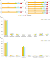
The changes in postsurgical stage by age are shown in Fig. 3B. In 2014, 76.4% of young patients aged 39 years or younger were classified as stage I, including 71.7% in stage IA and 4.6% in stage IB. On the other hand, 71.7% of the older patients aged 40 years or more were classified as stage I, including 53.9% in stage IA and 17.8% in stage IB, and the patients with stage IA was significantly more than that with stage IB (p<0.001). Advanced stage (stage III and IV) comprised 14.0% of the patients 39 years or younger but these advanced stages comprised 22.1% of those aged 40 years or more, which was significantly higher (p<0.001).
2. Histological types
The histological classifications of endometrial cancer are shown in Fig. 3C. Patients with endometrioid adenocarcinoma accounted for 82%–85% of the patients while those with serous adenocarcinoma were 4%–6%, and those with carcinosarcoma were 5%. Patients with endometrioid adenocarcinoma accounted for 84.8% of the patients in 2005 but significantly decreased to 81.7% in 2014. On the other hand, those with serous adenocarcinoma accounted for 3.8% in 2005 but significantly increased to 5.5% in 2014 (p<0.001).
3. Treatments
The changes in treatment methods for endometrial cancer are shown in Fig. 3D. Most patients (95%) underwent surgery as their main treatment. Chemotherapy was given to 2%–3% of the patients, and radiotherapy was given to 0.2%–0.6%. Most of the adjuvant therapy was chemotherapy, and 88%–96% of the patients who needed adjuvant therapy were treated with chemotherapy. In 2014, the number of patients who underwent chemotherapy was significantly higher than that in 2005 (p<0.001).
OVARIAN CANCER
1. Stage
The changes in postsurgical stage of ovarian malignant tumors are shown in Fig. 4A. Sixteen percents of the ovarian cancer patients were classified as stage IA, 0.9% as stage IB, 26.3% as stage IC, 1.3% as stage IIA, 1.6% as stage IIB, 6.2% as stage IIC, 1.0% as stage IIIA, 3.8% as stage IIIB, 22.8% as stage IIIC, 7.2% as stage IV, and 0.1% as unknown stage. Neoadjuvant chemotherapy (NAC) was given to 12.6% of the patients. The percentages of stage I, II, and IV patients in 2014 were almost the same as that in 2005, however, stage III patients decreased, and those treated with NAC increased (p<0.001, respectively).
Fig. 4
Characteristics of ovarian malignant tumor. (A) Distribution of post-surgical stages in ovarian malignant tumor. (B) Distribution of post-surgical stages in younger and older patients with ovarian malignant cancer. (C) Distribution of histological types in ovarian malignant cancer. (D) Distribution of treatment methods in ovarian malignant cancer.
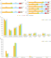
The changes in postsurgical stage by age are shown in Fig. 4B. Among the younger patients aged 39 years or less, stages I and II patients accounted for 68%–74% of the patients, and stages III and IV patients accounted for 24%–27%. Among the older patients aged 40 years or more, stages I and II patients accounted for 46%–51% of the patients, and stages III and IV patients accounted for 35%–44%, indicating that stages III and IV patients were significantly more prevalent. Therefore, there were significantly more patients aged 40 years or more who were treated with NAC than that aged 39 years or less (p<0.001).
2. Histological types
The histological types of ovarian malignant tumors are shown in Fig. 4C. Epithelial tumors were most common (94.8%–95.4%), while germ cell tumors were less abundant (3.0%–3.8%), and sex-cord tumors were very rare (0.3%–0.5%). Among the epithelial tumors, 33.0%–36.2% were classified as serous adenocarcinoma, 21.7%–24.8% as clear cell adenocarcinoma, 15.8%–17.5% as endometrioid adenocarcinoma, and 9.6%–12.8% as mucinous adenocarcinoma. The prevalence of serous adenocarcinoma remained constant over time, whereas clear cell adenocarcinoma and endometrioid adenocarcinoma significantly increased (p=0.001, p=0.030), and mucinous adenocarcinoma significantly decreased (p<0.001).
3. Treatments
The change in treatment methods for ovarian malignant tumors are shown in Fig. 4D. A combination of surgery and chemotherapy accounted for 62.5%–80.1% of the patients and this percentage significantly increased over time. On the other hand, surgery alone accounted for 17.8%–33.2% of the treatments and this percentage significantly decreased over time (p<0.001).
PROGNOSIS OF GYNECOLOGIC MALIGNANT TUMORS
The 5-year overall survival rates of cervical, endometrial, and ovarian cancer are shown in Tables 1, 2, 3, respectively. In all cancers, there were differences in prognosis between stages I and II, stage II and III, and stages III and IV.
Table 1
5-year overall survival in patients with cervical cancer (%)

| Stage | 2003 | 2006 | 2009 |
|---|---|---|---|
| I | 92.0 | 92.9 | 92.4 |
| II | 73.4 | 74.6 | 76.7 |
| III | 55.7* | 55.3 | 54.3 |
| IV | 24.3 | 24.3 | 25.2 |
Table 2
5-year overall survival in patients with endometrial cancer (%)

| Stage | 2003 | 2006 | 2009 |
|---|---|---|---|
| I | 95.4 | 96.3 | 94.6 |
| II | 86.8 | 92.7 | 89.4 |
| III | 75.4 | 80.6 | 78.3 |
| IV | 22.3 | 35.8 | 25.0 |
Table 3
5-year overall survival in patients with ovarian malignant tumor (%)

| Stage | 2003 | 2006 | 2009 |
|---|---|---|---|
| I | 90.3 | 90.6 | 90.5 |
| II | 76.3 | 82.9 | 78.8 |
| III | 42.4 | 48.7 | 46.0 |
| IV | 32.6 | 40.9 | 25.1 |
The 5-year overall survival rates in 2009 for cervical cancer were 98.0% for stage IA1 patients, 98.4% for stage IA2, 97.6% for stage IB1, 77.6% for stage IB2, 79.9% for stage IIA, 75.2% for stage IIB, 77.0% for stage IIIA, 52.5% for stage IIIB, 36.1% for stage IVA, and 19.5% for stage IVB. The 5-year overall survival rates for endometrial cancer were 97.1% for stage IA patients, 95.5% for stage IB, 88.9% for stage IC, 94.5% for stage IIA, 87.3% for stage IIB, 83.8% for stage IIIA, 60.0% for stage IIIB, 75.5% for stage IIIC, 16.4% for stage IVA, and 24.7% for stage IVB. The 5-year overall survival rates for ovarian cancer were 93.9% for stage IA patients, 96.0% for stage IB, 90.2% for stage IC (B), 84.6% for stage IC (A), 87.1% for Stage IC (1), 87.0% for stage IC (2), 83.1% for stage IIA, 89.3% for stage IIB, 74.2% for stage IIC (B), 82.6% for stage IIC (A), 66.7% for stage IIC (1), 72.6% for stage IIC (2), 57.1% for stage IIIA, 53.2% for stage IIIB, 44.2% for stage IIIC, and 25.1% for stage IV.
DISCUSSION
The changes in both the morbidity and mortality were analyzed using the population-based cancer registry which had the most complete records among the cancer registries in Japan. On the other hand, the actual number of patients was not comparable in the gynecologic cancer registry because it included 247 institutions in 2003 and 411 in 2014, which was approximately a 1.7-fold increase. Therefore, the gynecologic cancer registry was not able to be used to analyze the morbidity and mortality. The population-based cancer registry included less information, consequently, the gynecologic cancer registry was used for detailed analysis of clinicopathologic factors. In that analysis, percentages were used rather than the actual numbers.
The morbidity of cervical cancer had a tendency to decline but more recently has increased. The reasons are insufficient promotion of HPV vaccination and low rates of cancer screening. In contrast, the prevalence of endometrial cancer has significantly and consistently been increasing and represents the most common gynecologic malignant tumor in Japan. These changes are due to the changes in life style by Japanese women (e.g. westernized diet, delayed marriage, and low birth rate), and the increasing prevalence will likely continue. Ovarian cancer has also been increasing, but the main cause remains unclear. Recently, the mortality due to both cervical and ovarian cancer has remained flat. Molecular targeted therapy for both cervical and ovarian cancer were recently covered by insurance in Japan, however, the effects of these new drugs have not yet appeared in the registry. The mortality of endometrial cancer has also increased slowly with a rapid increase in the morbidity. This phenomenon seemed to reflect the good prognosis in endometrial cancer.
In cervical cancer, stage I accounted for approximately 75% of the cases and exceeded 90% in young patients. For the changes from 2005 to 2014 in both patient groups, stages I and IV were increasing, and stages II and III were decreasing in prevalence. Attention should be paid to the facts that advanced patients with distant metastasis are increasing while patients with cancer detected at an early stage are also increasing. Squamous cell carcinoma is the main histological type but adenocarcinoma and neuroendocrine tumors are gradually increasing. The patients underwent a combination of surgery and adjuvant therapy radiotherapy (including CCRT) is decreasing, while the patients underwent CCRT or a combination of surgery and adjuvant chemotherapy are increasing. There is little evidence for improved outcome with adjuvant chemotherapy for cervical cancer, and the treatment guidelines for cervical cancer treatment do not recommend it. However, because the severe adverse effects of adjuvant radiotherapy are known, and many gynecologic oncologists believe that radiotherapy which is recognized as a local treatment is not necessary after complete resection of cervical cancer, adjuvant radiotherapy is not so popular in Japanese gynecologic oncologists.
The prevalence of stage I endometrial cancer is increasing, and stages II and III are decreasing. The reason is that the FIGO 2008 staging system was adopted in 2012 in Japan, consequently, patients who had been previously diagnosed with stage II due to progression in the cervical mucosa and stage III due to positive ascitic cytology are currently diagnosed as stage I. The rate of patients with stage I endometrial cancer aged 39 years or younger had significantly more frequently than that of patients aged 40 years or older, however no marked differences like cervical and ovarian cancer were found. Endometrioid adenocarcinoma was the most common histological type, followed by carcinosarcoma, serous adenocarcinoma, and clear cell adenocarcinoma. The prevalence of serous adenocarcinoma is increasing. One reason of these findings is the possibility of bias since patients who were diagnosed with poorly-differentiated endometrioid adenocarcinoma before by hematoxilin-eosin staining alone are currently being diagnosed by immunohistochemical staining. In Japan, not only patients at high risk but also those at intermediate risk usually undergo adjuvant chemotherapy which is different from the treatment in Europe and in the United States. On the other hands, adjuvant radiotherapy is seldom performed for endometrial cancer patients because of same reason with cervical cancer.
Unlike uterine cancers, stages I and II accounted for only a half of the ovarian cancers but the percent of patients with advanced ovarian cancer is higher. It remains difficult to detect early-stage ovarian cancer. Most patients had epithelial malignant tumors, and only 3%–4% had germ cell tumors. Epithelial malignant tumors included serous, clear cell, endometrioid, and mucinous adenocarcinoma in order of prevalence. However, the prevalence of clear cell and endometrioid adenocarcinoma which is considered to develop from endometriosis is increasing. Since the treatment guidelines for ovarian cancer were established, surgery alone and chemotherapy alone have been decreasing and a combination of surgery and chemotherapy are now the most common treatment.
The prognosis of these 3 cancers in Japan has not dramatically changed although small changes were shown from 2003 to 2009. In particular, the overall survival of stage IV patients has shown the large annual change, probably because the number of stage IV patients is much smaller than that of other stage. Although there was no change in therapeutic method using approved drugs during these observation period in Japan, because molecular targeted therapy was approved by insurance recently, it might change treatment outcomes of cervical and ovarian cancer in the near future.
 XML Download