

 PDF
PDF Citation
 Print
Print


Abstract
Objective
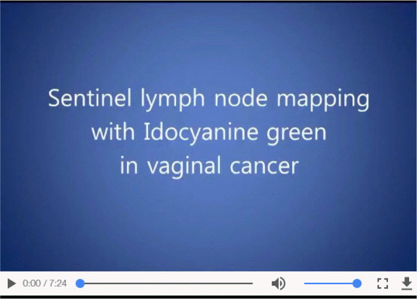
Sentinel lymph node (SLN) mapping is being adapted to gynecologic cancer. Higher SLN mapping rates were reported with indocyanine green (ICG) compared to other dyes. The aim of this film is to share our experience of SLN mapping with ICG in vaginal cancer.
Methods
A 40 year-old woman was diagnosed with squamous cell vaginal cancer. About 1.5 cm-sized tumor was located on the posterior vaginal fornix. Preoperatively she was assumed to be stage I vaginal cancer. Beginning of surgery, we performed SLN mapping by ICG injection into 3- and 9-o'clock positions of the vaginal tumor. Concentrated in 1.25 mg/mL, 1 mL of ICG solution was injected into deep stroma and another 1 mL submucosally in both sides. Bilateral SLN identification and lymphadenectomy were done. Afterward, laparoscopic Type C1 Querleu-Morrow radical hysterectomy with vaginectomy was done. A fluorescence endoscope produced by KARL STORZ (Tuttlingen, Germany) was used for ICG detection.
Results
To our knowledge, this is the first film report performing SLN mapping with ICG in vaginal cancer. The mapping was successful and we were able to recognize SLN of vaginal cancer. SLNs were located in the bilateral obturator fossa. According to the pathologic diagnosis, the mass size was 15 mm and invasion depth was 1 mm. Subvaginal tissue involvement and pelvic wall extension were absent. Resection margin of the vagina was free from carcinoma. No lymph node metastasis was reported including the bilateral SLNs.
Go to : 
 | undefined Video can be found with this article online at https://ejgo.org/src/sm/jgo-28-e29-s001.mp4. |
 XML Download