

 PDF
PDF Citation
 Print
Print


INTRODUCTION
Chylous or lymphatic ascites is defined as the pathologic accumulation of lymphatic fluid in the peritoneal cavity [1]. More specifically, nonmalignant ascites is categorized into two types: chylous and lymphatic, depending on the the anatomic location of lymphatic injury [1]. Lymphatic ascites can result from congenital lymphatic abnormalities, nephrotic syndrome, liver cirrhosis, and malignancy that occlude the lymphatic channels [2]. Postoperative lymphatic ascites is a rare complication, resulting from operative lymph node dissection or radiation [2]. Systematic pelvic and para-aortic lymphadenectomy is a common surgical procedure for the treatment of gynecologic malignancies. The incidence of lymphatic ascites in patients who received retroperitoneal lymphadenectomy was reported to be between 0.17% and 4% [134]. In particular, lymphatic ascites caused by retroperitoneal lymphadenectomy was associated more with para-aortic lymphadenectomy than with pelvic lymphadenectomy [4]. However, reports of massive lymphatic ascites caused by laparoscopic pelvic and para-aortic lymphadenectomy are rare.
Conservative treatment of lymphatic ascites is aimed at reducing lymph production, including low-fat diet with medium-chain triglycerides, total parenteral nutrition, octreotide, and paracentesis. With failure of conservative managements, lymphoscintigraphy and pedal or intranodal lymphangiography (INLAG) may localize the leak site and guide surgical intervention. Pedal lymphangiography (LAG) is commonly used as a means of evaluating and staging patients with disorders of the lymph nodes and lymphatic channels [5]. However, this procedure is both time-consuming and technically challenging. Several authors suggested that ultrasound-guided INLAG might be an alternative to the conventional pedal LAG [678]. The patients with chylothorax including iatrogenic, spontaneous, and postoperative etiologies were treated successfully using INLAG. In addition, this technique could be easily replicated by other interventional radiologist and N-butyl cyanoacrylate glue can be used to embolize the lymphatic leakage, which provides an additional mechanical obstruction. In June 2014, ultrasound-guided INLAG was first employed on a gynecologic cancer patient with massive lymphatic ascites after retroperitoneal lymphadenectomy.
To our knowledge, few reports in the English literature have evaluated risk factors of lymphatic ascites caused by laparoscopic pelvic and para-aortic lymphadenectomy. We initiated treatment of postoperative lymphatic leakage using ultrasound-guided INLAG with glue embolization. The purpose of this study was to evaluate the risk factor of massive lymphatic ascites after laparoscopic pelvic and para-aortic lymphadenectomy in patients with gynecologic cancers. In addition, we examined the feasibility of ultrasound-guided INLAG with N-butyl cyanoacrylate glue for the treatment of massive lymphatic ascites.
MATERIALS AND METHODS
Two hundred thirty-seven patients with gynecologic cancers underwent laparoscopic pelvic and para-aortic lymphadenectomy at Ajou University Hospital between April 2006 and November 2015. We excluded three patients who had a genitourinary injury during surgery, causing prolonged hospital stay. During the study period, we performed a retrospective analysis of 234 patients. Systematic pelvic and para-aortic lymphadenectomy were performed as described previously [9]. Briefly, four surgeons (HSR, SJC, JP, and TWK) performed laparoscopic/robotic pelvic and para-aortic lymphadenectomy using the same surgical devices. Graspers and suction irrigator were used to separate lymph node packets. Coagulation with Harmonic Ace Curved Shears (Ethicon Endo-Surgery, Cincinnati, OH, USA) was also performed. Para-aortic lymphadenectomy up to the level of the renal vessels was performed in endometrial cancer patients with preoperative tumor grade 2 or 3, elevated cancer antigen 125 level, or deep myometrial invasion by magnetic resonance imaging as described previously [10]. Also, we performed para-aortic lymphadenectomy up to the level of the renal vessels in early stage ovarian cancer patients. Pelvic drain was routinely placed into the peritoneal cavity. The pelvic drain was removed once the drainage volume has become less than 300 mL in 24 hours. All patients signed written informed consent. Clinicopathologic data were obtained from medical records after obtaining approval from the - Institutional Review Board.
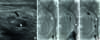
Postoperative massive lymphatic ascites was newly defined as follows: (1) the drainage fluid triglyceride content >110 mg/dL [11] and (2) the mean lymphatic leakage volume more than 500 mL/day and duration of pelvic drainage more than 4 weeks in a patient who underwent retroperitoneal lymphadenectomy despite conservative managements including a dietary modification and octreotide therapy. The patients with massive lymphatic ascites were managed with low-fat diet containing medium-chain triglycerides, fasting with total parenteral nutrition, octreotide therapy, and continuous drainage. A medium-chain triglyceride diet and total parenteral nutrition was continued for a minimum of 3 weeks. In addition, three doses of 100 µg octreotide acetate (Sandostatin, Novartis, Basel, Switzerland) were administered subcutaneously a day for a minimum of 7 days. In June 2014, we first performed INLAG with glue embolization to manage massive lymphatic ascites after failure of conservative measures. Briefly, after administration of local anesthesia, an inguinal lymph node was directly accessed using ultrasound guidance with a 22-gauge needle (Fig. 1A). Approximately 10 mL of ethiodized oil contrast medium (Lipiodol Ultra-Fluid, Laboratoire Andre Guerbet, Aulnay-sous-Bois, France) was slowly infused into the inguinal lymph node, resulting in opacification of the efferent lymphatics and leaking lymphatic channels (Fig. 1B). If extravasation was observed during injection, a 22-gauge needle was targeted to the site of pelvic lymphatic leakage under fluoroscopic guidance (Fig. 1C). After visualization of the pelvic lymphatic leakage, embolization of pelvic lymphatic leakage was performed using n-butyl cyanoacrylate glue (Histoacryl, B. Braun, Melsungen, Germany) (Fig. 1D). With no evidence of further lymphatic leakage, the procedure was terminated. The procedure time was approximately 20 to 90 minutes.
Fig. 1
(A) Ultrasound image shows the position of the needle (arrowhead) within the inguinal lymph node (arrow). Fluoroscopic images show (B) the extravasation of ethiodized oil (arrowhead), (C) the needle targeting (arrowhead), and (D) the embolization of the pelvic lymphatic leakage site using n-butyl cyanoacrylate glue (arrowhead).
After successful treatment for massive lymphatic ascites, we hoped that ultrasound-guided INLAG with glue embolization might reduce the duration of pelvic drain and hospital stay. We suggested a new indication of INLAG with glue embolization as follows: the mean lymphatic leakage exceeding 500 mL/day 7 to 14 days after retroperitonal lymphadenectomy. We performed INLAG in 17 patients with the mean lymphatic leakage exceeding 500 mL/day 7 to 14 days after retroperitonal lymphadenectomy based on a predefined indication in our institution.
All possible clinicopathologic factors related to massive lymphatic ascites were determined in pre-INLAG group (n=163). The incidence of leg lymphedema and infected lymphocele were evaluated. The Common Terminology Criteria for Adverse Events version 4.0 (CTCAE) were used to classify leg lymphedema. An infected lymphocele is defined as a large collection of protein-rich lymphatic fluid in the pelvic cavity, causing fever and abdominal pain [12]. In addition, we compared clinical courses between pre-INLAG group (n=163) and post-INLAG group (n=71).
Normality testing (Kolmogorov-Smirnov test) was performed to evaluate whether data were sampled from a Gaussian distribution. Pearson chi-square test and Fisher exact test were used for categorical variables, and the Student t-test and Mann-Whitney U statistics for continuous variables according to normality. Statistical analyses were performed using SPSS ver. 20.0 (IBM Co., Armonk, NY, USA). A two-sided p<0.05 was considered to indicate significant difference.
RESULTS
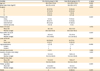
Table 1 shows the comparison of clinicopathologic features between non-massive and massive lymphatic ascites in 163 patients with gynecologic cancers who received laparoscopic pelvic and para-aortic lymphadenectomy before introducing INLAG. The median age of all patients was 49.0 years (range, 24.0 to 75.0 years). The indications for laparoscopic retroperitoneal lymphadenectomy included ovarian cancer (n=2), uterine cervical cancer (n=68), endometrial cancer (n=89), and uterine sarcoma (n=4). Microscopically, metastatic carcinoma in LNs was detected in four patients (2.5%) with uterine cervical cancer. In pre-INLAG group (n=163), four patients (2.5%) developed massive lymphatic ascites postoperatively. After starting an oral diet on the postoperative days 3 to 5, the drainage fluid became yellowish and milky and the amount of fluid drainage increased to 850 to 1,200 mL/day. All cases with massive lymphatic ascites were resolved by conservative treatments including a dietary modification, total parenteral nutrition, and octreotide therapy. The duration of pelvic drainage was 30, 32, 29, and 63 days, respectively. Overall, leg lymphedema (CTCAE grades 1 to 2) occurred in four patients (2.5%). Five patients (3.1%) required intravenous antibiotics and percutaneous catheter drainage due to infected lymphocele. Four patients (2.5%) developed postoperative massive lymphatic ascites. No significant differences were observed for age, body mass index, primary sites of gynecologic malignancies, the number of harvested pelvic and para-aortic LNs, LN metastasis, the level of para-aortic LN dissection, the incidence of leg lymphedema and infected lymphocele, and the duration of follow-up. There was no significant difference in the incidence of postoperative massive lymphatic ascites among the four surgeons. There were significant differences in the duration of pelvic drainage (median value, 11.0 days vs. 31.0 days, p=0.001) and hospital stay (median value, 13.0 days vs. 34.0 days, p=0.001). Postoperative massive lymphatic ascites was associated with three cases of liver cirrhosis (p<0.001).
Table 1
Comparison of clinicopathologic features between non-massive and massive lymphatic ascites in 163 patients with gynecologic cancers who received laparoscopic pelvic and para-aortic lymphadenectomy before introducing intranodal lymphangiography
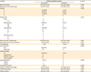
Table 2 shows the clinicopathologic characteristics of 71 patients with gynecologic cancers who received laparoscopic/robotic pelvic and para-aortic lymphadenectomy after introducing INLAG. The median age was 50.0 years (range, 22.0 to 72.0 years). Also, retroperitoneal lymphadenectomy was performed by the same four surgeons. The indications for laparoscopic/robotic retroperitoneal lymphadenectomy included ovarian cancer (n=11), uterine cervical cancer (n=12), endometrial cancer (n=47), and uterine sarcoma (n=1). The two patients (2.8%) had microscopic metastases in LNs. In June 2014, we first performed INLAG with glue embolization on postoperative day 28 in a patient with massive lymphatic ascites after failure of conservative measures. The interval between INLAG and pelvic drain removal was 6 days. After successful treatment for massive lymphatic ascites, the procedure was performed in 17 patients with the mean lymphatic leakage exceeding 500 mL/day 7 to 14 days after lymphadenectomy. The mean volume of the lymphatic leakage before INLAG ranged between 520 and 1,500 mL/day. After INLAG, the lymphatic drainage fluid appeared clear and the mean lymphatic drainage volume was <300 mL/day irrespective of oral diet (Fig. 2A). Among 18 patients who received INLAG, 17 patients had pelvic drain removed within 7 days after INLAG (Fig. 2B). Overall, 18 patients (25.4%) received INLAG with glue embolization. Among 18 patients who received INLAG, one patient had a persistent lymphatic leakage above 900 mL/day. She underwent repeat INLAG with ethiodized oil injection into the inguinal and pelvic lymphatics. The mean lymphatic drainage volume was <300 mL/day 3 days after repeat INLAG. There were no INLAG-related major complications including vessel or bowel injuries. Rescue analgesia such as ketorolac tromethamine (30 mg) was administered intravenously to three patients (16.7%) who received INLAG with glue embolization. There were one leg (1.4%) lymphedema (CTCAE grade 1). There was no infected lymphocele. The median duration of pelvic drainage and hospital stay was 10.0 days (range, 4.0 to 28.0 days) and 12.0 days (range, 5.0 to 35.0 days), respectively.
Table 2
Clinicopathologic characteristics of 71 patients with gynecologic cancers who received laparoscopic/robotic pelvic and para-aortic lymphadenectomy after introducing intranodal lymphangiography
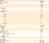
Values are presented as median (range) or number (%). Leg lymphedema was classified according to the CTCAE version 4.0.
FIGO, International Federation of Gynecology and Obstetrics; IMA, inferior mesenteric artery; INLAG, intranodal lymphangiography; LN, lymph node; PALND, para-arotic lymph node dissection.
Fig. 2
(A) The changes in the mean pelvic drainage volume before and after the intranodal lymphangiography (INLAG). (B) Clinical courses in 18 patients with lymphatic leakage who received intranodal lymphangiography.
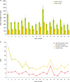
Table 3 presents the comparative data of surgical features and clinical courses between pre-INLAG group (n=163) and post-INLAG group (n=71). Although primary sites of gynecologic malignancies were significantly different (ovarian cancer, 1.2% vs. 15.5%; uterine cervical cancer, 41.7% vs. 16.9%; endometrial cancer, 54.6% vs. 66.2%; uterine sarcoma, 2.5% vs. 1.4%), para-aortic lymphadenectomy in post-INLAG group was performed at higher level of para-aortic LN (renal vein) and patients in post-INLAG group had more para-aortic LNs retrieved, compared to those in pre-INLAG group (level of renal vein, 22.5% vs. 6.1%, p<0.001; para-aortic LN count, 13.1 vs. 8.0, p=0.001). However, the mean duration of pelvic drainage and hospital stay decreased after the introduction of INLAG (13.2 days vs. 10.9 days, p=0.002; 15.2 days vs. 12.6 days, p=0.001). There was no recurrence of postoperative massive lymphatic ascites after this procedure.
Table 3
Comparison of surgical features and clinical courses between pre-intranodal lymphangiography group (n=163) and post-intranodal lymphangiography group (n=71)
DISCUSSIONS
Two reports evaluated the incidence and risk factors of lymphatic ascites in patients with gynecologic cancers who received pelvic and/or para-aortic lymphadenectomy [34]. Han et al. [3] calculated an incidence of 0.17% in 4,119 patients after laparotomic lymphadenectomy (0.32% in the para-aortic lymphadenectomy group vs. 0.077% in the pelvic lymphadenectomy group). Zhao et al. [4] found that the incidence of lymphatic ascites after laparoscopic lymphadenectomy was 0.9% (4.08% in the para-aortic lymphadenectomy group vs. 0.35% in the pelvic lymphadenectomy group). Both studies supported the view that the higher incidence of lymphatic ascites may be related to para-aortic lymph node dissection. However, few reports are available about massive lymphatic ascites after laparoscopic pelvic and para-aortic lymphadenectomy. We found an incidence of massive lymphatic ascites of 2.5%, which is higher than that of previous reports. The higher incidence may be associated with energy devices including monopolar coagulation and ultrasonically activated shears. In addition, all 163 patients in pre-INLAG group received para-aortic lymphadenectomy as well as pelvic lymphadenectomy.
We analyzed several risk factors associated with postoperative massive lymphatic ascites. There were no relationships between the number of retrieved LNs and the incidence of massive lymphatic ascites. Moreover, massive lymphatic ascites was not associated with the level of para-aortic lymphadenectomy. Postoperative massive lymphatic ascites was related to certain medical conditions including liver cirrhosis and heart failure. The majority of patients who present with ascites have underlying medical conditions including cirrhosis, heart failure, tuberculosis, and pancreatitis [2]. Thus, the major factors contributing to ascites formation are splanchnic vasodilatation with arterial underfilling in liver cirrhosis and reduced cardiac output in heart failure, and ascites may be exacerbated by lymphadenectomy in these patients. In pre-INLAG group, four patients presented with massive lymphatic ascites. Among these patients, three patients had chronic medical conditions (two hepatitis B virus related-cirrhosis and one alcoholic liver cirrhosis). In post-INLAG group, one patient with congestive heart failure also showed massive lymphatic ascites despite conservative managements for 4 weeks. Therefore, certain medical conditions that cause reduced effective circulating volume may be associated with massive lymphatic ascites after retroperitoneal lymphadenectomy.
Conservative treatment of lymphatic ascites is a dietary modification including low-fat diet with medium-chain triglycerides. The success rate of a dietary modification is reportedly 60% to 80% and treatment should be continued for at least 3 weeks if clinical improvement is acquired [2131415]. Moreover, this dietary modification should be maintained for several months after resolution of lymphatic ascites [1316]. Also, somatostatin and its analogue (octreotide) have been used to decrease lymph production. The responses were evident in under 72 hours. However, the optimal duration of somatostatin or octreotide therapy is considered to be 1 to 2 weeks and various success rates have been reported [171819]. In this study, a medium-chain triglyceride diet and total parenteral nutrition were continued for at least 3 weeks. In addition, three doses of 100 µg octreotide acetate were administered subcutaneously a day for a minimum of 7 days. Although the first three cases with massive lymphatic ascites were resolved by a dietary modification, total parenteral nutrition, and octreotide therapy, the duration of conservative managements was approximately 3 to 4 weeks. The fourth patient with severe liver cirrhosis did not respond to conservative treatments, developed a bacterial peritonitis, and was admitted to the intensive care unit. The amount of lymphatic ascites was more than >2,000 mL/day and the duration of hospital stay was 65 days. Before retroperitoneal lymphadenectomy, therefore, the underlying medical condition related to the reduced effective circulating volume, such as liver cirrhosis, nephrotic syndrome, and heart failure, should be meticulously evaluated.
In post-INLAG group, one patient with congestive heart failure showed massive lymphatic ascites. We first performed ultrasound-guided INLAG with glue embolization of pelvic lymphatic leakage site since conservative treatments were not effective. The interval between INLAG and pelvic drain removal was 7 days. Pedal LAG using ethiodized oil completely occluded lymphatic leakage in 70% of patients when the lymphatic drainage volume was <500 mL/day [20]. However, this procedure is both time-consuming and technically challenging and could not be easily replicated by other interventional radiologists. Recently, Rajebi et al. [6] showed that ultrasound-guided puncture of an inguinal lymph node could be used to treat the leaking lymphatic channels. Until now, ultrasound-guided INLAG with thoracic duct embolization has been used to treat postoperative chylothorax [678]. We employed this technique to treat massive lymphatic ascites after retroperitoneal lymphadenectomy. After successful treatment for massive lymphatic ascites, we performed INLAG in 17 patients with the mean lymphatic leakage exceeding 500 mL/day 7 to 14 days after retroperitonal lymphadenectomy. After INLAG with glue embolization, the lymphatic drainage fluid appeared clear and the mean lymphatic drainage volume was <300 mL/day irrespective of oral diet. Among 18 patients who received INLAG, 17 patients had pelvic drain removed within 7 days after INLAG. In addition, the mean duration of pelvic drainage and hospital stay decreased after the introduction of INLAG. Injured lymphatic channels during retroperitonal lymphadenectomy are transiently adhered and healed by the surrounding tissues. However, failure to heal these channels results in delayed leakage from them [2]. INLAG with glue embolization may enhance the closure of leaking lymphatic channels. Therefore, this procedure may decrease the incidence of lymphocele including infected lymphocele and be an alternative treatment options for massive lymphatic ascites after retroperitoneal lymphadenectomy. However, it is necessary to define the indication of this procedure through future studies on validating our initial experience of ultrasound-guided INLAG with glue embolization in patient with massive lymphatic ascites after retroperitoneal lymphadenectomy.
Lymph node status is important in determining prognosis in endometrial/cervical cancer and may influence the decision to administer adjuvant treatment [2122]. However, complete lymphadenectomy is associated with surgical morbidity including prolonged operative time, lymphatic ascites, vessel injury, and leg lymphedema [423]. Recently, several studies reported that sentinel node mapping with indocyanine green shows a consistently higher overall detection (94%) [2425262728]. In addition, the updated 2014 NCCN (National Comprehensive Cancer Network) guidelines state that sentinel lymph node mapping may be considered as part of the surgical staging in apparent stage I endometrial cancer. Thus, if sentinel node mapping becomes the standard of care in the management of endometrial/cervical cancer, lymphadenectomy-related complications will be reduced. However, we should perform systematic lymphadenectomy in some patients with presumed higher stage gynecologic cancers. Therefore, we suggest that INLAG with glue embolization may be a treatment option for managing massive lymphatic ascites after lymphadenectomy.
Our study had some limitations inherent in the retrospective nature of its design. First, multivariate analysis did not identify independent risk factors for postoperative massive lymphatic ascites because the sample size was small and the follow-up period was relatively short to make any conclusive remarks on these results. Secondly, INLAG cannot be used to treat massive lymphatic ascites when the lymphatic injury is at the level of cisterna chyli and above after para-aortic lymphadenectomy up to the level of renal vein since lymphatics act as a one-way drainage system and INLAG cannot access to the leaking site above the external iliac lymphatic channel. Thirdly, we could not evaluate the real incidence of massive lymphatic ascites in post-INLAG group because of early intervention. It is necessary to evaluate the indication and the timing of INLAG in patients who show postoperative massive lymphatic ascites. Lastly, meta-analysis findings showed that the pelvic drain placement is associated with a higher risk of symptomatic lymphocele formation when the pelvic peritoneum is left open [29]. In this study, however, pelvic drain was routinely placed into the peritoneal cavity, which may result in lymphocele formation and prolong the hospital stay. Also, lymphatic leakage may be spontaneously healed by the surrounding tissues even without a pelvic drain.
In conclusion, underlying medical conditions related to the reduced effective circulating volume, such as liver cirrhosis, nephrotic syndrome, and heart failure, should be meticulously evaluated before retroperitoneal lymphadenectomy in gynecologic cancer patients. INLAG with glue embolization can be used to identify and treat the leaking lymphatic channels, especially pelvic lymphatic leakage, in patients with massive lymphatic ascites after retroperitoneal lymphadenectomy in gynecologic cancers. Therefore, this procedure may be an alternative treatment options for massive lymphatic ascites after retroperitoneal lymphadenectomy. However, future studies are needed to determine the indication of this procedure.
 XML Download