

 PDF
PDF Citation
 Print
Print


INTRODUCTION
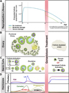
During embryonic development, germ cells proliferate and reach a maximal number (6–7 million) at 20 weeks of gestation [12]. This number decreases due to atresia to 1 million at birth and to 400,000 oocytes around puberty. During the reproductive lifespan, this finite number of oocytes—called the ovarian reserve—further decreases with each menstrual cycle, ultimately reaching 1,000 at the time of menopause (Fig. 1A) [34]. Endocrine function and fertility depend on the cyclical recruitment and development of oocytes from the ovarian reserve. The ovarian follicle secretes hormones that are necessary for not only oocyte development and fertility, but also endocrine function that impacts other systems, such as the cardiovascular system and bone. Thus, preservation of the ovarian reserve is necessary to maintain overall women’s health (Fig. 1B). Unfortunately, chemo- and radiation therapies that are effective in eliminating cancer cells also destroy growing oocytes in the ovary (Fig. 1B). The toxic environment also negatively affects the oocytes in the ovarian reserve, causing death or abnormal activation leading to atresia (Fig. 1C), all of which results in depletion of ovarian follicles and hormonal imbalance (Fig. 1D) [56].
Fig. 1
Consequences of gonadotoxic treatment in reproductive-aged women. (A) The primordial follicle population—the ovarian reserve—can be represented by a parabolic curve across the female lifespan in which activation or death of primordial ovarian follicles occurs progressively with each menstrual cycle, from puberty to the menopause. Reprinted from Wallace and Kelsey [4]. (B–D) The growth of follicles with each cycle maintains hormonal balance necessary for overall women’s health. Gonadotoxic stress or treatment, such as chemotherapy or radiation therapy (red bar across all panels), induces a rapid decrease in the highly sensitive primordial follicles of the ovarian reserve (A, C), resulting in a follicle-depleted ovary (B) and premature ovarian failure (POF). Depletion of the ovarian reserve disrupts normal endocrine function and the production of hormones such as follicle-stimulating hormone (FSH), luteinizing hormone (LH), estradiol, progesterone, inhibin B, and inhibin A (D), leading to hormonal imbalance similar to that seen in postmenopausal women.
The degree of depletion of ovarian follicles caused by cancer treatment depends on the dose and type of chemotherapy and radiation, fractionation scheme, and irradiation field used [789]. Oocytes are one of the most sensitive cells in the human body to radiation; indeed, 50% of immature human oocytes can be destroyed by exposure to 2 Gy of radiation [810]. Exposure to abdominal radiation of 20 to 30 Gy in childhood or total-body irradiation of 15 Gy leads to loss of ovarian function (premature ovarian failure, POF) and increased risk of adverse pregnancy outcomes in up to 90% of patients [1112]. Chemotherapy that includes alkylating agents such as cyclophosphamide (Cy) or busulfan affects somatic cells in other tissues as well as the oocyte and the ovarian follicle.
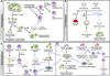
In men with cancer, current options for preserving fertility prior to or during chemo- and radiation therapy include gonadal shielding and sperm banking (semen cryopreservation). Testis-sparing surgery can also preserve fertility in male cancer patients. In women with cancer, fertility preservation strategies include controlled ovarian stimulation (COS) followed by in vitro maturation (IVM) of the retrieved oocytes, which can then be either cryopreserved or fertilized in vitro (IVF) to produce embryos for cryopreservation [13]. Ovarian tissue cryopreservation is an experimental option, with the thawed tissue used for transplantation at later date. Several emerging technologies available in the near future include in vitro ovarian follicle culture, ovarian follicle transplantation (artificial ovary), in vitro activation of ovarian follicles, and the use of fertoprotective agents (Fig. 2). In this review, the various existing and emerging technologies for the preservation of fertility and restoration of reproductive function in women with cancer will be discussed, with the aim of illustrating how cancer treatments and fertility preservation options can be tailored to meet the needs and desires of individual patients.
Fig. 2
Schema of fertility preservation approaches in cancer patients. Each procedure is marked with a line, either black (established) or blue (investigational). (A) Patients undergoing a natural or hyperstimulated cycle produce mature eggs that can be matured in vitro (IVM) and either cryopreserved or fertilized by in vitro fertilization (IVF)/intracytoplasmic sperm injection (ICSI) to produce embryos that are cryopreserved. In patients who cannot undergo controlled ovarian stimulation (e.g., prepubertal girls or women with hormone-sensitive cancers) or in those who must start cancer treatment immediately, strips of ovarian cortex can be removed and cryopreserved, or individual follicles can be isolated from the tissues and cryopreserved. Investigational methods are focusing on use of thawed ovarian tissues for transplantation or in vitro growth of follicles, followed by IVM and IVF/ICSI. (B) During treatment, gonadotoxicity can be mitigated by the use of fertoprotective reagents that induce death in tumor cells while preventing off-target effects in other tissues, resulting in preserved fertility and endocrine function. (C) After cancer treatment, cryopreserved embryos can be thawed and transferred into the uterus, or cryopreserved eggs can be thawed and used for IVF/ICSI and the resulting embryos transferred to the patient. On the investigational side (blue arrows), cryopreserved follicles can be grown in culture, matured in vitro, and fertilized to produce embryos for transfer. Ovarian tissue can be transplanted back into the patient, or follicles can be isolated and used to produce embryos or cultured on a three-dimensional bioplotted scaffold as an artificial ovary for transplantation back into patients. Transplantation of ovarian tissue or follicles within an artificial ovary has the potential of restoring fertility as well as endocrine function after cancer treatment. (D) In patients who were unable to cryopreserve eggs, embryos, or ovarian tissue prior to treatment, researchers are now investigating the possibility of using oogonial stem cells (OSCs) to repopulate follicle-depleted ovaries, or differentiating follicle somatic cells and oocytes from embryonic stem (ES) cells or induced pluripotent stem (iPS) cells to assemble follicles de novo for transplantation or IVM and IVF/ICSI to create embryos for transfer.
EXISTING TECHNOLOGIES
In women diagnosed with cancer, there are two standard options for fertility preservation: cryopreservation of mature oocytes or embryos (Fig. 2A). Ovarian tissue cryopreservation for later transplantation is still considered to be experimental and can only be performed under IRB approval. Selecting the optimal method for fertility preservation depends on timing of cancer treatment, treatment regimen, cancer type, age of the patient, and presence or absence of a partner.
1. Embryo cryopreservation
Embryo cryopreservation is the most widely used method in infertility clinics, resulting in thousands of births each year from frozen embryos (Fig. 2A). Human embryos are highly resistant to damage by freezing, with reported survival and implantation rates of over 90% and 30%, respectively [14]. Moreover, the live birth rate per embryo transfer has been reported to be 20%, while the accumulated pregnancy rate from cryopreservation of multiple embryos has been reported to be more than 60% in infertility patients [15]. Since the entire embryo cryopreservation procedure requires approximately 2 weeks, it is not an option for women who have aggressive cancers and must start cancer treatment immediately. Because women must undergo COS to produce mature eggs, this option is also not recommended for women who have hormone-sensitive cancers and is not possible in prepubertal girls. For women without a partner, donated sperm can be used to create embryos.
2. Oocyte cryopreservation
In 2013, the American Society for Reproductive Medicine and the Society for Assisted Reproductive Technology announced that modern procedures for oocyte cryopreservation should no longer be considered experimental [16]. This method provides an alternative to embryo cryopreservation for those women who do not have a partner or do not want to use donated sperm. In addition, the cryopreserved oocyte is the legal property of the woman, which avoids the complex custody issues often associated with cryopreserved embryos. Like embryo cryopreservation, however, cryopreservation of oocytes involves hormonal stimulation of the ovary (COS), so this method is not available to women with aggressive cancers that require immediate treatment or hormone-sensitive cancers, or for prepubertal girls. Collected oocytes can be cryopreserved as mature MII eggs or as immature germinal vesicle (GV) oocytes.
1) Mature oocyte cryopreservation
The first live birth from a cryopreserved oocyte was reported by Chen [17] in 1986. Due to improved freezing and thawing techniques, the pregnancy rate with cryopreserved oocytes has improved considerably. Recently, high survival rates (≥90%) and pregnancy rates (40% to 55%) have been reported in egg donation programs [18]. A number of studies have reported no differences in the pregnancy rate or implantation rate of embryos obtained from cryopreserved mature oocytes compared with those from fresh oocytes, and further, no noticeable differences in chromosomal abnormalities have been reported [192021]. At the present time, mature oocyte cryopreservation is a preferred fertility preservation method if the individual is postpubertal and if chemotherapy or radiation treatment can be delayed to allow time for the full procedure.
2) Immature oocyte cryopreservation and in vitro maturation
Aspiration of immature oocytes followed by IVM is an option for patients who are unable to undergo ovarian stimulation, including prepubertal girls, women with hormone-sensitive cancers, and patients with polycystic ovarian syndrome (PCOS) who cannot undergo stimulation due to ovarian hyperstimulation syndrome. Aspiration of immature oocytes offers the advantage of allowing immediate cancer treatment, since COS is not required. In addition, it is thought that less damage is caused by cryopreservation of immature oocytes than mature oocytes, as immature oocytes lack the spindle and nuclear membrane that protect the chromatin. However, studies have reported that cryopreserved mature oocytes achieved better outcomes compared to IVM of cryopreserved immature oocytes [22]. For this reason, immature oocytes are often matured by IVM and then cryopreserved as mature oocytes [2223]. There have been only a few reports of successful birth using cryopreserved oocytes or embryos from immature oocytes that were matured in vitro [24]. So far, this technique has mainly been used in PCOS patients, and data on efficacy in cancer patients are not available.
3. Ovarian stimulation protocols used for embryo or mature oocyte cryopreservation
1) Random-start COS
In a conventional COS protocol for embryo or oocyte cryopreservation, cycles require up to 5 weeks, since ovarian stimulation must be started on day 2 to 3 of the menstrual cycle (Fig. 2A). In many cases, this time frame is appropriate, since many patients undergo tumor resection prior to chemo- or radiation therapy. That said, some patients need to start cancer treatment sooner and therefore random start COS protocols have been developed. In this procedure, ovarian stimulation is initiated irrespective of the phase of the cycle [252627]. This random start protocol is based on the recent concept that there are multiple waves of follicle recruitment within a single interovulatory period [28]. Moreover, this approach is possible because the purpose of the protocol is to retrieve mature eggs from the ovary and does not require the full maturation of the uterus. Several case reports and comparative studies have shown that the number of total and mature oocytes retrieved, oocyte maturity rates, and fertilization rates were not compromised in random-start COS cycles compared to conventional cycles [2930]. This protocol minimizes delays in treatment and enables more patients to pursue fertility preservation with egg or embryo cryopreservation prior to chemo- or radiation therapy.
2) The use aromatase inhibitors or tamoxifen in patients with hormone-sensitive cancer
For women who have hormone-sensitive cancers, such as breast cancer, mature oocyte retrieval can be attempted during a natural cycle to avoid exposure to supraphysiological levels of estrogen during COS. However, the number of oocytes collected is reported to be low. Recently, daily letrozole administration during COS has been used to prevent high levels of circulating estradiol in patients with estrogen-sensitive tumors. A commonly used protocol involves the use of 5 mg of letrozole from the 2nd to 3rd day of the cycle, followed by initiation of gonadotropins. The use of letrozole did not affect the number of oocytes retrieved, but a lower oocyte maturity rate in letrozole cycles was reported in some studies [3031]. This protocol is used only to obtain oocytes for cryopreservation, after which patients can then proceed to their planned cancer treatment. Some studies have examined the use of letrozole immediately prior to embryo transfer; in a study of 911 infants, there was no difference in the incidence of birth defects in children when letrozole versus clomiphene was used during ovulation stimulation [32]. Thus far, prospective data regarding the risk of breast cancer recurrence in patients undergoing COS with the letrozole protocol are reassuring when compared to the risk of recurrence in women with breast cancer who did not undergo COS [33].
4. Ovarian tissue cryopreservation
Ovarian tissue cryopreservation—as opposed to oocyte or embryo cryopreservation—does not require COS or a delay the start of cancer therapy, and there is no need for a partner or sperm donor. Ovarian tissue cryopreservation may be the best option for preserving fertility for single women who do not wish to used donated sperm and prepubertal girls. However, this procedure is invasive, and requires general anesthesia and surgical removal of ovarian tissue. In addition, there are relatively few medical centers with experience in this procedure. Because the ovarian reserve—the number of immature oocytes enclosed in follicles within the ovary—is age-dependent, the procedure should not be offered to women after the age of 39 years (Oncofertility Consortium) [3435]. Once removed, the ovarian tissue, which contains multiple follicles, is cryopreserved, and can potentially be used later to restore fertility in two main ways: (1) transplantation of the thawed tissue back into the cancer survivor or (2) isolation of follicles from the thawed tissue for in vitro growth, maturation, and fertilization (Fig. 2C). The main causes of low follicular survival rate after transplantation are cryo-induced injury and ischemic damage. Preventing cryo-injury and reducing the duration of ischemia from transplantation to angiogenesis is essential for preservation of follicles and extension of function and lifespan of the transplanted tissue [36].
1) Cryopreservation of ovarian cortical tissue
In 2004, Donnez et al. [37] reported the first successful pregnancy and birth from transplantation of strips of cryopreserved ovarian cortex (Fig. 2C). To date, at least 60 live births from either natural conception or IVF have been achieved after re-implantation of cryopreserve ovarian tissue [38]. Women who receive an ovarian tissue transplant are generally poor responders to COS, since of the 500 to 1,000 primordial follicles that are present in the transplanted ovarian tissue, more than 50% are lost [37]. Restoration of ovarian activity is higher if primordial follicles are present in the transplanted tissue and the chance of live birth after replacement of ovarian tissue is roughly 20% [39]. Recently, the first case was reported of successful fertility restoration after transplantation of ovarian tissue cryopreserved before menarche [40]. According to studies published to date, it takes approximately 4 to 6 months after transplantation of ovarian cortex tissue for follicular development to resume, with a rise in estradiol [41]. This is consistent with the 4- to 6-month period required for a primordial follicle to progress to the large antral follicle stage with a fully mature oocyte. Follicle-stimulating hormone values during the follicular phase were relatively higher after transplantation, which can be explained by the small number of primordial follicles that survived in the transplanted tissues. Following transplantation, ovarian function continues for approximately 5 years on average [3839].
2) Cryopreservation of whole ovary
Theoretically, whole ovary transplantation with vascular anastomosis is the most ideal method for minimizing duration of ischemia from transplantation to angiogenesis because it allows immediate blood circulation after the transplantation. However, adequate dispersion of cryoprotectant throughout the ovary is difficult in large mammals or humans due to the size of the tissue. Vascular damage from formation of ice crystals in the blood vessels can also occur. Recently, Martinez-Madrid et al. [42] proposed a method involving cryopreservation of the whole ovary together with blood vessels, which showed high post-thawing survival rate (75.1%) of follicles, blood vessels, and stromal structures. Bedaiwy et al. [43] also achieved a high follicle survival rate after freezing and thawing of the whole ovary, using a method that involved dispersing cryoprotectant into the ovary parenchyma by injection through the ovarian artery. Further studies on whole-ovary cryopreservation and transplantation are necessary for this technique to be applied in clinical practice.
3) Aspiration of immature oocytes during ovarian tissue cryopreservation
Oocytes collected from ovarian tissue prior to ovarian tissue cryopreservation could be cryopreserved or undergo IVM to produce mature oocytes for cryopreservation. Oocytes can be collected from antral follicles within the ovarian tissue or from the dissection medium. There is only one report of a live birth resulting from cryopreserved embryos that were created using IVM oocytes retrieved from ovarian tissue after oophorectomy [44].
4) Practical considerations with ovarian tissue cryopreservation
(1) Freezing methods
Rapid advances in vitrification methods have led to successful cryopreservation of embryos and mature oocytes. However, unlike embryo and oocyte cryopreservation, studies comparing slow freezing and vitrification of ovarian tissue have presented conflicting results [454647]. Most reported live human births were achieved after transplantation of ovarian tissue that had been slow-frozen [39] and there are only a few reports of live births from transplantation of vitrified human ovarian tissues [4849]. An optimal protocol for the vitrification of ovarian tissue has been investigated [365051]. The results of recent studies show that both vitrification and slow freezing are able to preserve follicle and stromal morphology.
(2) Transplantation sites
Orthotopic reimplantation of cryopreserved ovarian tissue onto the ovary has the advantage of allowing for resumption of natural cycling within an appropriate environment for follicular development. When the ovary remains in the pelvic cavity, the ovarian tissue is transplanted below the cortical capsule. When no ovary remains on which to transplant, the tissue is instead transplanted into the peritoneal window. Heterotopic transplantation, in which the ovarian tissue is transplanted to sites other than the pelvic cavity, includes the rectum, pectoralis muscle, abdominal wall, and forearm [5253545556]. Most reported live births using cryopreserved ovarian tissue were from orthotopic transplantation, with only one published case report of a live birth after heterotopic transplantation [57].
Recurrence of the primary cancer or malignant transformation of transplanted ovarian tissue are possible outcomes of ovarian tissue transplantation. Although most cancers that occur in reproductive-age women rarely metastasize to the ovary, lobular tumors in the breast, leukemia, and Burkitt’s lymphoma are known to frequently metastasize to the ovary. Moreover, the risk of malignant transformation of the ovarian tissue may be increased by the rapid temperature changes that occur during the cryopreservation process, exposure to cryoprotectant, and incomplete methylation during the IVM process [58596061]. Therefore, thorough examination of the ovarian tissue to detect residual malignant disease needs to be performed prior to both cryopreservation and transplantation.
5. Fertoprotective adjuvant therapy
Another approach to preventing ovarian damage by cancer treatment is to mitigate gonadotoxicity by administrating fertoprotective agents (Fig. 2B). Recently, gonadotropin-releasing hormone (GnRH) agonists have been used during chemotherapy as a way to reduce the toxic effects of cancer treatment on growing ovarian follicles and their enclosed oocytes. GnRH agonists interfere with the hypothalamic-pituitary-gonadal axis and block ovarian function by suppressing gonadotropin levels to prepubertal levels. GnRH agonists are administered at least 10 days before the beginning of chemotherapy because of the initial flare-up effect and treatment continues to 2 weeks after the end of chemotherapy. It is still unclear how GnRH agonists protect the ovary from cancer treatment-induced damage or preserve fertility after treatment [62]. Nevertheless, recent studies have demonstrated protective effects of GnRH agonists, with a highly significant reduction in amenorrhea and ovarian insufficiency [6364].
EMERGING TECHNOLOGIES
1. Ovarian follicle culture in vitro
Some cancer patients, including those who have been diagnosed with acute lymphoblastic leukemia or acute myeloblastic leukemia (AML), cannot delay cancer treatment and thus cannot utilize embryo or oocyte cryopreservation methods to preserve fertility. Ovarian tissue cryopreservation, as described above, has been investigated as an alternative option; however, the cryopreserved ovarian tissue might contain lingering malignant tumor cells such as leukemic cells [65], which could be reintroduced into patients when the tissue is transplanted [6667]. Cancer recurrence after transplantation of cryopreserved ovarian tissue was recently reported [68]. Some studies in male mice have demonstrated that re-introduction of as few as 10 AML cells was able to induce leukemia [69]. Therefore, careful consideration should be given before the transplantation of frozen-thawed ovarian tissues. Moreover, breast cancer patients who carry BRAC1 and BRAC2 mutations have an increased potential of developing ovarian cancer [70]. To mitigate the risks of reintroducing cancer cells with transplanted ovarian tissue, methods to isolate and culture individual follicles from banked tissue are practical approaches. The goal of these methods are to be able to retrieve fully developed oocytes from cultured follicles that could be matured in vitro and fertilized, producing viable embryos for transfer to the uterus (Fig. 2C).
Techniques to mechanically isolate ovarian follicles were first established in the 1990s and have greatly improved since then. Isolated single follicles can be cultured in two-dimensional (2D) [71] or three-dimensional (3D) systems (Fig. 2A, C) [97273]. For example, bovine follicles in 2D culture successfully produce estradiol and form antral cavities, suggesting that 2D culture is capable of supporting follicular viability and function [74]. Recent reports using murine follicles have found that 3D culture systems are more appropriate than 2D culture systems with regard to maintaining the spatial morphology, growth rate, and gene expression patterns associated with normal oocyte development [75]. Three-dimensional in vitro culture methods have been developed that mimic the native physiological environment of the ovary in vivo by maintaining a spherical morphology and the cell-cell and cell-matrix interactions between the oocyte and somatic cells of the of follicle. These 3D systems have been shown to achieve greater follicular viability, follicle and oocyte diameters, and steroid hormone production [767778798081], and have been successfully used to culture follicles from mice [76], buffalo [82], rhesus monkeys [83], and humans [778485].
2. Ovarian follicle transplantation (artificial ovary)
While in vitro follicle culture presents an alternative to ovarian tissue transplantation that avoids the risk of reintroducing cancer cells, researchers have also been working on the development of an engineered ‘artificial ovary’ for transplantation. This artificial ovary comprises follicles isolated from a patient’s cryopreserved ovarian tissue, along with other ovarian cells, which are assembled on a 3D matrix scaffold; the resulting structure would allow follicles to grow within an ovary-like environment and would potentially restore both fertility and endocrine function once transplanted into the patient [86878889]. Researchers have successfully grown ovarian follicles on a bioplotted scaffold seeded with isolated mouse follicles [89]. Evidence of vascularization has been observed in fibrin clots containing ovarian cells grafted into mice [90]. Recent studies have shown that an artificial ovary using alginate microcapsules with granulosa and theca cells from rats was able to produce the sex hormones estrogen and progesterone [91] and that primary ovarian cells seeded onto decellularized scaffolds successfully produced estradiol [92]. These studies reflect promising progress in the development of an artificial ovary, not only for applications in restoring fertility of cancer patients, but also for providing a possible alternative to hormone replacement therapy, since artificial ovaries can potentially restore a functional endocrine system.
3. Oogonial stem cells
When healthy follicles—for either in vitro follicle growth or for building an artificial ovary—are not able to be retrieved from the ovarian tissue of cancer patients, other sources of cells carrying the patient’s genetic material may be considered, including oogonial stem cells (OSCs) (Fig. 2D) [93]. The existence of OSCs remains controversial, however [94]. Another potential source may be oocytes derived from embryonic stem (ES) cells or induced pluripotent stem (iPS) cells. This method has been successful in male mice, in which normal sperm was produced from ES cells [95]. A recent study reported successful production of oocytes from ES cells, also in mice [96]. These findings provide proof-of-concept support for future methods that may be able to produce new oocytes for cancer patients who have lost all of their oocytes as a result of chemo- or radiation therapy. These methods might also have applications in the treatment of POF.
4. In vitro activation of ovarian follicles
Ovarian tissues collected from prepubertal cancer patients contain immature primordial follicles that must be activated in order to enter the growing pool and produce mature oocytes that can be fertilized. The same is true for ovarian tissues from patients with primary ovarian insufficiency who have a reduced ovarian reserve [97]. Researchers have been successful in inducing primordial follicle activation using ovarian fragmentation, drilling, and laser techniques [98]. These approaches disrupt the ovarian Hippo signaling pathway and promote actin polymerization in somatic and granulosa cells, which activates the growth of primordial follicles in the ovarian reserve [4899]. This work supports the hypothesis that primordial ovarian follicles are held by surrounding cells in a state of restricted growth, and that local Hippo signaling regulates the location of growing follicles and interfollicle communication [100]. Once activated, the follicles are treated with Akt stimulators, PI3K activators, and PTEN inhibitors before used in auto-transplantation [49101102103]. This approach has resulted in a successful live birth.
5. Administration of drugs for Specific target tissues
1) Use of nanoparticles
Researchers are continuing to focus on finding better ways to protect oocytes from the toxic effects of cancer treatments. One approach is to encapsulate chemotherapeutic reagents inside nanoparticles that specifically target cancer cells [104105], thereby decreasing the plasma levels and toxicity of these drugs into non-target tissues like ovary. This strategy can be applied to other chemotherapeutics to more specifically target cancer cells while also protecting the gonads.
2) Use of fertoprotective reagents
Other studies aim to develop fertoprotective agents that can kill cancer cells but also protect oocytes against chemo- and radiation treatment (Fig. 2B). Several studies have explored the development of such fertoprotective reagents, including the apoptosis inhibitors imatinib, AS101, T3, S1P, G-CSF, and tamoxifen. It has been suggested that imatinib mesylate (Gleevec, Novartis, Basel, Switzerland) protects oocytes through binding to c-Abl [106107], a protein tyrosine kinase. It was also proposed that c-Abl is upregulated by DNA damage induced by radiation or cisplatin and stimulates apoptotic pathways. TAp63 lies downstream of c-Abl and is a main factor involved in primordial oocyte death [108], indicating that c-Abl may be a target for blocking apoptotic pathways in oocytes using imatinib mesylate. Although c-Abl is upregulated by DNA damage [109], further studies are needed to understand the relevance of c-Abl in oocyte death or for the direct regulatory relationship between c-Abl and TAp63.
AS101 has been identified as an immunomodulator that protects oocytes in large growing follicles co-treated with Cy, through reduction of apoptosis and down-regulation of the PI3K/PTEN/AKT pathway, resulting in a significantly higher numbers of pups from mice treated with Cy and AS101 than with Cy alone [110]. This finding suggests that AS101 can block the abnormal activation and growth of follicles induced by Cy treatment. More importantly, clinical trials have shown that AS101 is not harmful to humans. However, further research is needed to determine how Cy activates primordial follicles without inducing apoptosis. 3,5,3’-Triiodothyronine (thyroid hormone, T3) has anti-apoptotic effects in various cell lines and protects granulosa cells against paclitaxel-induced apoptosis [111]. However, there is no clear evidence of a fertoprotective effect of T3 against chemotherapeutics, requiring further investigation. Sphingoshine-1-phosphate (S1P) is an inhibitor of sphingomyelin hydrolysis-induced apoptosis in cells. S1P (FTY720) pretreatment has protective effects against both radiation treatment and chemotherapy agents including dacarbazine (DIC or DTIC) [112]. This effect was confirmed in human ovarian tissue with S1P pretreatment followed by Cy and doxorubicin (DXR) treatment [113114]. These promising results were replicated in macaques, which had live births after co-treatment of S1P with radiation exposure [115]. However, the mechanism by which S1P protects primate ovaries from cytotoxic drug-induced damage in vivo still remains to be determined. Moreover, S1P must be administered directly into the ovary, making clinical translation a challenge. Granulocyte colony-stimulating factor (G-CSF) has shown to reduce the loss of primordial follicles induced by Cy, busulfan, and cisplatin through protection of the ovarian blood vessels [116]. Despite optimistic results showing a protective effect against chemotherapy, the mechanism by which G-CSF protects blood vessels is still not known. Finally, tamoxifen also showed a protective effect against follicular loss caused by Cy, 7,12-dimethylbenzanthracene, and DXR in rat ovaries [117] and against radiation in oocytes [118]. In summary, these data suggest several feasible fertoprotective agents with the potential to protect oocytes against chemo- or radiation therapy may be available in the near future.
CONCLUSIONS
As the number of cancer patients who survive from their disease increases, concerns about quality of life are becoming more important. Young women diagnosed with cancer may be particularly concerned about their ability to have a family in the future as a part of good quality of life as well as to maintain endocrine function for overall their health. There are many options to help patients confront the threat that cancer treatment poses to their fertility, using both existing technologies that are widely available, as well as investigational techniques. New techniques for mitigating the negative effect of cancer treatments, protecting ovarian function, and preserving fertility are constantly being developed. A ‘precision medicine’ approach should be taken with regard to addressing the fertility concerns of women diagnosed with cancer, with the risks of cancer treatment, and with the best and the most appropriate options for fertility preservation communicated to each patient in a collaborative discussion that includes patients, family members, counselors, and health care providers.
 XML Download