Journal List > J Gynecol Oncol > v.27(2) > 1093747

Choi and Park: Clinical significance of human papillomavirus genotyping

Abstract

Cervical cancer is the fourth most common cancer in women worldwide, and the human papillomavirus (HPV) is the main causative agent for its development. HPV is a heterogeneous virus, and a persistent infection with a high-risk HPV contributes to the development of cancer. In recent decades, great advances have been made in understanding the molecular biology of HPV, and HPV’s significance in cervical cancer prevention and management has received increased attention. In this review, we discuss the role of HPV genotyping in cervical cancer by addressing: clinically important issues in HPV virology; the current application of HPV genotyping in clinical medicine; and potential future uses for HPV genotyping.

INTRODUCTION

Human papillomavirus (HPV) is the causative agent for cervical cancer [1]. This heterogenous virus family includes more than 200 genotypes [2]; among which, more than 40 HPV types can easily spread through genital tract [3]. Fourteen HPV genotypes (HPV 16, 18, 31, 33, 35, 39, 45, 51, 52, 56, 58, 59, 66, and 68) are considered pathogenic or “high-risk” for causing the development of cervical cancer [45]. Although most sexually active females become infected with HPV once in their lifetime [6], less than 10% of women become persistently infected [7], and it is the ‘persistent’ infection with a high-risk genotype HPV that contributes to cervical cancer development [789].
In recent decades, great advances have been made in understanding the molecular biology of HPV, and the importance of HPV genotyping, a method that identifies specific HPV genotypes, has become more widely recognized (Fig. 1). HPV particles were first visualized in the mid-1900s, and in the late 1990s, high-risk HPV genotypes were revealed to be the main risk factor for development of cervical cancer. During the last two decades, a number of HPV genotyping tests have been developed, and three types of vaccines designed to prevent HPV infection have been approved by the U.S. Food and Drug Administration (FDA). HPV genotyping is essential for preventing cervical cancer, and investigations concerning the therapeutic use of genotyping tests are in progress. To elucidate the role of HPV genotyping in cervical cancer, the following subjects are addressed in this review: (1) clinically important issues in HPV virology; (2) the current application of HPV genotyping in clinical medicine; and (3) potential future uses for HPV genotyping.
Fig. 1
Timeline of human papillomavirus (HPV) research. In the last two decades, the numbers of studies investigating the human papillomavirus have greatly increased. ACS, American Cancer Society; ASCCP, American Society for Colposcopy and Cervical Pathology; FDA, US Food and Drug Administration.
jgo-27-e21-g001

CLINICALLY IMPORTANT HPV VIROLOGY

Knowledge of HPV virology is essential for understanding cervical cancer development. The structure of the HPV genome was characterized in in 1965 (Fig. 1) [10], and this information greatly contributed to understanding the role of HPV in cervical cancer [111213].

1. HPV genome

The HPV is a small (50 to 55 nm in diameter and ~8 kb in length) double stranded DNA virus that exhibits tropism toward epithelial cells, and infects the skin and mucosa (Fig. 2) [1415]. It consists of icosahedral capsid composed of 72 capsomers that have shapes resembling five-pointed stars. A virus genome exists inside the capsid, and harbors eight partially overlapping open reading frames. The genome is divided into three regions: an early region (E), late region (L), and a long control region (LCR) [516].
Fig. 2
The human papillomavirus (HPV) genome and a schematic view of HPV-mediated cervical cancer progression. HPV consists of six early genes (E), two late genes, and a long control region (L). HPV virions infect the cervical basal epithelial cells and contribute to cervical cancer development. CIN, cervical intraepithelial neoplasia; CIS, carcinoma in situ; LCR, long coding region.
jgo-27-e21-g002
The early region of the genome encodes early genes E1, E2, E4, E5, E6, and E7 (Fig. 2), which are involved in viral replication, transcription regulation, and oncogenesis (Table 1) [5]. The products of early genes E6 and E7 are oncoproteins that play important roles in cancer progression. The oncoprotein from E6 binds to tumor suppressor gene p53, where it impairs DNA repair, inhibits apoptosis, and destabilizes the chromosome. Oncoprotein E7 binds to retinoblastoma (Rb) protein, which is a well known tumor suppressor, and promotes dysregulation of the cell cycle. Additionally, oncoprotein E7 also interacts with several cellular proteins other than Rb protein, and is involved in apoptosis inhibition and evasion of immune surveillance functions [51517].
Table 1

Functions of human papillomavirus genes

jgo-27-e21-i001
Region Gene Function
Coding region
 Early region (E) E1 · Enables episomal replication and acts as a replicative helicase
E2 · Regulates viral transcription; particularly inhibits E6 and E7
E4 · Binds to cytoskeletal proteins and breaks the cytoskeletal network, contributing to the deformation of infected cells (koilocytosis)
E5 · Inhibits apoptosis and exposure of histocompatibility complex types I and II; thus preventing a T-cell mediated response
· Interacts with growth factor receptors
E6 · Binds to tumor suppressor gene, p53
E7 · Binds to tumor suppressor gene, retinoblastoma (Rb)
 Late region (L) L1 · Encodes a major structural capsid protein (55 kDa in size)
L2 · Encodes a minor structural capsid protein (70 kDa in size)
Noncoding region
 Long control region LCR · Involved in viral replication and transcription
The late region of the genome encodes structural proteins L1 and L2 (Fig. 2). These proteins comprise the capsid protein, protect the viral genome located inside (Table 1), and are expressed in the upper layer of the epithelium [5]. Additionally, L1 protein can assemble itself into empty capsid-like structures, and its immunogenicity is similar to that of infectious virions. Hence, current vaccines used to prevent HPV infection include L1 protein as a main constituent [1819]. L2 protein is necessary for allowing viral entry into cells, the transport of viral components into the nucleus, and their binding with DNA. L2 protein evokes production of a broad spectrum of neutralizing antibodies against different types of HPV. These antibodies are more cross-reactive between HPV genotypes when compared to the antibodies provoked by L1. As a result, L2 protein is considered to be an important component of future vaccines [20].
Unlike the other two regions (early and late regions), the long control region (LCR) is a noncoding upstream regulatory region located between E6 and L1 (Fig. 2). It contains a core promoter sequence, as well as enhancer and silencer sequences, and is necessary for viral replication and transcription (Table 1). The size and nucleotide composition of the LCR display considerable variation among different HPV genotypes [521].

2. HPV classification

HPV belongs to the family papillomaviridae, and its classification is clinically significant for the following reasons: (1) only one specific HPV genus is associated with cervical cancer; (2) the pathogenicity of HPV varies according to genotype. HPV is grouped into five genera (alpha, beta, gamma, mu, and nu), and the genus Alphapapillomavirus includes HPV genotypes that infect both genital and oral mucosa [14]. Additionally, HPV can be classified based on its L1 genome sequence, type, intratypic lineage, and sublineage. Different types, intratypic lineages, and sublineages of HPV are defined by having L1 sequences that differ by at least 10%, more than 1%, and less than 1%, respectively [322]. HPV can also be grouped into categories of “high-risk” and “low-risk” based on their oncogenic potential. Among 14 high-risk HPV genotypes (HPV 16, 18, 31, 33, 35, 39, 45, 51, 52, 56, 58, 59, 66, and 68), the two most common (HPV 16 and 18) are the causative factors for 71% of cervical cancers [23]. Two low-risk HPV genotypes (HPV 6 and 11) contribute to the formation of genital warts, most of which require treatment [14].

3. Life cycle of HPV

The transformation of HPV infected cells to cancer cells is a multi-step process [924]. HPV infects basal cells located in the epithelial transformation zone (Fig. 2). This transformation zone exists between the stratified squamous epithelium of the ectocervix and the columnar epithelium of the endocervix, and provides an entry site for HPV [25]. The viral replication process begins shortly after the virus enters a host cell [26]. Initial viral replication is tightly linked to the epithelial cell differentiation cycle. HPV infects only dividing basal epithelial cells; thus HPV DNA replicates only when basal cell DNA is replicated [27]. Genes E1 and E2 are required for the maintenance of viral genomes in host cells, as they serve as the initial sites for replication of viral DNA, and also recruit cellular DNA polymerase needed for replication [28]. E6 and E7 oncoproteins act to enhance cellular proliferation, resulting in increased numbers of infected cells and infectious virions [2829]. In summary, carcinogenesis is a multi-step process, not only because viral genes take various actions to transform a normal cervical cell into a cervical cancer cell, but also because cervical epithelial tissue progresses through phases of being normal epithelium, cervical intraepithelial neoplasia tissue (CIN; CIN 1, CIN 2, and CIN 3), and carcinoma in situ, when developing into cervical cancer (Fig. 2). Overexpression of viral genes results in the transformation of HPV infected cells to malignant cells [172729].

CURRENT APPLICATION OF HPV GENOTYPING IN CLINICAL FIELD

1. Cervical cancer screening

Cervical sampling is used to detect HPV infection after amplifying expression of the viral genome or mRNA [30], and several FDA-approved HPV tests are commercially available (Table 2) [31323334353637]. Traditional Papanicolaou (Pap) screening [38] was first implemented 50 years ago. In 2012, American Cancer Society guidelines for the early detection of cervical cancer began including HPV DNA testing as a method to be used in conjunction with cytology or part of a triage of tests that can be employed to further investigate abnormal cytology findings [39]. This recommended use of HPV DNA testing has been incorporated into current clinical practice. According to the National Comprehensive Cancer Network guideline, co-testing with the Pap and HPV tests is a first-line cervical cancer screening method, and it is recommended that women aged 30 to 65 years have these tests performed every 5 years [40]. Its main advantage is that it provides improved sensitivity for detecting CIN 2 lesions [41]. The best method for managing women with normal cytology findings but who test positive for HPV has been a subject for debate. The current recommendation is to perform either a follow-up test 12 months later or genotyping of HPV 16 and 18. If the genotyping of HPV 16 and 18 is ‘negative,’ co-testing after 12 months is recommended; if it is ‘positive,’ further examination with colposcopy is recommended [42].
Table 2

Food and Drug Administration-approved HPV tests

jgo-27-e21-i002
Molecular target Principle Name Remark
DNA based assay
 High-risk HPV DNA test Full genome Hybridization Hybrid capture 2 (HC2) HPV DNA test Not designed for genotyping individual HPV types
L1 Invader assay Cervista HPV HR test High sensitivity in the detection of CIN 2+ [31]
 High-risk HPV DNA tests with partial genotyping for the main high-risk HPV types L1 Real-time PCR Cobas 4800 HPV test Approved for HPV primary screening [32]
Hybridization Cervista HPV 16/18 test Low false positive rate with high sensitivity and specificity to genotyping HPV 16 and 18 [3334]
L1 Real-time PCR assay Abbot RealTime HR HPV test High specificity with no cross reaction with low-risk HPV types [35]
mRNA based assay
 High-risk HPV mRNA test E6/E7 Transcription mediated amplification APTIMA HPV test No cross reaction with low-risk HPV types [36]
 High-risk HPV mRNA test with partial genotyping for the main high-risk HPV types E6/E7 Transcription mediated amplification APTIMA HPV 16, 18/45 test Includes HPV 45 to identify more women at risk for adenocarcinoma, in addition to HPV 16 and 18 [37]
CIN, cervical intraepithelial neoplasia; HPV, human papillomavirus; HR, high-risk; PCR, polymerase chain reaction.
The National Cancer Institute conducted the ASC-US-LSIL Triage study (ALTS), and the results supported the use of triage for women with atypical squamous cells of undetermined significance (ASC-US) [43]. When used for diagnosing women aged >21 years with ASC-US, the triage method identified 96% of CIN 3+ cases, and only 56% of cases were referred for colposcopy; indicating that triage testing has sensitivity comparable to that of colposcopy. In addition, the ALTS study led to the conclusion that the low-risk HPV types are less likely to contribute cervical cancer development, thus testing for such is not clinically valuable. Subsequently, triage platforms with genotyping of high-risk HPV types and/ or genotyping of HPV 16 and 18 have been established with success [4445]. However, the prevalence of high-risk HPV infections was too high to permit an accurate evaluation of triage for diagnosing low-grade squamous intraepithelial lesions (LSIL).
HPV testing is used in follow-up after CIN treatment in order to monitor possible recurrence [4647] and a recent data reported type-specific HPV genotyping improves the prediction of CIN recurrence as compared to HPV testing [48]. It is known that ~10% of women treated for CIN 3+ develop residual/ recurrent disease [49]. High sensitivity is essential in detecting HPV infection in residual/recurrent disease [50] and a recent meta-analysis reported that HPV testing improved the sensitivity (85% to 97%) as compared to the conventional cytology [51].

FUTURE CLINICAL APPLICATION OF HPV GENOTYPING

1. New era of cervical cancer screening

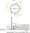
As guidelines for cervical cancer screening continue to evolve, HPV genotyping has assumed a significant role in newly published recommendations. In 2014, the FDA approved an HPV test for use in primary screening for cervical cancer [52]. Additionally, the Society of Gynecologic Oncology and the American Society for Colposcopy and Cervical Pathology recently released interim guidelines that endorsed screening with an HPV test alone (without cytology) every 3 years for women ≥25 years old [32]. Based on previous data [53], the guidelines recommended using different clinical approaches for managing patients infected with different HPV genotypes identified by HPV genotyping (Supplementary Fig. 1). Patients positive for HPV 16/18 would undergo colposcopy, while patients infected with any of 12 other high-risk HPV types would be tested by reflex cytology. Although issues regarding patient preferences and reimbursement remain unresolved, most health practitioners support the effectiveness of HPV genotyping as a primary screening modality [54]. Recent findings suggest that the use of HPV genotype testing as a primary screening tool will increase, and thus play a more important role in cervical cancer prevention.
An HPV test is more sensitive than cytology for detecting cervical precancerous cervical lesions, and less affected by the individual who collects the specimen [5355565758]. Therefore, HPV genotyping using self-collected samples might be an option to reduce costs and increase patient participation in HPV screening programs. Studies comparing the results of HPV tests performed using self-and clinician-collected samples showed equivalent HPV genotype distributions and prevalence [5960]. HPV genotyping using self-collected samples was feasible and well accepted, and showed sensitivity and specificity comparable to those achieved using clinician-collected samples [61]. Moreover, self-testing detected precancerous cervical lesions even earlier than cytology [62]. Home-based HPV testing is a good alternative not only for people residing in developed countries, but also for people living in developing countries where clinics are not easily accessible. Previous observations showed that a home-based HPV genotyping test identified sufficient numbers of women at risk for cervical cancer to produce a reduction in morality [63]. However, the test displayed limited specificity, and thus might require the use of additional triage tests (e.g., triage with cytology or methylation-marker testing) when used to confirm HPV positive results [64].

2. Therapeutic HPV vaccines

HPV genotyping is required to select individuals eligible to receive vaccines being studied for safety and efficacy in clinical trials [6566]. Vaccines that prevent HPV infection provide little protection in women with a pre-existing HPV infection [6768]; therefore, therapeutic vaccines that might control an existing infection are currently under investigation. Moreover, therapeutic vaccines should not only manage HPV-related lesions, but also establish a systemic immunological memory to help prevent disease recurrence [69]. Many of the therapeutic vaccines currently being studied contain the E6/E7 oncogenes of specific high-risk HPV genotypes (particularly HPV 16 and 18), and work by inducing a robust cellular immune response that eradicates HPV-related lesions [65707172]. For example, Kim et al. [65] developed a therapeutic vaccine using HPV 16-specific CD8+ cytotoxic T-lymphocyte responses that stimulates the expansion of CD8α+ lymphoid dentritic cells and facilitated the expression of HPV antigen through the major histocompatibility complex class I pathway. Therefore, HPV genotyping must be conducted to identify individuals with high-risk HPV types (particularly HPV 16 and 18) who would benefit from receiving a therapeutic vaccine.

CONCLUSIONS

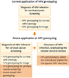
This review examined the role of HPV genotyping in the prevention and treatment of a cervical cancer or precancerous lesion. We elucidated the structure of the HPV genome, the functions of each gene during the HPV life-cycle, and how they relate to development of cervical cancer (Table 1). The history of HPV research shows that the field has achieved great scientific advances during the past two decades (Fig. 1). And although, previous data reported that HPV genotyping has limitations in its suboptimal specificity [73], it has significantly reduced the burden of HPV-related cervical lesions. HPV genotyping is changing from being a supporting method used to help prevent cervical cancer to a main method that also assists in managing pre-existing cervical lesions (Fig. 3). Although persistent infection with a high-risk HPV genotype is known to be a major carcinogenic factor, the various high-risk HPV genotypes have different carcinogenic potentials [23]. Therefore, an understanding of the genotype-specific aspects of HPV infection would facilitate the development of better strategies to prevent and manage cervical cancer.
Fig. 3
Schematic representation showing changes in the application of human papillomavirus (HPV) genotyping. HPV genotyping is currently perceived as a supporting method used in cervical cancer screening, but it will become a main method in the future. The yellow box denotes future applications of HPV genotyping in cervical cancer prevention and management.
jgo-27-e21-g003

Notes

Conflict of Interest No potential conflict of interest relevant to this article was reported.

References

1. zur Hausen H. Papillomaviruses in the causation of human cancers: a brief historical account. Virology. 2009; 384:260–265.
2. International Human Papillomavirus Reference Center. Human papillomavirus reference clones [Internet]. Stockholm, SE: International Human Papillomavirus Reference Center;c2015. cited 2015 Nov 6. Available from: http://www.hpvcenter.se/html/refclones.html.
3. de Villiers EM, Fauquet C, Broker TR, Bernard HU, zur Hausen H. Classification of papillomaviruses. Virology. 2004; 324:17–27.
4. Kjaer SK, van den Brule AJ, Paull G, Svare EI, Sherman ME, Thomsen BL, et al. Type specific persistence of high risk human papillomavirus (HPV) as indicator of high grade cervical squamous intraepithelial lesions in young women: population based prospective follow up study. BMJ. 2002; 325:572.
5. Burd EM. Human papillomavirus and cervical cancer. Clin Microbiol Rev. 2003; 16:1–17.
6. Satterwhite CL, Torrone E, Meites E, Dunne EF, Mahajan R, Ocfemia MC, et al. Sexually transmitted infections among US women and men: prevalence and incidence estimates, 2008. Sex Transm Dis. 2013; 40:187–193.
7. Ho GY, Bierman R, Beardsley L, Chang CJ, Burk RD. Natural history of cervicovaginal papillomavirus infection in young women. N Engl J Med. 1998; 338:423–428.
8. Rodríguez AC, Schiffman M, Herrero R, Wacholder S, Hildesheim A, Castle PE, et al. Rapid clearance of human papillomavirus and implications for clinical focus on persistent infections. J Natl Cancer Inst. 2008; 100:513–517.
9. Egawa N, Egawa K, Griffin H, Doorbar J. Human papillomaviruses: epithelial tropisms, and the development of neoplasia. Viruses. 2015; 7:3863–3890.
10. Crawford LV. A study of human papilloma virus DNA. J Mol Biol. 1965; 13:362–372.
11. zur Hausen H, Meinhof W, Scheiber W, Bornkamm GW. Attempts to detect virus-secific DNA in human tumors. I. Nucleic acid hybridizations with complementary RNA of human wart virus. Int J Cancer. 1974; 13:650–656.
12. zur Hausen H. Condylomata acuminata and human genital cancer. Cancer Res. 1976; 36(2 pt 2):794.
13. zur Hausen H. Human papillomaviruses and their possible role in squamous cell carcinomas. Curr Top Microbiol Immunol. 1977; 78:1–30.
14. Stanley M. Pathology and epidemiology of HPV infection in females. Gynecol Oncol. 2010; 117:2 Suppl. S5–S10.
15. Palefsky JM, Holly EA. Molecular virology and epidemiology of human papillomavirus and cervical cancer. Cancer Epidemiol Biomarkers Prev. 1995; 4:415–428.
16. Zheng ZM, Baker CC. Papillomavirus genome structure, expression, and post-transcriptional regulation. Front Biosci. 2006; 11:2286–2302.
17. Doorbar J. Molecular biology of human papillomavirus infection and cervical cancer. Clin Sci (Lond). 2006; 110:525–541.
18. Kirnbauer R, Booy F, Cheng N, Lowy DR, Schiller JT. Papillomavirus L1 major capsid protein self-assembles into virus-like particles that are highly immunogenic. Proc Natl Acad Sci U S A. 1992; 89:12180–12184.
19. Chen J, Ni G, Liu XS. Papillomavirus virus like particle-based therapeutic vaccine against human papillomavirus infection related diseases: immunological problems and future directions. Cell Immunol. 2011; 269:5–9.
20. Pereira R, Hitzeroth II, Rybicki EP. Insights into the role and function of L2, the minor capsid protein of papillomaviruses. Arch Virol. 2009; 154:187–197.
21. Dell G, Gaston K. Human papillomaviruses and their role in cervical cancer. Cell Mol Life Sci. 2001; 58:1923–1942.
22. Bernard HU, Burk RD, Chen Z, van Doorslaer K, zur Hausen H, de Villiers EM. Classification of papillomaviruses (PVs) based on 189 PV types and proposal of taxonomic amendments. Virology. 2010; 401:70–79.
23. de Sanjose S, Quint WG, Alemany L, Geraets DT, Klaustermeier JE, Lloveras B, et al. Human papillomavirus genotype attribution in invasive cervical cancer: a retrospective cross-sectional worldwide study. Lancet Oncol. 2010; 11:1048–1056.
24. Bosch FX, Burchell AN, Schiffman M, Giuliano AR, de Sanjose S, Bruni L, et al. Epidemiology and natural history of human papillomavirus infections and type-specific implications in cervical neoplasia. Vaccine. 2008; 26:Suppl 10. K1–K16.
25. von Knebel Doeberitz M. New markers for cervical dysplasia to visualise the genomic chaos created by aberrant oncogenic papillomavirus infections. Eur J Cancer. 2002; 38:2229–2242.
26. Fehrmann F, Laimins LA. Human papillomaviruses: targeting differentiating epithelial cells for malignant transformation. Oncogene. 2003; 22:5201–5207.
27. Münger K, Baldwin A, Edwards KM, Hayakawa H, Nguyen CL, Owens M, et al. Mechanisms of human papillomavirus-induced oncogenesis. J Virol. 2004; 78:11451–11460.
28. Hamid NA, Brown C, Gaston K. The regulation of cell proliferation by the papillomavirus early proteins. Cell Mol Life Sci. 2009; 66:1700–1717.
29. Doorbar J. The papillomavirus life cycle. J Clin Virol. 2005; 32:Suppl 1. S7–S15.
30. Fontaine V, Mascaux C, Weyn C, Bernis A, Celio N, Lefèvre P, et al. Evaluation of combined general primer-mediated PCR sequencing and type-specific PCR strategies for determination of human papillomavirus genotypes in cervical cell specimens. J Clin Microbiol. 2007; 45:928–934.
31. Johnson LR, Starkey CR, Palmer J, Taylor J, Stout S, Holt S, et al. A comparison of two methods to determine the presence of high-risk HPV cervical infections. Am J Clin Pathol. 2008; 130:401–408.
32. Huh WK, Ault KA, Chelmow D, Davey DD, Goulart RA, Garcia FA, et al. Use of primary high-risk human papillomavirus testing for cervical cancer screening: interim clinical guidance. Gynecol Oncol. 2015; 136:178–182.
33. Einstein MH, Martens MG, Garcia FA, Ferris DG, Mitchell AL, Day SP, et al. Clinical validation of the Cervista HPV HR and 16/18 genotyping tests for use in women with ASC-US cytology. Gynecol Oncol. 2010; 118:116–122.
34. Bartholomew DA, Luff RD, Quigley NB, Curtis M, Olson MC. Analytical performance of Cervista HPV 16/18 genotyping test for cervical cytology samples. J Clin Virol. 2011; 51:38–43.
35. Poljak M, Kovanda A, Kocjan BJ, Seme K, Jancar N, Vrtacnik-Bokal E. The Abbott RealTime High Risk HPV test: comparative evaluation of analytical specificity and clinical sensitivity for cervical carcinoma and CIN 3 lesions with the Hybrid Capture 2 HPV DNA test. Acta Dermatovenerol Alp Pannonica Adriat. 2009; 18:94–103.
36. Dockter J, Schroder A, Eaton B, Wang A, Sikhamsay N, Morales L, et al. Analytical characterization of the APTIMA HPV Assay. J Clin Virol. 2009; 45:Suppl 1. S39–S47.
37. Castle PE, Reid J, Dockter J, Getman D. The reliability of high-risk human papillomavirus detection by Aptima HPV assay in women with ASC-US cytology. J Clin Virol. 2015; 69:52–55.
38. Saslow D, Castle PE, Cox JT, Davey DD, Einstein MH, Ferris DG, et al. American Cancer Society Guideline for human papillomavirus (HPV) vaccine use to prevent cervical cancer and its precursors. CA Cancer J Clin. 2007; 57:7–28.
39. Saslow D, Runowicz CD, Solomon D, Moscicki AB, Smith RA, Eyre HJ, et al. American Cancer Society guideline for the early detection of cervical neoplasia and cancer. CA Cancer J Clin. 2002; 52:342–362.
40. Saslow D, Solomon D, Lawson HW, Killackey M, Kulasingam SL, Cain J, et al. American Cancer Society, American Society for Colposcopy and Cervical Pathology, and American Society for Clinical Pathology screening guidelines for the prevention and early detection of cervical cancer. Am J Clin Pathol. 2012; 137:516–542.
41. Dillner J, Rebolj M, Birembaut P, Petry KU, Szarewski A, Munk C, et al. Long term predictive values of cytology and human papillomavirus testing in cervical cancer screening: joint European cohort study. BMJ. 2008; 337:a1754.
42. Wright TC Jr, Stoler MH, Sharma A, Zhang G, Behrens C, Wright TL, et al. Evaluation of HPV-16 and HPV-18 genotyping for the triage of women with high-risk HPV+ cytology-negative results. Am J Clin Pathol. 2011; 136:578–586.
43. Schiffman M, Solomon D. Findings to date from the ASCUS-LSIL Triage Study (ALTS). Arch Pathol Lab Med. 2003; 127:946–949.
44. Castle PE, Cuzick J, Stoler MH, Wright TC Jr, Reid JL, Dockter J, et al. Detection of human papillomavirus 16, 18, and 45 in women with ASC-US cytology and the risk of cervical precancer: results from the CLEAR HPV study. Am J Clin Pathol. 2015; 143:160–167.
45. Stoler MH, Wright TC Jr, Cuzick J, Dockter J, Reid JL, Getman D, et al. APTIMA HPV assay performance in women with atypical squamous cells of undetermined significance cytology results. Am J Obstet Gynecol. 2013; 208:144.e1–144.e8.
46. Paraskevaidis E, Arbyn M, Sotiriadis A, Diakomanolis E, Martin-Hirsch P, Koliopoulos G, et al. The role of HPV DNA testing in the follow-up period after treatment for CIN: a systematic review of the literature. Cancer Treat Rev. 2004; 30:205–211.
47. Jones J, Saleem A, Rai N, Shylasree TS, Ashman S, Gregory K, et al. Human papillomavirus genotype testing combined with cytology as a ‘test of cure’ post treatment: the importance of a persistent viral infection. J Clin Virol. 2011; 52:88–92.
48. Heymans J, Benoy IH, Poppe W, Depuydt CE. Type-specific HPV geno-typing improves detection of recurrent high-grade cervical neoplasia after conisation. Int J Cancer. 2011; 129:903–909.
49. Kreimer AR, Guido RS, Solomon D, Schiffman M, Wacholder S, Jeronimo J, et al. Human papillomavirus testing following loop electrosurgical excision procedure identifies women at risk for posttreatment cervical intraepithelial neoplasia grade 2 or 3 disease. Cancer Epidemiol Biomarkers Prev. 2006; 15:908–914.
50. Hoste G, Vossaert K, Poppe WA. The clinical role of HPV testing in primary and secondary cervical cancer screening. Obstet Gynecol Int. 2013; 2013:610373.
51. Arbyn M, Ronco G, Anttila A, Meijer CJ, Poljak M, Ogilvie G, et al. Evidence regarding human papillomavirus testing in secondary prevention of cervical cancer. Vaccine. 2012; 30:Suppl 5. F88–F99.
52. U.S. Food and Drug Administration. FDA news release [Internet]. Silver Spring, MD: U.S. Food and Drug Administration;c2015. cited 2015 Nov 6. Available from: http://www.fda.gov/NewsEvents/Newsroom/PressAnnouncements/ucm394773.htm.
53. Rijkaart DC, Berkhof J, van Kemenade FJ, Coupe VM, Rozendaal L, Heideman DA, et al. HPV DNA testing in population-based cervical screening (VUSA-Screen study): results and implications. Br J Cancer. 2012; 106:975–981.
54. Cooper CP, Saraiya M. Perceived effectiveness of HPV test as a primary screening modality among US providers. Prev Med. 2015; 78:33–37.
55. Nanda K, McCrory DC, Myers ER, Bastian LA, Hasselblad V, Hickey JD, et al. Accuracy of the Papanicolaou test in screening for and follow-up of cervical cytologic abnormalities: a systematic review. Ann Intern Med. 2000; 132:810–819.
56. Bulkmans NW, Berkhof J, Rozendaal L, van Kemenade FJ, Boeke AJ, Bulk S, et al. Human papillomavirus DNA testing for the detection of cervical intraepithelial neoplasia grade 3 and cancer: 5-year follow-up of a randomised controlled implementation trial. Lancet. 2007; 370:1764–1772.
57. Castle PE, Stoler MH, Wright TC Jr, Sharma A, Wright TL, Behrens CM. Performance of carcinogenic human papillomavirus (HPV) testing and HPV16 or HPV18 genotyping for cervical cancer screening of women aged 25 years and older: a subanalysis of the ATHENA study. Lancet Oncol. 2011; 12:880–890.
58. Rijkaart DC, Berkhof J, Rozendaal L, van Kemenade FJ, Bulkmans NW, Heideman DA, et al. Human papillomavirus testing for the detection of high-grade cervical intraepithelial neoplasia and cancer: final results of the POBASCAM randomised controlled trial. Lancet Oncol. 2012; 13:78–88.
59. Gravitt PE, Belinson JL, Salmeron J, Shah KV. Looking ahead: a case for human papillomavirus testing of self-sampled vaginal specimens as a cervical cancer screening strategy. Int J Cancer. 2011; 129:517–527.
60. Petignat P, Faltin DL, Bruchim I, Tramèr MR, Franco EL, Coutlée F. Are self-collected samples comparable to physician-collected cervical specimens for human papillomavirus DNA testing? A systematic review and meta-analysis. Gynecol Oncol. 2007; 105:530–535.
61. Dijkstra MG, Heideman DA, van Kemenade FJ, Hogewoning KJ, Hesselink AT, Verkuijten MC, et al. Brush-based self-sampling in combination with GP5+/6+-PCR-based hrHPV testing: high concordance with physician-taken cervical scrapes for HPV genotyping and detection of high-grade CIN. J Clin Virol. 2012; 54:147–151.
62. Porras C, Hildesheim A, González P, Schiffman M, Rodríguez AC, Wacholder S, et al. Performance of self-collected cervical samples in screening for future precancer using human papillomavirus DNA testing. J Natl Cancer Inst. 2014; 107:400.
63. Gage JC, Ajenifuja KO, Wentzensen N, Adepiti AC, Stoler M, Eder PS, et al. Effectiveness of a simple rapid human papillomavirus DNA test in rural Nigeria. Int J Cancer. 2012; 131:2903–2909.
64. Verhoef VM, Bosgraaf RP, van Kemenade FJ, Rozendaal L, Heideman DA, Hesselink AT, et al. Triage by methylation-marker testing versus cytology in women who test HPV-positive on self-collected cervicovaginal specimens (PROHTECT-3): a randomised controlled non-inferiority trial. Lancet Oncol. 2014; 15:315–322.
65. Kim TJ, Jin HT, Hur SY, Yang HG, Seo YB, Hong SR, et al. Clearance of persistent HPV infection and cervical lesion by therapeutic DNA vaccine in CIN3 patients. Nat Commun. 2014; 5:5317.
66. Bissa M, Illiano E, Pacchioni S, Paolini F, Zanotto C, De Giuli Morghen C, et al. A prime/boost strategy using DNA/fowlpox recombinants expressing the genetically attenuated E6 protein as a putative vaccine against HPV-16-associated cancers. J Transl Med. 2015; 13:80.
67. Muñoz N, Kjaer SK, Sigurdsson K, Iversen OE, Hernandez-Avila M, Wheeler CM, et al. Impact of human papillomavirus (HPV)-6/11/16/18 vaccine on all HPV-associated genital diseases in young women. J Natl Cancer Inst. 2010; 102:325–339.
68. Lehtinen M, Paavonen J, Wheeler CM, Jaisamrarn U, Garland SM, Castellsagué X, et al. Overall efficacy of HPV-16/18 AS04-adjuvanted vaccine against grade 3 or greater cervical intraepithelial neoplasia: 4-year end-of-study analysis of the randomised, double-blind PATRICIA trial. Lancet Oncol. 2012; 13:89–99.
69. McKee SJ, Bergot AS, Leggatt GR. Recent progress in vaccination against human papillomavirus-mediated cervical cancer. Rev Med Virol. 2015; 25:Suppl 1. 54–71.
70. Kenter GG, Welters MJ, Valentijn AR, Lowik MJ, Berends-van der Meer DM, Vloon AP, et al. Vaccination against HPV-16 oncoproteins for vulvar intraepithelial neoplasia. N Engl J Med. 2009; 361:1838–1847.
71. Zong J, Wang C, Liu B, Liu M, Cao Y, Sun X, et al. Human hsp70 and HPV16 oE7 fusion protein vaccine induces an effective antitumor efficacy. Oncol Rep. 2013; 30:407–412.
72. Sadraeian M, Rasoul-Amini S, Mansoorkhani MJ, Mohkam M, Ghoshoon MB, Ghasemi Y. Induction of antitumor immunity against cervical cancer by protein HPV-16 E7 in fusion with ricin B chain in tumor-bearing mice. Int J Gynecol Cancer. 2013; 23:809–814.
73. Eklund C, Forslund O, Wallin KL, Zhou T, Dillner J. WHO Human Papillomavirus Laboratory Network. The 2010 global proficiency study of human papillomavirus genotyping in vaccinology. J Clin Microbiol. 2012; 50:2289–2298.
Supplementary Materials

Supplementary Fig. 1

Human papillomavirus (HPV) genotyping for primary cervical cancer screening, recommended by American Society for Colposcopy and Cervical Pathology (ASCCP) interim guideline, in 2015 [32]. The guideline is recommended for the women of ≥25 years old. ASC-US, atypical squamous cells of undetermined significance.
TOOLS
Similar articles