

 PDF
PDF Citation
 Print
Print


INTRODUCTION
Epithelial ovarian cancer (EOC) remains the most lethal gynecologic malignancy despite recent advances in treatment [1]. Surgical management of EOC consists of complete surgical staging, including hysterectomy, bilateral salpingo-oophorectomy, omentectomy, peritoneal washing cytology, multiple biopsies, and pelvic and para-aortic lymph node dissection [2]. However, the radicality of extensive surgery can cause irreversible infertility and hormonal deficiency in women younger than 40 years old, who account for 3% to 17% of all EOCs [134567].
In borderline and non-epithelial ovarian tumors, fertility-sparing surgery (FSS), which preserves the uterus and at least one ovary with adnexa is a current recommendation for those under 40 [8910]. However, the procedure has not yet been examined specifically in women with early-stage EOC, although several retrospective studies have reported postoperative outcomes of FSS in patients with early-stage EOC [11121314]. A recent review article demonstrated that women with grade 1-2, non-clear cell histology, and the International Federation of Gynecology and Obstetrics (FIGO) stage I or with clear cell histology and FIGO stage IA could safely undergo FSS [15]. However, the results of these studies are controversial, stemming from a concern about disease relapse in EOC patients with uterus and adnexa [16].
Despite the concern, fertility sparing is an important issue for young women with early-stage EOC. With the advancement of laparoscopic equipment and surgical skills, several studies reported the feasibility of laparoscopic surgery in gynecologic malignancies, including EOC [17181920]. Furthermore, recent reports of laparoscopic staging surgery for EOC showed both safety and efficacy compared to laparotomy [1921222324]. The objective of this study was to evaluate the perioperative, oncological, and obstetric outcomes of laparoscopic FSS to determine its feasibility and safety in presumed clinically early-stage EOC.
MATERIALS AND METHODS
We retrospectively reviewed the electronic medical records of patients who had undergone laparoscopic FSS for EOC between January 1999 and December 2012 in the Department of Obstetrics and Gynecology, Samsung Medical Center, Seoul, Korea with IRB approval (IRB No.: 2014-05-083-002). Patients aged 40 years or younger were included. Patients who were presumed to have clinically stage I disease and who strongly desired fertility preservation were included. The range of surgical procedures was within the surgeon’s discretion, spanning from unilateral salpingo-oophorectomy to complete surgical staging. Complete surgical staging included a complete exploration of the whole peritoneum with multiple biopsies, washing cytology, omentectomy, bilateral pelvic lymph node dissection, and para-aortic lymph node dissection in addition to ovarian tumor removal. The tumor histology of mucinous, serous, endometrioid, and clear cell adenocarcinoma were included. Patients with non-epithelial or borderline ovarian tumors were excluded. Patients who switched to laparotomy procedures were also excluded.
After FSS, patients received adjuvant chemotherapy according to their surgeons’ discretion. During the follow-up period after the primary treatment, patients were scheduled to visit an outpatient clinic every 3 months for the first 2 years, every 6 months for the next 3 years, then once in a year as long as there was no evidence of disease recurrence. Patients underwent physical examinations, transvaginal ultrasonography, and serum tumor marker evaluation at every visit to the outpatient clinic. Imaging studies, such as computed tomography (CT), magnetic resonance imaging, and positron emission tomogram (PET) or PET/CT were performed every 6 months to a year.
Perioperative and postoperative long term results including obstetrical and oncological outcomes were evaluated. The rate of recurrence, recurrent interval from the primary treatment, and recurrent site were also reviewed. Data were reported as absolute numbers of patients with percentage or median (range). Statistical analysis was performed using PASW ver. 18 (SPSS Inc., Chicago, IL, USA).
RESULTS
1. Baseline characteristics
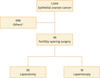
Of 1,044 patients with EOC, we excluded women who were older than 40 years old. Those who did not want to preserve fertility or received primary debulking surgery, neoadjuvant chemotherapy followed by debulking surgery, or palliative surgery followed by chemotherapy were also excluded. Excluding the 28 patients who received open surgery, the remaining 18 patients who underwent laparoscopic FSS were evaluated (Fig. 1).
Fig. 1
Diagram of patient selection. *Age>40 years, primary debulking surgery, neoadjuvant chemotherapy followed by debulking surgery, or biopsy or palliative surgery followed by chemotherapy.
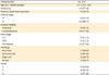
Baseline characteristics of patients are shown in Table 1. Of the total 18 patients, nine were married. Two patients were presumed to be clinically stage IA before and during the operation. However, they were upstaged to FIGO stage IIIA1 due to the presence of pelvic or para-aortic lymph-node micrometastasis on the final pathologic report. There were no patients with omental or peritoneal metastasis during the operation or at the final pathology. The mucinous type of tumor was the most common (38.9%) histologic type.
Table 1
Baseline characteristics of participants (n=18)
2. Perioperative and long-term outcomes of participants
Perioperative and long-term outcomes of participants are summarized in Table 2. There were no perioperative complications and no gross residual tumors after the primary surgery. During the median follow-up of 47.3 months (range, 11.5 to 195.3 months), there were no postoperative long-term complications. Patients with high-risk factor, including high grade, clear cell histologic type, tumor growth through capsule, surface excrescences, malignant cells in ascites or peritoneal washings, preoperative rupture, dense adhesion and FIGO stage higher than or equal to IC, received adjuvant chemotherapy. However, two patients who were at clinical stage IA and grade 1 received adjuvant chemotherapy according to the surgeon’s discretion.
Table 2
Perioperative and long-term outcomes of participants (n=18)
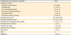
Although all patient had apparently early-stage disease during initial staging, two were upstaged to FIGO stage IIIA1 due to microscopic metastatic pelvic (n=1) and para-aortic (n=1) lymph nodes after complete staging surgery. The radical surgery including hysterectomy, salpingo-oophorectomy of contralateral ovary, omentectomy, pelvic and para-aortic lymphadenectomy and multiple peritoneal biopsy, was mandatory in this two patients, but they were unmarried with strong desire to preserve their fertility. One of them who had FIGO stage IIIA1 disease because the tumor was involved in the removed ovary with one pelvic lymph node. The histologic type of the tumor was serous adenocarcinoma and the grade was 2. She was treated to 6 cycles of platinum-based combination chemotherapy after primary surgery and was closely monitored during a follow-up period of 41 months. Recently, she was alive with no evidence of disease. Another one who had FIGO stage IIIA1 disease had disease recurrence at 14.9 months after the primary treatment. Preoperative cancer antigen 125 (CA-125) was 21.4 U/mL. No tumor involvement was found except in one ovary in a preoperative imaging study. She received complete staging operation and was upstaged to FIGO stage IIIA1 because the tumor was involved in the removed ovary with one para-aortic lymph node. The histologic type of the tumor was serous adenocarcinoma and the grade was 3. She received 6 cycles of platinum-based combination chemotherapy after the primary surgery. She had no signs or symptoms related to tumor recurrence. However, she had an increased level of tumor marker (CA-125, 73.1 U/mL) with abnormal findings on a follow-up PET/CT. She had multiple recurrent tumors in the left ovary, the hilum of spleen, the surface of the diaphragm and liver, the serosa of sigmoid colon, multiple tumors in the pelvic and abdominal peritoneum, and multiple tumors in the retroperitoneal lymph nodes. After recurrence, she received debulking surgery, including hysterectomy and salpingo-oophorectomy, followed by platinum-based chemotherapy. The patient’s surgical outcome was successfully achieved as no residual disease state after debulking surgery. Although she received adjuvant chemotherapy after the debulking surgery, she suffered a relapse of the cancer. Currently, she was alive with multiple metastases of the stable disease. All other patients had no disease recurrence. No one died of disease, including the patient who had the recurrence (Table 2).
3. Laparoendoscopic single site surgery for FSS
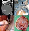
We demonstrated one case of laparoendoscopic single site (LESS) surgery in Fig. 2. The patient was a 36-year-old nullipara. Her preoperative CA-125 was 42.3 U/mL. Except for the 23 cm sized left ovarian tumor with a solid portion, no other lesion was suspected to be involved in the tumor based on the preoperative abdominopelvic CT scans. Before performing laparoscopic salpingo-oophorectomy, the surgeon aspirated cystic fluid of the tumor through an umbilical incision. The aspirated and sutured cyst was reinserted into the abdominal cavity. The surgical procedure included a left salpingo-oophrectomy, right ovarian wedge biopsy, omentectomy, and exploration of the entire peritoneal cavity. She was presumed to be stage IC1, grade 1, mucinous EOC. She did not receive adjuvant chemotherapy. Disease recurrence was not found during a follow-up period of 39.1 months.
Fig. 2
A case of laparoendoscopic single site surgery showing post-contrast computed tomography image, operative procedures, and specimen findings from the patient. (A) An ovarian tumor of about 23 cm containing a solid mass (white arrow) is shown. (B) Aspiration of the ovarian tumor through single umbilical incision was performed. The black arrow indicates the suction aspirator. (C) The reinserted left ovary was ligated in the abdominal cavity. (D) Gross findings of the specimen are shown. The blue arrow indicates the solid portion of the epithelial ovarian cancer.
4. Pregnancy outcome after FSS
The clinical characteristics of four women (22.2%) who conceived after primary treatment are shown in Table 3. Two patients received in vitro fertilization and embryo transfer. The other two became pregnant spontaneously after the primary treatment. All patients had received comprehensive surgery and 3 to 6 cycles of adjuvant platinum-based chemotherapy. None of them had disease recurrence (Table 3). The clinicopathologic information of whole study population (n=18) including last disease status and pregnancy outcome, was shown in Table 4.
Table 3
Clinical characteristics of patients who conceived after treatment

BOC, bilateral ovarian cystectomy; LOC, left ovarian cystectomy; LOWR, left ovary wedge resection; LSO, left salpingo-oophorectomy; NED, no evidence of disease; OMT, omentectomy; PALND, para-aortic lymph node dissection; PLND, pelvic lymph node dissection; RSO, right salpingo-oophorectomy.
*Interval, time from cessation of treatment to conception.
Table 4
Clinicopathologic information of participants (n=18)
FIGO, International Federation of Gynecology and Obstetrics; F/U, follow-up; LOC, left ovarian cystectomy; LOWR, left ovary wedge resection; LSO, left salpingo-oophorectomy; NA, not available; NED, no evidence of disease; OMT, omentectomy; PALND, para-aortic lymph node dissection; PLND, pelvic lymph node dissection; ROC, right ovarian cystectomy; ROWR, right ovary wedge resection; RS, right salpingectomy; RSO, right salpingo-oophorectomy; SD, stable disease.
DISCUSSION
In this study, one out of 18 patients who received laparoscopic FSS for clinically early-stage EOC had disease recurrence. However, no one died of the disease during the median follow-up of 41.3 months. Four out of the 18 patients had successful pregnancies after laparoscopic FSS. There were no perioperative or long term complications after the laparoscopic FSS for clinically early-stage EOC. Up to now, few studies have shown the operative, oncological, or obstetrical outcomes of laparoscopic FSS for EOC. Although a small number of patients were included in this study, it provides operative, oncologic and pregnancy outcomes of laparoscopic FSS for clinically early-stage EOC.
Selecting appropriate candidates for FSS is a fundamental but complicated problem in treating young EOC patients. The American College of Obstetrics and Gynecology (2007) recommended FSS to reproductive-age EOC patients with grade 1–2 and FIGO stage IA with non-clear cell histology [25]. In 2008, the European Society for Medical Oncology reported that patients with FIGO stage I EOC with grade 1–2 and non-clear cell histology without dense adhesions were optimal candidates for FSS [26]. However, the number of previous reports regarding FSS in EOC patients is limited. In addition, the retrospective design and small sample size of most studies makes it difficult to draw a unanimous consensus for the selection of candidates for FSS.
There are several retrospective studies with relatively larger sample sizes regarding this issue. Schilder et al. [27] reported an excellent survival of conservatively-treated FIGO stage IA or IC EOC patients whose 5-year survival and 10-year survival were 98% and 93%, respectively. Kajiyama et al. [28] reported no difference in 5-year overall survival and disease-free survival between EOC patients who received radical surgery and those who received FSS. They also reported that FIGO stage IC did not affect the prognosis of patients with FSS compared to patients who underwent radical surgery. In this study, the proportion of patients with clinically stage IC EOC was over 60%. None of the patients with stage IC had disease recurrence. However, Satoh et al. [13] suggested that FSS should not be performed in patients with FIGO stage IC and clear cell histology because they found a 5-year recurrence-free survival of only 66.0%, even though 73.3% patients were treated with platinum-based adjuvant chemotherapy. Many previous reports have suggested that FSS should not be undertaken for EOC patients who have FIGO stage IC and grade 3 due to a high recurrence rate [12132729]. In this study, one out of three patients with grade 3 had disease recurrence. The tumor involvement was found in the removed ovary and one retroperitoneal lymph node. Even though the patient had 6 cycles of platinum-based adjuvant chemotherapy, she had multiple peritoneal and retroperitoneal recurrences. As shown in this case, grade 3 disease carries a potential risk of micrometastasis with high recurrence, even though preoperative evaluations represent early stage disease.
Because the upstaging rate after a staging operation is nearly 30% in presumed clinically early-stage EOC, complete surgical staging is also important in patients who want to preserve fertility [3031]. In this study, 10 and 11 patients received pelvic lymphadenectomy and omentectomy, respectively. However, complete surgical staging was done in only four patients (22.2%). Disease recurrence occurred in one patient who received complete surgical staging and was upstaged to FIGO stage IIIA1 with satellite para-aortic lymph node metastasis in final pathology. If she had not received complete surgical staging, she would have been misdiagnosed clinically as stage IA disease. This case suggested that FSS in EOC should be accompanied by complete surgical staging as the first step of treatment.
This study has some limitations other than its low rate of complete surgical staging. Adjuvant chemotherapy was administered even in patients who received comprehensive surgery with clinically stage IA and grade 1 disease. In addition, the grade 1 mucinous type tumor procedure was unecessary due to the surgeon’s concern of hidden metastasis after performing incomplete surgical staging. Furthermore, the high rate (94.4%) of adjuvant chemotherapy might have influeced the recurrence rate. There was no definite criteria in our instituion for selecting appropriate candidates for FSS. Therefore, the decision to perform FSS was largely discretionary, and depended on the surgeon. Because this study was conducted retrospectively, selection bias could not be completely excluded. The small number of study participants and short follow-up period are also limitations of this study. The pregnancy rate in this study is lower (22.2%) compared to previous review articles (50% to 100%) [1532]. In other articles, the pregnancy rate was calculated among patients who wanted to conceive. However, the number of patient who wanted to conceive was not clearly recorded in the medical records of patients in this study. Despite these limitations, this study has an interesting point because only laparoscopic FSS, including LESS was included. If comparable perioperative, oncologic, and obstetric outcomes are expected, laparoscopic FSS for presumed clinically early-stage EOC can be an alternative procedure to reduce surgical complications associated with laparotomy.
In conclusion, FSS in young patients with presumed clinically early-stage EOC is a challenging and cautious procedure. World-wide indications for selecting optimal candidates for FSS in EOC patients are not established yet. The ethical and practical problems made by a randomized clinical trial of FSS for EOC are difficult. However, laparoscopic FSS with complete surgical staging in young patients with presumed clinically early-stage EOC could be a treatment options in selected patients in accordance with minimally invasive concepts. The accumulation of retrospective studies in the future could determine the safety and feasibility of laparoscopic FSS in young patients with presumed clinically early-stage EOC.
 XML Download