

 PDF
PDF ePub
ePub Citation
 Print
Print


I. INTRODUCTION
Only recently have we become familiar with the striking phenomenon associated with determination of the form and location of certain organs. It has been discovered by the author that if at the stage of the earliest appearance of the neural folds the ectoderm of the presumptive right ear region be transplanted to the left side it forms a left ear in all cases which means that only the general frame work of the ear was at this stage determined. On the other hand, at the stage of the closure of the neural folds the same ectoderm forms a right ear in all cases. At this stage the laterality of the bilateral organ viz. of the ear, was already determined. To distinguish these grades of determination I have called the former 'Gross-determination' and the later 'Particular determination.' Lateralization is not apparently included in gross determination of the ear but belongs to particular determination and the actual moment of the lateralization lies between the stage of the appearance and closure of the neural folds. The author further has attempted to investigate exactly when the posture of a bilateral organ such as the ear is determined and in what relations it stands to other determination.
II. EXPERIMENTAL METHOD AND MATERIALS
All the experiments were carried out on the embryo of Rana temporaria at two stages as follows;
1. The FIRST STAGE is that of the earliest appearance of the neural folds. The ectoderm of the ear region consists of two layer of epithelial cells and no difference from the neighbouring ectoderm is apparent.
2. SECOND STAGE is that of closure of the neural folds. The ectoderm of the ear region is slightly thickened as a few layers of epithelial cells and is thus differentiated from the neighbouring ectoderm.
Operative work was done in 0.2% saline solution, under the binocular microscope using fine needles for dissection. A piece of ectoderm 0.5 mm square was removed from the left ear region and replaced after rotating 180 degrees. The anterior part of the piece comes to lie posteriorly and the superior part of the piece comes to lie at the lowest part of the graft. The specimens were allowed to go on with their development for from 42 to 45 days, at the end of which time they were preserved in bichloride-picric acid solution. The preserved specimens were embedded in paraffin, cut in serial sections and stained with eosin-haematoxylin. Reconstruction wax-plate models after Born's method were made. The ear region of each specimen examined as to form, position, posture, size, semicircular canals, endolymphatic appendage, lagena, accompanying nerves and ganglia and the cartilaginous capsule.
III. TRANSPLANTATION OF LEFT EAR REGION ECTODERM AFTER ROTATING 180 DEGREES INTO ITS ORIGINAL SITE (FIRST STAGE)
Thirty four embryoes were used. In 13 cases normal ears were formed on the site of the grafted area, 2 cases were almost normal and the remaining ears developed in a defective manner. In 13 cases the size of the ear was normal as compared with the normal of the ear of the opposite side, 2 cases was very small and all others were smaller than the normal. In 13 cases perfect semicircular canals were formed in a normal location and connected with a more or less developed sacculus and utriculus. All the rest were not definite but did show at least a membranous labyrinth. In 17 cases the ear was in a normal position, 7 cases were a little dorso-anterior, 10 cases were situated dorso-laterally to the eye and in close contact with the eye ball. In 18 cases the lagena projected on the postero-medial part of the labyrinth and others were not distinct. In 10 cases an endolymphatic appendage was formed on the dorso-anterior part of the labyrinth and the others were not distinct. In all cases a cartilaginous capsule was formed. The ganglion acustica was also present in all cases on the wall of the labyrinth and was connected with the sensory epithelium of the postero-ventro-medial part of the labyrinth. In 13 cases complete left side ears were formed, others were so abnormal as to make classification difficult. In 13 cases a definite left ear was formed from the ectoderm rotated 180 degrees. The posture of others was not clear enough for classification.
Thus the grafted ectoderm of the ear region can form a complete well differentiated ear in a new environment, namely autotansplantation of the ectoderm of the left ear region which had been rotated 180 degrees resulted in the formation of a normal left side ear. Consequently in presumptive ear region ectoderm at the stage of the earliest appearance of the neural folds the posture of the ear is not determined. The surrounding nerves and ganglia are well developed in the normal location in spite of the transplantation.
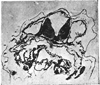
Description of wax-plate model, Op No. I. 6-8. 100 X. The general shape of the ear is incomplete ovaL Three incomplete semicircular canals are shown as well as both utriculus and sacculus on the medio-ventral part of the labyrinth connected with the three canals. The endolymphatic appendage projects from the medial part of the dorso-anterior part of the labyrinth while the lagena projects from the ventral wall of the postero-medial part. The position of the experimental ear is a little anterior while the size of the ear and its posture are normal. In short we have a difinite normal left side ear [Fig. No. I, II.].
IV. TRANSPLANTATION OF LEFT EAR REGION ECTODERM AFTER ROTATING 180 DEGREES INTO ITS ORIGINAL SITE (SECOND STAGE)
Twenty embryoes were used. In all cases a more or less complete membranous labyrinth was formed on the site of the grafted area. In 5 cases a comparatively complete ears were formed while the others were developed abnormally. In 8 cases the size was smaller than normal, 2 cases were larger than normal and the others were approximately of normal size. In 3 cases the ear was in the normal position, a few were situated dorso-anteriorly, and in some cases the ear was situated dorsolaterally to the eye ball and closely connected with it. In no cases were three complete semicircular canals formed. In 5 cases the anterior canal was posterior, the posterior canal was antero-inferior while the lateral canal was dorso-lateral. Namely the posture of the whole ear was rotated 180 degrees. In 10 cases the posture was not clear due to defective development. In 5 cases the lagena was projected on the antero-lateral part of the labyrinth and all others were indefinite as to anatomical relations but the wall was consisted in parts of high columnar ciliated epithelium connected with the neighbouring nerve fibers especially at the dorso-medioanterior, or postero-ventro-medial part of the labyrinth. In no cases did the endolymphatic appendage appear on the proper part of the labyrinth nor was it found at the normal site on the brain. In all cases cartilaginous capsules were present around the labyrinth. The nerve and ganglion acustica usually present on the proper part of the brain and connected with some part of the labyrinth but in some cases the labyrinth was located abnormally although the nerve and ganglia were independently present on the normal region of the brain but not linked up to the labyrinth. In 5 cases comparatively complete left side ear were formed. All others showed marked abnormalities.
Thus the ectoderm of the ear region rotated 180 degrees before transplantation on the same site finally resulted in the formation of a reversed left side ear. Consequently in presumptive ear region ectoderm at the stage of closure of the neural folds the posture of the ear was well determined.
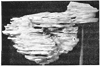
Description of wax-plate model, Op. No. II. 7-4. 100 X. The ear showed three incomplete semicircular canals. The whole ear was reversed. The anterior pole was posteriorly directed, the posterior canal anteriorly and the lateral canal was of course still lateral. The incomplete endolymphatic appendage was situated on the ventro-medio-anterior part of the labyrinth. A lagena was not formed but a certain part of the labyrinth (dorso-anterior, or ventro-medio-posterior part of the labyrinth) consisted of high columnar ciliated epithelium. The cartilaginous capsule was comparatively well developed. The position and size are both normal in short we have here a definite reversed left side ear [Fig. No. III, IV.].
V. ROTATION THROUGH 180 DEGREES AND TRANSPLANTATION OF THE ECTODERM OF THE LEFT EAR REGION TO THE REGION OF THE MIDDLE PART OF THE LATERAL LINE (SECOND STAGE)
Ten embryoes were used. The same method being used as previously described, the left ear region ectoderm was rotated and grafted upon the middle part of the left lateral line region. In 4 cases more or less complete ear was formed not far from the left pronephros. In other embryoes a very defective labyrinth was formed in the same region. In 4 cases the ears were almost of normal size and the others were much smaller than normal. In 4 cases three more or less incomplete semicircular canals were formed and chambers of the labyrinth representing the utriculus and sacculus were formed. In no cases was the lagena and endolymphatic appendage definite but a certain part of the labyrinth was consisted of high columnar ciliated epithelium like the macula and was connected with the neighbouring spinal nerves. In all cases incomplete cartilaginous capsules were formed. In 4 cases the posture of the ear was incompletely reversed through the axis of the vertically. The other ears were all very defective.
Thus the ectoderm of the presumptive ear region when grafted and rotated to 180 degrees on an indifferent region still develops a reversed ear.
Description of wax-plate model, Op. No. IV. 2-7. 100 x . The whole labyrinth is incomplete oval, the posterior pole being more prominent than the anterior and the medial side being thicker than the lateral. There is no endolymphatic appendage nor lagena. Three incomplete semicircular canals and both utriculus and sacculus are visible on the part of the labyrinth. The anterior canal being posterior and the posterior canal being anterior, the lateral canal is of course still lateral.
Therefore the ear is a reversed left side ear. Consequently the posture of the ear developing from the ectoderm of second stage embryoes is comparatively more or less influenced by the surrounding tissues [Fig. No. V, VI.].
VI. DISCUSSION AND CONCLUSION
It has been found by the author ('031) that if at the stage of the earliest appearance of the neural folds, the ectoderm of the presumptive right ear region be transplanted to the left side it forms a left ear. On the other hand, at the stage of closure of the neural folds the same region ectoderm forms a right ear. Consequently the frame work of the ear was determined even at the stage of the earliest appearance of the neural folds but the nature of the side was determined only between the stage of the appearance and closure of the neural folds. Therefore lateralization is not included in gross determination of the whole ear, but belongs to particular determination. Similarly if at the stage of the earliest appearance of the neural folds the ectoderm of the presumptive ear region be rotated 180 degrees and grafted it forms a normal postured ear. On the other hand, at the stage of the closure of the neural folds the reversed ectoderm forms a reversed ear. Namely the reversed ear showed an anterior canal posteriorly situated and a posterior canal anteriorly located while the lateral canal of course remained lateral. Therefore the determination of the posture of an ear is not included in gross determination of the whole ear, but belongs to particular determination.
Streeter ('09) stated that the right or left characteristics of the ear vesicle is not controlled by the environment but in determined by some intrinsic power of its constituent cells. He took out the left ear cup from larvae of Rana pipiens then grafted it closely against the front of the right side ear region. He found that the left side ear cup developed left ear on the right side ear region. Therefore the ear cup of the left side already had determined its side characteristics and the posture of the ear had also been determined at the stage of the ear cup.
Spemann ('010) removed the ear vesicle at the stage of the earpit (Hörgrübchen) in Rana esculenta larvae, inverted it, and rotated it 180 degrees to an oblique-transverse location and watched development. Out of 12 operations 9 cases retained the inverted posture, 2 cases showed partial inversion and 1 cases rotated back to the normal position. He surmised that it had not been sufficiently turned, and had slipped back into its normal position. The result therefore agree with my results though he used the earpit stage's embryoes and I used ear region ectoderm which represents considerable difference in the developmental stage of the materials.
The author has described ('031) that at an indifferent area there occurred after grafting the formation of about 65% normal ears while normal ears were formed in almost 100% at the ear area itself. Similarly the posture of the ear was very erratic at the indifferent area while more or less normal posture of the ear was noted on the ear region itself. Therefore the tissue surrounding the ear region has more determining force for the posture of the ear than the indifferent area of the other part of the body. We are already known that the ear vesicle, capable of marked power of self-differentiation, apparently was not entirely independent of the surrounding structures. Specifically the posture of the developed labyrinth, the situation of its canals, and various chambers seems to be controlled to some extent by the environment.
1. In presumptive ear region ectoderm at the stage of the earliest appearance of the neural folds the posture of the ear is not yet determined.
2. After rotation through 180 degrees and autotransplantation of the ectoderm of the ear region at the stage of closure of the neural folds the posture had already been determined.
3. The presumptive ear region ectoderm at the stage of the closure of the neural folds can develop and differentiate further in an indifferent area and form a abnormal reversed posture ear. Therefore the characteristic development of the posture of the presumptive ear region ectoderm is dependent to some extent on the changed environment.
 XML Download