

 PDF
PDF ePub
ePub Citation
 Print
Print


INTRODUCTION
Postoperative intraperitoneal adhesions (PIAs) are one of the most important problems surgeons have to face after laparotomies. PIAs may form after many abdominal surgeries. Prevalence is around 67%–93%. However, the percentage of total formations necessitating surgical operation caused by PIAs is around 15%–18% [1]. These percentages are lowered on account of development and common usage of minimally invasive techniques such as laparoscopy [2]. PIAs can cause hospitalization and surgical operations due to mechanic ileus or pelvic pain. Additionally, PIAs may cause secondary infertility in women in the reproductive age group. Iatrogenic injuries are very common in operations needed on the grounds of adherence because exploration is extremely hard. These complications on the other hand may cause an increase in mortality and morbidity. Additionally, one of the most important consequences of ileus caused by PIAs is the increase in workforce loss and patient costs. For example, a study conducted in the United States shows that the cost of operations due to PIAs is around 1,3 billion United States dollars [3].
Steroids and nonsteroidal anti-inflammatory drugs, immunosuppressive drugs, ClinOleic, high molecular weight hyaluronic acid and low molecular weight hydroxypropyl methylcellulose, fibrinogen degradation products, recombinant tissue plasminogen activator, ankaferd blood stopper, Vitamin-E and carboxymethyl-cellulose was used as adhesion barrier in order to decrease and prevent adherence [45]. Different results were obtained in each of these agents. The agents, except adhesion barrier, could not be applied clinically. This is the main reason the prevention of PIAs and fighting off adhesions are always an important subject for surgeons, especially those working in abdominal surgery.
It was determined that postoperational inflammatory response plays a great role in the formation of PIAs [6]. A study examining the formation of peritoneal adhesion formation at the molecular level indicates that TGF-β1 and interleukin-6 (IL-6), which are pleiotropic molecules, shows an increase in peritoneal fluid during and after abdominal operations. It was asserted that TGF-β1 and IL-6 interacted with each other, promoted mesothelial to mesenchymal transition and facilitated peritoneal adhesion formation. This is why the inflammatory response is thought to be efficient in decreasing PIAs, especially in repressing TGF-β1 and IL-6 [7]. Thus, it is important that the agent used for preventing PIAs contain anti-inflammatory features. It was also reported that adhesion molecules such as E-cadherin play a role in intra-abdominal adhesion formation. Thus, anti-inflammatory agents effecting adhesion molecules can be seen as ideal agents in preventing PIAs.
It was also determined that botulinum toxin type A (BoNT-A) disturbs the bond between cells on the epithelial barrier by binding epithelial molecules, and has anti-inflammatory effectiveness [8]. In this study, the hypothesis that BoNT-A may decrease and even prevent PIAs was emphasized due to its anti-inflammatory effectiveness. There are no studies examining the effect of BoNT-A on intra-abdominal adhesions.
The aim of this study was to examine the effectiveness of local application of BoNT-A in various dosages on the prevention of intra-abdominal adhesions on rats with experimental intra-abdominal adhesions.
METHODS
Rat groups
Forty Wistar Albino female rats weighing 260–280 g were randomly separated into 4 groups, 10 rats in each group; Control (group 1), sham (group 2), 10-µg/kg low-dose BoNT-A (group 3), and 30-µg/kg high-dose BoNT-a (group 4). Subserosal injuries were created on the group 1 rats by rubbing sterile gauze pads on the caecum serosa without applying any drugs. Group 2 rats were also wounded with caecum subserosal injuries with an application of 1 mL 0.09% NaCl into the peritoneal cavity. Ten- and 30-µg/kg BoNT-A (BOTOX, Allergan, Dublin, Ireland) with 1 mL 0.09% NaCl into the peritoneal cavity were respectively applied on groups 3 and 4 rats.
Anesthesia and surgical technique
Rats were kept in a room with a fixed temperature (18℃–24℃) and light (12-hour light-dark cycle), 4–6 rats in a single cage, with free water and feed supplies. Rats were given standard rat feed and normal water. Rats were left hungry for 6 hours before surgery. As anesthetics, 75-mg/kg ketamine (Ketalar, Pfizer, Istanbul, Turkey) and 10-mg/kg xylazine (Rompun, Bayer AG, Leverkusen, Germany) were applied intraperitoneally, respectively. Polyvinylpyrolidone iodine (polividon-iodine) 10% was applied for asepsis. 100-mg/kg ceftriaxone was applied for intramuscular prophylaxis purposes. All rats were given 0.25% bupivacaine on the incision for postoperative analgesia control purposes. Postoperative analgesia was performed with 3-mg/kg ketoprofen (Profenid, Sanofi Aventis, Paris, France) subcutaneously, daily for 3 days. Each rat was given 10 mL 0.09% NaCl subcutaneously for dehydration. The rats were placed in different cages postsurgery and followed up. The rats were given free feed and water 5 hours after surgery.
A 6-cm abdominal midline incision was applied on all rats after anesthesia. Additionally, subserosal injuries were created on the all rats by rubbing sterile gauze pads on the caecum serosa. After the experimental processes were applied on each group, rat derma and subcutaneous tissue were closed by 4/0 polyglactin continuous suture.
Evaluation of adhesions and tissue sampling
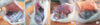
The rats were operated on once more on the 15th day under ketamine and 10-mg/kg xylazine intra-peritoneal anesthesia. In order to prevent injuries due to adhesions and to evaluate the adhesions due to incision, laparotomy with left paramedian incision was applied on all rats. A blinded researcher was brought in and asked to evaluate the intra-abdominal adhesion levels between 0 and 9 according to the Linsky Scale [9]. Adhesion severity, degree, and adhesion rates in the defected area were evaluated (Fig. 1A–D). A general score was also obtained from the total of these three parameters. After the evaluation, tissue samples were taken from caecum and surrounding area for histopathology. Samples were determined in formaldehyde for histopathologic evaluation. After the operation, all rats under anesthesia were euthanized through decortication.
Histopathologic examination
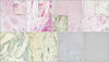
The same pathologist performed all histopathologic examinations. Samples were prepared on paraffin-embedded blocks and thin sections painted with hematoxylin and eosin stains were examined under light microscope. Images were saved to a computer. Histopathologic staging was done in accordance with Ehrlich-Hunt Model [10] (Table 1).
Evaluation criteria in this model are determined as following: inflammatory cell, fibroblast, neovascularization, and collagen amount. Cellular and histopathologic scoring is evaluated in 4 stages semiquantitatively. Different calculations were made for inflammatory cell, fibroblast proliferation, neovascularization, and collagen deposition.
Immunohistochemical examination
Sample tissues were taken from all rats in study groups and control group for immunohistochemical examination. The following processes were applied on samples respectively: formalin fixation, paraffin application, blocking and immunohistochemical dyeing. E-cadherin protein amounts were semiquantitatively evaluated on the samples belonging to different groups. (absent: 0, slight [1]: up to 20% positive, moderate 21%–50% positive, potent 51%–100%).
Statistical analysis
The SPSS ver. 15.0 (SPSS Inc., Chicago, IL, USA) was used for statistical analysis. Kruskal-Wallis test was used for group comparisons. Kruskal-Wallis post hoc tests and Sidak-Dunn test were used for pair comparisons. Results were expressed with a confidence interval of 95%. Results were given as mean ± standard error. Values of P < 0.05 were considered significant. 25th and 75th percentiles were calculated from definitive statistics. Group size calculation was performed by the resource equation method.
RESULTS
Adhesion scores
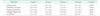
General adhesion scores for each group was determined to be as follows; 6,2 ± 1.17 for group 1, 5.8 ± 0.49 for group 2, 4.2 ± 0,7 for group 3 and 2,4 ± 0,6 for group 4. General adhesion scores for groups 1 and 2 were determined to be significantly high when compared to group 4 (P < 0.001). There was no significant difference in general adhesion scores between groups 1 and 2 (P > 0.05). There was also no significant difference in general adhesion scores of group 3 and groups 1 and 2. A significant difference was also determined between groups 3 and 4 in terms of general adhesion scores (P < 0.05) (Table 2).
Histopathological examination results
A significant difference was determined in the comparisons between groups in terms of neovascularization, fibroblast density, collagen deposition, and inflammatory cell (P < 0.001). In pair comparisons, a significant decrease in high-dose BoNT-A group (group 4) when compared to groups 1 and 2 in terms of neovascularization, fibroblast density, collagen deposition, and inflammatory cell was determined (P < 0.05). (Table 3; Fig. 2A–D).
DISCUSSION
PIAs are one of the most important problems surgeons have to face with [3]. Clinical problems caused by adhesions cause an increase in mortality and morbidity. Thus, finding a solution against PIAs is an important subject for surgeons. Many agents were used in clinical and experimental studies in order to prevent PIA formation [45]. However, the search for an effective agent still continues. Additionally, there is no common approach for PIA treatment.
Even though the physiopathological mechanism of PIA formation is not fully explained, it has been indicated that the inflammatory reaction in the defected tissue plays an important role [6]. Macrophages and fibroblasts are active for the treatment of wounded tissue. Collagens and matrix proteins also play a role in tissue repair. As a result of the physiopathological events started by inflammatory reaction, fibrous adhesions are formed. This is the main idea behind using anti-inflammatory agents to prevent PIAs.
BoNT-A is a neurotoxin with a neurotoxic protein structure produced by Clostridium botulinum bacteria [11]. It is used for many diseases such as BoNT-A upper motor neuron syndrome, focal hyperhydrosis, blepharospasm, strabismus, chronic migraine, and anal fissure. In most of the clinical cases in which BoNT-A is used, BoNT-A's anti-inflammatory effectiveness is beneficial [12]. BoNT-A demonstrates its anti-inflammatory effectiveness through macrophages, which are the main cells of the immune system [8]. Macrophages are also responsible for secretion of many pro-inflammatory mediators and small molecules playing part in the inflammation such as nitric oxide. BoNT-A mainly affects proteins such as nuclear factor-κB in the immune system signal pathway, transcription factors and mitogen-activated protein kinases including c-Jun N-terminal kinase, p38, and extracellular signal-regulated kinase [13]. As a result, these proteins affect the genes responsible for the secretion of pro- and anti-inflammatory cytokine and chemicals. Ultimately, BoNT-A receives anti-inflammatory features. Our study presents the anti-inflammatory effectiveness of BoNT-A and focuses on the hypothesis that BoNT-A may decrease and even prevent intra-abdominal adhesions.
During the comparisons in our study, it was determined that the inflammatory cell count significantly decreased in high-dose BoNT-A group when compared to control and sham groups. Recent studies have suggested that BoNT-A repressed inflammation and presented an anti-inflammatory effect [14]. Anti-inflammatory effects of BoNT-A in Sjögren Syndrome and chronic inflammatory pain treatments were also reported [15]. A study indicates that dural neurogenic inflammation plays a role in migraine pathophysiology and also that BoNT-A may have a positive effect on eliminating migraine and other headaches by decreasing dural neurogenic inflammation with its anti-inflammatory effects [16]. BoNT-A causes a decrease in inflammatory cell count as a result of its anti-inflammatory effect [17]. In a study conducted by Kim et al. [18] on flap necrosis, it was shown that the inflammatory cell count decreased in the group with BoNT-A applied. In another study conducted on the effect of BoNT-A on a rat surgical wound model, it was indicated that BoNT-A causes a decrease in inflammatory cell count. The same study also shows that TGF-β1 expression, which is known to be active in inflammatory processes and peritoneal adhesion formation, also decreases as a result of BoNT-A application [19]. Our study also showed that the inflammatory cell count in the high-dose BoNT-A group decreased and created a correspondence with the existing literature. Even though the inflammatory cell counts in the low-dose BoNT-A group decreased, no significant difference was determined when compared to control and sham groups. This result proves that the anti-inflammatory effect of BoNT-A in the peritoneum is highly dependent on the dosage and increases accordingly.
Our study showed a significant decrease in neovascularization in high-dose BoNT-A group when compared to control and sham groups. Neovascularization plays an important role in adhesion formation development. An increasing neovascularization also causes an increase in adhesion formation [20]. The significantly decreasing adhesion score in the high-dose BoNT-A group that was determined in our study may be related to decreasing neovascularization. The significant decrease in neovascularization was determined to be dependent on BoNT-A application dosage. The effect was more significant in higher dosages.
Fibroblasts are responsible of the secretion of extra matrix proteins and collagen. Increasing fibroblast density also causes an increase in collagen secretion, granulation tissue and adhesion [21]. In our study, a significant decrease in fibroblast density in high-dose BoNT-A group was determined when compared to control and sham groups. In the study conducted by Xiao et al. [22] on hypertrophic scars, a decrease in fibroblast count in the groups applied with BoNT-A was reported. In the study conducted by Oh et al. [23] on human dermal fibroblasts, BoNT-A was determined to cause a decrease in fibroblast count and collagen amount. Decreasing fibroblast count in the high-dose BoNT-A group detected in our study was determined to be in correspondence with existing literature data.
In this study, it was determined that the collagen count in high-dose BoNT-A group significantly decreased when compared with sham and control groups. Collagen is the main protein of extracellular matrix and secreted by fibroblasts. Collagen takes an active part in wound healing processes. Our study determined that high-dose intraperitoneal BoNT-A application causes a significant decrease in fibroblast count. Decreasing fibroblast count may be responsible for the decrease in collagen density. The in vitro study suggested that BoNT-A application caused a decrease in collagen expression levels [24]. In the study conducted by Kim et al. [25], a decrease in collagen levels in tissues was determined in the BoNT-A group. Additionally, in the study conducted by Sahinkanat et al. [26] on the effects of BoNT-A on urethral wound healing, a significant decrease in collagen count was determined in BoNT-A group. On the other hand, in a study on BoNT-A's effect on muscle contractile and structural properties, a significant increase in the collagen content of tissues in the BoNT-A group was reported [27]. The increase in collagen deposition after BoNT-A induced muscle fiber atrophy was responsible for collagen increase. Another study failed to show an effect of BoNT-A on collagen synthesis [28]. Data obtained from the current literature can be summarized to have different effects on different tissues in terms of collagen synthesis and count.
E-cadherin is one of the molecules that are responsible for intracellular adhesion and communication. The increase in E-cadherin expression leads to peritoneal adhesion formation [29]. No significant difference between groups in terms of E-cadherin expression was determined in our study. Even though molecular studies regarding the relationship between E-cadherin and BoNT-A were performed, no significant result could be obtained [30].
Our study included several limitations. This study is the first study to work on the effects of BoNT-A on postoperative intra-peritoneal adhesion formation. Thus, this study can be considered a pilot study. Therefore, a detailed ultra-structural examination of the histopathological changes BoNT-A causes on the peritoneum is necessary. Gene studies and electron microscopy studies can be developed on the subject. The relationship between Desmocollin 3, Cadherin 3, ARHGDIA, CYFIP2, and LAMA4 gene expressions and α5β1 proteins and BoNT-A can be examined in detail. This study examined peritoneal adhesion formation in a single timeframe. New examinations on different timeframes and different doses can be performed.
In conclusion, it can be said that a significant decrease was observed only in postoperative intra-peritoneal adhesions in the high-dose BoNT-A group between all 4 rat-groups with experimentally created postoperative intraperitoneal adhesions. Neovascularization, fibroblast count, collagen deposition, and inflammatory cell count significantly decreased in high-dose BoNT-A group. BoNT-A is determined to be an effective agent in preventing postoperative intraperitoneal adhesions. A further ultrastructural examination on the histopathologic changes BoNT-A causes on the peritoneum is necessary.
 XML Download