

 PDF
PDF ePub
ePub Citation
 Print
Print


INTRODUCTION
Appendectomy is one of the most common abdominal operations performed by general surgeons. Today, in developed countries, approximately 8% of the population has undergone appendectomy for acute appendicitis during their lifetime [1]. Although the severity of acute appendicitis is defined and classified by both surgeons and pathologists, the postoperative management depends primarily on the surgeons' intraoperative findings [2]. As the diagnosis made by pathologists is typically decided several days after the operation, it usually does not influence the patients' postoperative management unless the diagnosis is an abnormality other than inflammatory change [3].
In South Korea, appendicitis has been included obligatorily in the Disease Related Group reimbursement system since July 2013. The government's health insurance review and assessment service approves only two diagnostic categories of appendicitis for the payment of medical care expenditure: simple or complicated. Because there have been no established regulations about the determination of whether the appendicitis is simple or complicated, the identification depends on the International Classification of Diseases (ICD)-10 code, which is assigned according to the pathologic diagnosis. Therefore, the question about the correlation of pathologic diagnosis with surgical findings has been increasingly raised [4].
In this retrospective observational study, we aimed to investigate the discrepancies between the surgical and pathologic diagnoses of appendicitis, and to identify the clinical significance of these diagnoses.
METHODS
Patient selection and treatment protocol
Between January 2010 and December 2013, 1,817 patients who underwent 3-port laparoscopic appendectomy for the final diagnosis of appendicitis were included in this study.
The details of our protocol have been previously reported [5]. Briefly, appendicitis was diagnosed on the basis of the patients' medical history, physical examinations, and radiological findings. The surgeons described their operative findings in accordance with the required format. All patients received postoperative treatment, including intravenous antibiotics treatment, until they showed clinical improvement after receiving enteral feeding and there were no signs of infection such as fever or abdominal pain.
After hospital discharge, all patients were observed postoperatively and followed by their surgeons at an outpatient clinic. If the patients complained of any symptoms such as fever or abdominal pain during the follow-up period, a diagnostic evaluation was performed to identify postoperative complications.
This study was reviewed and approved by the Institutional Review Board of Bucheon St. Mary's Hospital, The Catholic University of Korea College of Medicine (HC16RISE0001).
Study design
On the basis of our previous research [5] and other literature reports, we selected clinical variables, from the medical records, that could estimate the severity of appendicitis and the intensity of the treatment pathway, as follows: (1) duration of symptoms, WBC count, CRP level, presence of diabetes mellitus (DM), operation time, and incidence of postoperative intraabdominal abscess (IAA) for the severity; and (2) performance of intraoperative peritoneal irrigation or drainage and the length of postoperative antibiotics treatment for the treatment intensity. We hypothesized that an operation time could reflect disease severity because the more intraperaitoneal inflammation is severe, the more intraoperative procedures, such as removal of remaining abscess and phlegmon, peritoneal irrigation, suction, cleansing procedure, and placement of peritoneal drainage, would be required. These variables were reviewed, compared, and analyzed according to the surgical and pathologic diagnoses. Furthermore, we selected specific patient groups and analyzed their correlation with disease severity and treatment intensity: patients with surgically and pathologically simple appendicitis (group A); surgically simple and pathologically complicated appendicitis (group B); surgically complicated and pathologically simple appendicitis (group C); and, surgically and pathologically complicated appendicitis (group D).
We classified the surgical diagnosis of appendicitis into 2 categories on the basis of the operation record. A simple appendicitis was defined as an inflamed appendix without evidence of perforation, periappendiceal abscess, localized purulent fluid collection, and generalized peritonitis. On the other hand, a complicated appendicitis was defined as any case that could not be included in the simple appendicitis group. It seems that surgeons usually decided appendicitis to be “gangrenous” if a darkish gray or purple change was noticed on the surface of the appendix. However, as there has been no consensus about the gross morphology of gangrenous appendicitis, there might be a controversy about the surgeons' judgments on whether the appendicitis is simply inflamed or gangrenous. Accordingly, in this study, a suspicious gangrenous appendicitis without evidence of definite perforation, periappendiceal abscess, localized purulent fluid collection, and generalized peritonitis was considered as simple appendicitis.
The pathologic diagnoses at our hospital are as follows: (1) acute appendicitis shows neutrophil infiltration in the muscularis propria layer without evidence of a purulent exudate; (2) acute suppurative appendicitis shows a purulent exudate in the appendiceal lumen with or without abscess formation in the appendiceal wall; (3) acute gangrenous appendicitis presents with gangrenous necrosis of the entire wall without evidence of a hole; and (4) perforated appendicitis shows frank holes that can be identified grossly. An official ICD-10 code is decided on the basis of the final pathologic diagnoses and used for billing purposes: (1) pathologically acute or suppurative appendicitis is considered as simple, and (2) gangrenous or perforated appendicitis is considered as complicated. Accordingly, in this study, the pathologic diagnoses of appendicitis were decided according to this classification.
Statistical analysis
The summary statistics are presented as number (%) for categorical variables, and as mean ± standard deviation or median (interquartile range) for continuous variables. The chi-square and Fisher exact test were used to compare categorical variables. The Wilcoxon signed-rank test was used to compare continuous variables. Analysis between patient groups was performed by multiple comparisons from Kruskal-Wallis test, followed by post hoc Dwass, Steel, Critchlow-Fligner Method. All results were analyzed by using SAS 9.4 (SAS Institute Inc., Cary, NC, USA). A P-value of <0.05 was considered statistically significant.
RESULTS
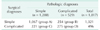
Table 1 presents the discordances between the surgical and pathologic diagnoses of the entire cohort. Of 1,321 cases of surgically simple appendicitis, 254 (29.3%) were found to be complicated appendicitis pathologically (group B). On the other hand, 221 (44.5%) of 496 cases of surgically complicated appendicitis were judged to be pathologically simple (group C). In group C patients, the surgeons' judgment was complicated appendicitis for the following reasons: (1) grossly purulent fluid accumulation in the peritoneal cavity (102 cases; 46.15%), (2) purulent material or pus spillage during appendectomy (68 cases; 30.77%), (3) periappendiceal abscess formation (26 cases; 11.76%), and (4) a large amount of phlegmon observed around the appendix (25 cases; 11.35%). In group B patients, the pathologic diagnosis was complicated for the following reasons: (1) histologically perforated appendicitis (175 cases; 68.9%) and (2) histologically gangrenous appendicitis (79 cases; 31.1%).
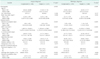
Table 2 demonstrates the correlation of the surgical and pathologic diagnoses with disease severity and treatment intensity. Neither the surgical nor pathologic diagnosis of appendicitis affected the development of postoperative IAA (P = 0.079 for surgical diagnosis; P = 0.288 for pathologic diagnosis). The variables that showed significant differences in both types of diagnosis were age, body mass index, duration of symptoms, WBC count, CRP level, operation time, incidence of peritoneal irrigation, incidence of peritoneal drainage after irrigation, and duration of postoperative antibiotics treatment. In surgically complicated appendicitis, the proportion of DM cases was significantly higher than in surgically simple appendicitis; however, this correlation was not observed in the analysis of the pathologic diagnosis.
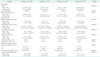
In the analysis of the patient groups (Table 3), the CRP level was higher, the duration of symptoms was longer, and the operation time was longer in groups C and D than in groups A and B (CRP level: group D > group C > group B > group A, P = 0.0001; duration of symptoms: groups C and D > group B > group A, P = 0.0001; operation time: groups C and D > groups A and B, P = 0.0001). Patients of groups C and D were older than patients of groups A and B (age: groups C and D > group B > group A, P = 0.0001), and the proportion of DM cases showed differences between groups (P = 0.0001). More patients in groups C and D received intensive treatment, including peritoneal irrigation, subsequent peritoneal drainage after peritoneal irrigation, and a longer duration of postoperative antibiotics treatment, than patients in groups A and B. The diffenreces in treatment intensity between groups C and D or groups A and B were negligible. There was no difference in the incidence of postoperative IAA between the groups.
DISCUSSION
In this study, there were discrepancies between the surgical and pathologic diagnoses of appendicitis, which is an occasionally reported condition [267]. The clinical significance of these findings remains unclear. Previously, it has been reported that a surgical grading system [8] or the intraoperative diagnosis [6910] could predict the development of postoperative infectious complications; however, no recent studies have demonstrated these features in the pathologic diagnosis. As we reported in our recent article [5], the surgical diagnosis of appendicitis alone seems inappropriate for predicting the development of postappendectomy IAA. Rather, the adequacy of intraoperative infectious source control and postoperative antimicrobial treatment were more predictive of the outcome than the gross morphology of appendicitis itself [11]. In the present study, as shown in Table 2, most IAA cases occurred in surgically or pathologically simple appendicitis. Therefore, the risk of postoperative complications seems inappropriate for determining the clinical significance of both the surgical and pathologic diagnoses of appendicitis. This conflicting result seems to arise from our treatment protocol in which surgically complicated appendicitis diagnosed at the time of the operation triggers an intensive treatment pathway (more peritoneal drainage, a longer duration of postoperative antibiotics treatment, and more antimicrobial combination regimen), which, in turn, can reduce the incidence of postoperative IAA. This finding might be extrapolated to any center in which postoperative treatment is decided on the basis of the surgeons' intraoperative view.
As shown in Table 2, both the surgical and pathologic diagnoses of appendicitis showed a correlation with disease severity and treatment intensity, to some degree. We found that more intensive postoperative treatment was delivered in surgically and pathologically complicated appendicitis and that the severity of appendicitis was well correlated with both diagnoses. However, there were significant differences between groups C and B: clinically, more patients in group C exhibited severe appendicitis than patients in group B; more patients in group C received intensive postoperative treatment than patients in group B. We also found that the appendicitis severity and treatment intensity could be largely divided into 2 categories: (1) more severe appendicitis with more intensive post-operative treatment (groups C and D); and, (2) less severe appendicitis with less intensive postoperative treatment (groups A and B). These results could suggest that the surgical diagnosis might have more corelations with disease severity and postoperative treatment intensity than the pathologic diagnosis. Additionally, we found that there was some disagreement between the actual cost of treating the patients in hospitals and the final payment of the patients, as the official ICD-10 code was determined according to the final pathologic diagnosis, whereas the post-operative treatment was decided according to the surgical diagnosis. In fact, according to our experiences, there could be a large amount of phlegmon around the appendix, cecum, and terminal ileum, and there might be localized exudate in Douglas' pouch or the right paracolic gutter, although the appendix did not show any obvious perforation morphologically. Moreover, appendicolith or pus might have spilled during the resection of the appendiceal base. In these cases, the surgical diagnosis was complicated appendicitis, whereas the pathologic diagnosis was simple. Accordingly, it seems obvious that the 2 diagnostic criteria—surgical or pathologic diagnosis—would differ because the criteria differ. Therefore, we suggest that an intensive postoperative treatment might be required occasionally to prevent postappendectomy infectious complications in some cases of pathologically simple appendicitis. The surgeon's assessment would be more predictive of the postoperative outcome than the pathologist's because surgeons can assess the presence of peritonitis, purulent fluid and pus accumulation, and gross spillage of purulent material during the operation in addition to the gross morphology of the inflamed appendix.
On the contrary, the limited accuracy of surgical diagnosis might cause unnecessary postoperative intensive treatment and excessive medical expenses. The accuracy of surgical diagnosis for the assessment of appendicitis remains debatable. Several studies reported that the laparoscopic grading system showed good accuracy for the diagnosis of acute appendicitis [12], and that the surgical grading system was well correlated with adverse postoperative outcomes [8]. Comparatively, there can be variations among surgeons in the intraoperative assessment of appendicitis, according to their career experience or ongoing frequency of performing appendectomies [713]. Therefore, it might be helpful in predicting the surgical outcome if a quantitative and objective surgical grading system can be established. We suggest that it should include not only the morphology of inflamed appendix but also the degree of peritonitis such as followings: grade I, no fluid collection or serous fluid collection; and grade II, grossly purulent fluid collection or peritoneal contamination during the operation. The morphology of inflamed appendix can be graded according to the currently using grading system [8]: grade 0, normal appearance; grade 1, inflamed without perforation; grade 2, gangrenous, without perforation; grade 3, perforated with localized fluid; grade 4, perforated with a regional abscess; and grade 5, perforated with diffuse peritonitis. We also recommend that objective guidelines for the grossly gangrenous appendicitis would be required to reduce inter-observer variations among surgeons.
In the present study, we found discrepancies between the surgeons' intraoperative assessment and the pathologists' final histologic diagnosis of appendicitis. Although neither of the 2 diagnosis types affected the development of postoperative IAA directly, it might cause a disagreement between the actual expense of medical resources and the final payment of the patients. We suggest that the surgeon's classification is more predictive of the outcome than the pathologist's because only the surgeon's findings are available immediately after surgery when decisions have to be made. Therefore, a reliable surgical grading system should be established and used nationwide by surgeons to increase the accuracy and cost-effectiveness of postoperative treatment.
 XML Download