

 PDF
PDF ePub
ePub Citation
 Print
Print


INTRODUCTION
Laparoscopic cholecystectomy is one of the most frequently performed operations globally. Bile duct injury that could lead to serious complications, such as biliary sepsis, remains an unsolved problem for the surgeon. The incidence of biliary injury has remained steady at 0.15%–0.6% [1]. In most cases, misinterpretation of biliary anatomy is the main cause of the iatrogenic biliary injury. The incidence of aberrant biliary anatomy, which hinders location of the ligation point and compromises safety, has been reported in up to 23% of cases. Several studies reported that 34%–49% of surgeons could experience a bile duct injury during their career [12]. Therefore, many efforts have attempted to reduce the risk of biliary injury, such as meticulous dissection of Calot triangle for critical view of safety, preoperative magnetic resonance cholangiopancreaticography (MRCP), intraoperative cholangiography (IOC) and laparoscopic ultrasonography (LUS) [345].
NIRFC after intravenous (IV) injection of indocyanine green (ICG) has been suggested as an alternative technique for easy intraoperative recognition of biliary anatomy. Previous studies have shown the feasibility of NIRFC for safe exploration of biliary anatomy. It provides the real time assessment of extrahepatic biliary anatomy and can be done quickly without radiation exposure [6].
Herein, we present a case of cystic duct variation found by NIRFC having a possibility of biliary injury during dissection of Calot triangle. We review the relevant literature concentrating in particular on the clinical efficacy of fluorescent cholangiography.
CASE REPORT
A 32-year-old female was admitted to Seoul St. Mary's Hospital for surgery on a gallbladder polyp that was incidentally diagnosed during a periodic examination. The physical examination revealed no abnormal findings. Laboratory results were all within normal limits. Abdominal ultrasonography revealed a gallbladder polyp. Informed consent was obtained prior to the operation and laparoscopic cholecystectomy with NIRFC was performed
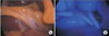
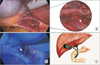
In the operating room, a single dose of ICG (0.05 mg/kg) was injected IV one hour prior to making a surgical incision. We used a specially designed laparoscopic system for fluorescence imaging (KARL STORZ Near Infrared System, Tuttlingen, Germany). Under general anesthesia, a 10-mm trocar was inserted through the umbilicus. The patient was positioned to reverse Trendelenburg with left tilting. Establishment of pneumoperitoneum was executed and intraabdominal pressure was applied and maintained at 12 mmHg during the operation. Two other trocars were introduced under direct visualization at the right flank and epigastric areas. The gallbladder was then visualized, grasped and elevated to expose Calot triangle. Before dissection of the Calot triangle, the display mode was altered to the fluorescent imaging. A long cystic duct was fluorescent (Fig. 1). Dissection progressed carefully after the display mode was changed to conventional imaging (Fig. 2A). Before ligation of the cystic duct, the display mode was changed to fluorescent imaging for confirmation of the anatomic variation. Then another tubular structure was fluorescent imaged. Partial dissection of Calot triangle was performed carefully. After the fluorescence mode was applied again, the accessory hepatic duct joining the cystic duct was observed (Fig. 2B and C). The cystic duct was ligated using a metallic clip with the preservation of the patency of the accessory hepatic duct joining to the cystic duct.
The operation time was 15 minutes. The patient was able to eat a soft diet on postoperative day 1. Postoperative laboratory tests were all within the normal limits and the patient was discharged on postoperative day 2. Final histopathologic report revealed cholesterol polyps. After a week the patient showed a tolerable status without any complications.
DISCUSSION
The incidence rate of bile duct injury during cholecystectomy has not changed for several decades. In 1995, Strasberg [7] first suggested a strategy called critical view of safety to minimize the risk. However, the critical view of safety was too difficult to routinely use for safety during dissection of Calot triangle and surgery took a long time. Therefore, a number of novel methods have been introduced to prevent such injuries. IOC during laparoscopic cholecystectomy has shown promising results in decreasing incidence of biliary injury. Regardless of this advantage of IOC, its' routine use remains controversial because of limitations that include radiation exposure, increased operative time and technical difficulties [3]. Although LUS has been considered, it is not routinely used because of its inaccuracy and lengthy learning curve [4]. Preoperative MRCP is seldom used for confirmation of anatomic variation due to increased cost.
Recently, NIRFC after IV injection of ICG has been presented as a safe and the effective technique that enables the real-time visualization of the biliary anatomy during laparoscopic cholecystectomy. ICG is swiftly removed from the blood by the liver with a plasma half-life of 3–5 minutes and is excreted into the bile within 10–15 minutes of administration. It emits light with a peak wavelength of 800–810 nm that can be displayed on a fluorescence imaging system. The display mode can be interchanged between conventional and fluorescence imaging in real-time. Several advantages of the approach include realtime visualization, time-saving and lack of radiation exposure. NIRFC may also be less expensive to perform compared to IOC, with a reported average cost of $778 for IOC, compared to $14.10 for NIRFC [8].
From the surgical point of view, NIRFC after IV injection of ICG is a very attractive tool for reducing biliary injury. With this technology, the biliary anomaly was found to be much better than conventional laparoscopic system during the dissection of Calot triangle. In this case, by using NIRFC, we could recognize the accessory hepatic duct to joining the cystic duct during dissection of Calot triangle and prevent the unexpected biliary injury. NIRFC provided additional information about the biliary anatomy during surgery and helped to lessen the risk of iatrogenic biliary injury.
However, NIRFC during laparoscopic cholecystectomy has some limitations. First, the tissue penetration capability of near-infrared light is limited to 5–10 mm. Thus, the detection rate of biliary structure is low in cases featuring abundant fatty tissue or severe inflammation around the gallbladder and Calot triangle. A previous study reported that the identification rate before dissection of Calot triangle in noninflammatory status differed from acute cholecystitis [6]. In the absence of inflammation, the rate of identification of the cystic duct, common hepatic duct and common bile duct (CBD) was 93%, 88%, and 91%, respectively. In comparison, the identification rate in patients with acute cholecystitis was 91.6%, 75%, and 79.1% in the same respective order. This highlights the necessity of additional methods to enhance the rate of successful identification. In a bid to minimize the risk of biliary injury, some articles have reported that direct injection of ICG into the gallbladder improves visualization of biliary structures [9]. Second, in contrast to IOC, NIRFC is not sufficient to detect CBD stones in most cases because the fluorescent light cannot be detected from the intrapancreatic portion of CBD and the refraction of light could confuse determination of small CBD stones. Thus, patients with suspected CBD stones should receive an additional examination, such as preoperative MRCP. Finally, although ICG has been generally reported to be safe, some cases of anaphylactic reaction following ICG administration have been reported [10]. Therefore, in cases of NIRFC after IV injection of ICG, the surgeon should use a proper amount (0.05 mg/kg) and the patient must be monitored for allergic reaction.
Regardless of these limitations, NIRFC during laparoscopic cholecystectomy could be a safe and effective procedure that enables the real-time visualization of the biliary anatomy. We expect that it could help to avoid biliary injury during laparoscopic cholecystectomy.
 XML Download