

 PDF
PDF ePub
ePub Citation
 Print
Print


INTRODUCTION
Isolated internal iliac artery aneurysms without an accompanying aortic aneurysm are rare. They are usually seen in common iliac arteries. Internal iliac artery (IIA) aneurysm accounts for 0.3% to 0.5% of abdominal aneurysm [1].
Due to their location, IIA aneurysms are not directly accessible for stent-graft placement. Thus, endovascular treatment includes excluding the vessel from the circulation by coiling the side-branches of the IIA and placing a covered stent in the ipsilateral common iliac artery (CIA) and external iliac artery (EIA) covering the origin of the IIA. Open surgical repair remains the standard treatment option for IIA. It provides excellent results, particularly in elective interventions [2]. Lately, stenting and coiling are regarded as equivalent to proximal ligation and distal ligation, respectively, in the treatment of internal iliac aneurysm [2].
Complications of coil embolization technique include inadvertent occlusion of the wrong vessels, resulting in infarction of normal structures, abscess formation, dislodgement, and migration of coil. Here we report a case of colon penetration caused by the embolization coil placed for internal iliac aneurysm.
CASE REPORT
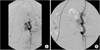
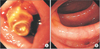
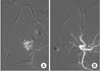
A 64-year-old man visited the Emergency Department for hematochezia that had persisted for 3 days. The pain was mild in intensity. It did not radiate to the back. The patient denied having fever, anorexia, nausea, vomiting, diarrhea, constipation, or any hemodynamic symptoms. Stent insertion and coil embolization of the left IIA aneurysm had been performed on the patient 18 months ago (first temporarily by placing the coils and an angioplasty balloon; later definitively by placing a stent-graft in the CIA and EIA across the ostium of the IIA; Fig. 1). He had no prior abdominal operation. In the Emergency Department, the patient's vital signs were normal. He was in no apparent distress. His physical examination results were normal except for mild tenderness in the left lower abdomen without rebound tenderness or pulsating mass. Bowel sounds were normal as well. Laboratory findings were also normal. At that time, contrast enhanced abdominal computed tomogram and colonoscopy were performed as part of the diagnostic work up. CT scan showed multiple diverticulosis and colitis involving the sigmoid colon. Colonoscopy suggested penetration of sigmoid colon by the embolization coil and diverticulitis (Fig. 2). Because the patient's general condition and vital signs were stable, intervention radiology rather than surgery was selected. Angiography revealed extravasation of contrast media at the left IIA. Therefore, covered stent deployment was done in the left IIA (Fig. 3). After the intervention, hematochezia was consistent. However, the amount of hematochezia was markedly decreased.
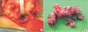
One week after the stent insertion, operation was performed. During the operation, the patient was found to have a penetrated embolization coil across the left internal iliac aneurysm. They severely adhered to each other (Fig. 4). The patient underwent anterior resection, aneurysm resection, and coil removal. The patient recovered without any complications. He was discharged at 14 days after the operation.
DISCUSSION
Isolated aneurysms of the IIA are rare and asymptomatic. When initially detected, aneurysms are usually relatively large (with a median size of 7.7 cm) [1]. Symptoms at presentation may consist of rupture with pain and hemodynamic instability or compression of either the genitourinary system or the lumbosacral nerves. The incidence of rupture is reported to be as high as 33% at initial presentation with a mortality rate of 33%–50% at open surgical repair [34]. Open surgical repair remains the standard treatment option for IIA. It provides excellent results, particularly in elective interventions. During the last decade, several reports on endovascular treatment of the IIA have appeared. Endovascular techniques have been used to treat these lesions and described to be safe and effective in short- and midterm period with satisfactory rates of aneurysm exclusion [56].
Complications of coil embolization technique include inadvertent occlusion of the wrong vessels, resulting in infarction of normal structures, abscess formation, dislodgement, and migration of coil [7].
In the present patient, exclusion procedure performed 18 months ago used palmatz and coil. It is extremely rare for coil of migration following endovascular exclusion to the GI tract. Only one case report has described the migration of coil through the celiac aneurysm to stomach with inflammation in autopsy. It is the seventh to describe coil migration following embolization of a visceral artery pseudoaneurysm [78].
The fistula formed between the iliac artery and the sigmoid colon might have been caused by diverticulitis considering the patient's history of several episodes of abdominal pain and the fact that multiple diverticula were detected in the sigmoid colon. Complications of diverticulitis include bowel obstruction, abscess formation, perforation, peritonitis, and fistula [9]. Colovesical, colocutaneous, and coloenteric fistulas are the most common ones. Colovenous fistulas make up only 2.5% of the fistulas. They are quite uncommon. They are caused by intramesocolic perforation associated with microabscesses in the mesocolon [10].
In extremely rare cases, fistula might be formed with urachus and vein. However, there has been no case report about fistula formed between arteries. In case of this patient, after performing coil embolization, migration of embolic coil might have been caused by consistent blood flow, leading to irritation by pulsation and interaction with diverticulitis.
Therefore, coil migration combined with diverticulitis of sigmoid colon might be responsible for microabscess formation and subsequent development of arteriocolic fistula. Anatomical evaluation for the surrounding structures is required for this type of aneurysm.
 XML Download