

 PDF
PDF ePub
ePub Citation
 Print
Print


INTRODUCTION
Medullary thyroid cancer (MTC) is a rare malignancy. It accounts for 3%–10% of all thyroid carcinomas [1]. The majority of MTC cases are sporadic and present with a thyroid nodule. Cervical lymph node metastases are common at initial presentation and 10%–22% of patients will demonstrate distant metastases, at the time of diagnosis, even when the primary tumor is still quite small [12]. Early diagnosis and radical surgical treatment are the keys to improve the morbidity and mortality associated with MTCs. Nevertheless, MTC is unique, since the cancer can be detected by the measurement of serum calcitonin (CT), which is accepted universally as a specific and sensitive marker [3].
Over 20 years, several studies have been conducted to assess the value of CT screening during the work-up of thyroid nodule cases to diagnose MTC [3456]. The American Thyroid Association cannot recommend either for or against routine measurement of serum CT in patients with thyroid nodules, based on the retrospective nature of the survival data, lack of availability of pentagastrin (PG) and potential biases in cost-effective analysis [7]. The American Association of Clinical Endocrinologists (AACE), Associazione Medici Endocrinologi (AME), and European Thyroid Association (ETA) Thyroid Nodule Guidelines clearly states that measurement of basal serum CT level may be a useful test in the initial evaluation of thyroid nodule and should be considered before thyroid surgery for nodular goiter [8].
The aim of the study presented here was to evaluate the diagnostic accuracy of CT measurement routinely performed among patients with newly diagnosed nodular thyroid disease.
METHODS
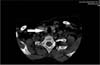
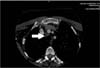
The design of the study is a retrospective chart review of 640 consecutive patients with nodular thyroid disease admitted to the Endocrine Surgery Outpatient Clinic. A duly constituted local research ethics committee approved the research. We reviewed all the patients' charts and recorded the results of the routine laboratory tests that were performed upon admission. Diagnostic work-up included thyroid examination, ultrasonography (US) of the neck, measurement of basal free T3, basal free T4, thyroid-stimulating hormone and CT levels; and fine needle aspiration biopsy (FNAB). Computed tomography was performed (Figs. 1, 2). Cases with previously diagnosed medullary cancer and those with a family history of MTC or multiple endocrine neoplasia - II were excluded.
Serum CT levels were measured by using IMMULITE 2000 automated chemiluminescent assays (Siemens Healthcare Diagnostics GmbH, Istanbul, Turkey); a solid-phase, enzyme-labeled, 2-site immunometric assay. The assay received regulatory approval based on data showing an analytical sensitivity of 2 pg/mL. Subsequent testing by the manufacturer has shown an analytical sensitivity of 0.34 pg/mL and a functional sensitivity of 0.7 pg/mL.
When basal values resulted between 10–100 pg/mL, testing was repeated with a PG stimulation test. This test was administered as an intravenous injection of PG at a dose of 0.5 µg/kg of body weight. Blood samples for CT evaluation were taken at 0, 2, 3, 5, and 11 mininutes after the PG injection. The test was considered positive if the peak serum CT concentration after stimulation was ≥100 pg/mL. FNAB was performed in a standardized fashion in all the patients whose nodular formation(s) was accessible to this procedure. Unaware of the serum CT results, the same pathologist examined the specimens. Surgery was recommended in patients with elevated basal CT levels (confirmed by a positive PG stimulation test), suspicious findings on US, and/or positive FNAB results. Cases undergoing surgery received total thyroidectomy, including central lymph node clearance. Histopathologically, serial sections throughout the thyroid specimen were obtained to detect MTC or C-cell hyperplasia. Immunohistochemistry was performed.
The direct cost of CT (±PG stimulation test) spent to diagnose one MTC case has been calculated. The result has been presented in euros (€).
RESULTS
Among the patients, 503 (78%) were female and 137 (22%) male patients. The mean ± standard deviaion age was 49.5 ± 13.5 years. Basal CT levels exceeded 10 pg/mL in 25 patients (4%). In 21 patients, the basal CT level was between 10–100 pg/mL. In 4 out of the 21 patients with basal CT level between 10–100 pg/mL, PG stimulation resulted in elevation. One patient declined the PG test and any further investigation. The histological diagnosis was MTC in only 1 patient in this subgroup. Furthermore, plasma basal CT levels exceeded 100 pg/mL in 4 patients (0.6%). One case refused surgery. MTC was diagnosed in 3 cases in this subgroup. In total, 4 cases (0.62%) of MTC were identified. Table 1 shows the clinical details regarding the MTC patients. Cases with TNM stage III cancer had no cervical lymph nodes that were visible by US or positive by palpation.
Of the remaining 16 out of 21 patients with basal CT level between 10–100 pg/mL, 12 underwent surgery and 4 were advised follow-up, based on FNAB and US reports. Histological report of 2 of the patients undergoing surgery revealed papillary carcinoma, while the remaining 10 patients received a diagnosis of multinodular goiter. No cases of C-cell hyperplasia were identified. Neither permanent recurrent nerve palsy, nor permanent hypocalcemia was observed in cases receiving surgical treatment. Postoperatively, 3 patients with MTC presented basal CT levels <2 pg/mL and remain stable. One patient, with a preoperative basal CT level of 1,258 pg/mL remains stable with basal CT levels between 50–100 pg/mL, without proven locoregional disease or distant metastasis.
Positive predictive value (PPV) for basal CT levels in the preoperative diagnosis of MTC was 5% for values between 10–100 pg/mL; and 100% for values >100 pg/mL. PPV for the PG test (>100 pg/mL) was 25% in the entire series. Possible reasons for false positivity were papillary thyroid cancer in 17%, renal insufficiency in 8%, Hashimoto thyroiditis in 17%, and use of β-blockers in 33%.
The direct cost spent to diagnose MTC was estimated as €912.68 per case, based on the cost of basal CT measurement and PG stimulation test.
DISCUSSION
In this series of 640 patients with nodular thyroid diseases, routine measurement of CT resulted in 4 newly diagnosed MTC cases. This results in a frequency of 0.62%. Other series reported a frequency between 0.4%–1.37% [4569101112]. Different from the ATA's conflicting and unclear recommendations [7], the ETA consensus report clearly states that routine measurement of CT should be applied in the management of thyroid nodule cases, since it is more sensitive than FNAB [13]. Besides, the AACE, AME, and ETA's common document declared that routine measurement of basal CT may be useful, and strongly recommended its measurement in certain high-risk groups [8]. The finding of elevated serum CT modifies the extent of the surgery, from total-/hemithyroidectomy to total thyroidectomy, including central lymph node clearance. The objective behind this philosophy is that this approach enables effective treatment even after the first intervention [9].
The usual argument against routine testing for CT is that most would have otherwise been diagnosed by family history or by FNA. In the current study, results of FNAB revealed 1 malignant and 1 suspicious result out of 4 patients who were diagnosed with MTC. It can be criticized that based on these clinical grounds alone, surgical treatment would already have been advised to 2 of them. However, the extent of surgery could be inadequate, since FNAB of one patient indicated nuclear features mimicking papillary carcinoma, and in the other FNAB was inconclusive. Recently, it has been clearly demonstrated that FNAB and US are not accurate for the detection of MTC and may record false-negative results [14]. The usefulness of cytology has been questioned: cytology results of MTC may be interpreted as papillary carcinoma, indeterminate, or even benign [151617].
Although measurement of serum CT is a diagnostic tool of utmost importance for MTC, routine screening of thyroid nodule cases with CT is inconclusive, due to the low prevalence of the disease and increased cost of routine assessment. The improvement of the specificity of CT with the use of a PG stimulation test is well known and accepted, although there is a wide spectrum of PPV, ranging between 25%–100% [141517]. In the present study, PPV for basal CT levels in the preoperative diagnosis of MTC was as low as 5% for values between 10–100 pg/mL, while it increased to 100% for values >100 pg/mL. PPV for the PG test (>100 pg/mL) was 25% in the entire series. Unfortunately, PG is currently seldom available throughout the world. Nevertheless, we know well that calcium infusion instead of PG has overcome the lack of a stimulation test, but the problem of the stimulated CT cutoff values remains unsolved [13]. Elisei et al. [17] demonstrated in their study with more than 10,000 patients that routine measurement of serum CT in thyroid nodules is the most informative test for the early diagnosis and treatment of sporadic MTC. They recommend serum routine CT measurement in the clinical work-up of thyroid nodules, followed by a PG stimulation test in all cases of detectable basal CT levels to be performed preferably in a referred center. Pacini et al. [12] reported that preoperative measurement of basal CT increased the diagnostic accuracy and helped the surgeon perform more radical treatment. In Costante et al. [18]'s study, basal CT levels displayed a PPV of 33.8% for C-cell hyperplasia and MTC. They recommended PG test confirmation of basal values between 20–100 pg/mL, reaching a PPV of 90% for the diagnosis of C-cell hyperplasia or MTC. It was seen to be less helpful in identifying MTC alone (PPV = 40%). In Herrmann et al. [19]'s study, it has been shown that with the adoption of routine CT measurement, MTC cases could be detected in early tumor stage. Niccoli et al. [11] found that the sensitivity and specificity of basal CT measurement for the diagnosis of MTC was 69.9% and 97.6%, respectively. Routine CT measurement allowed them to detect MTC at an early stage as well. Radical surgery allowed them to normalize CT levels in these patients, and thus is expected to improve the prognosis and survival of sporadic MTC. Vierhapper et al. [5] reported a 3-fold increase of MTC cases after they started routine screening of basal CT in thyroid nodules in their prospective series.
In this paper, we do not insist that basal CT cutoff level should be above 100 pg/mL. We included patients who had a basal CT level >10 pg/mL in this study. And those basal values resulting between 10–100 pg/mL underwent a repeated testing with a PG stimulation test. If it was >100 pg/mL, the test was accepted as positive. We accepted the cutoff value as 100 pg/mL, because of previous studies [78181920]. There is one recent Korean study addressing the CT cutoff value, which compared 2 different cutoff values: 100 pg/mL and 65 pg/mL [21]. It was expressed that 65 pg/mL was more sensitive and accurate than 100 pg/mL, even though values below 65 pg/mL could not exclude the presence of MTC such as small volume MTC or premalignant C-cell hyperplasia.
Although an elevated level of serum CT is a highly sensitive marker for MTC, possible false positive reasons may be responsible for high basal CT levels [22]. These are use of proton pump inhibitors, β-blockers and glucocorticoids; Hashimoto thyroiditis; renal insufficiency; follicular and papillary thyroid carcinomas; C-cell hyperplasia and neuroendocrine tumors. Nine patients in our series having a basal CT level of >10 pg/mL had false positive results: Two had papillary thyroid cancer, 4 were on β-blockers, 2 had Hashimoto thyroiditis and 1 suffered from chronic renal failure.
Several reports from Europe and United States have considered the cost of routine CT screening [561417232425]. It has been reported that the measurement of serum CT costs between €15–37 [56142324] differing between countries. In the current series, the cost of the tool was €4.63 per CT measurement. A PG stimulation test, which includes up to 5 determinations of serum CT, is more expensive. The results of the study of Cheun et al. [23] from United States suggest that CT screening appears to be cost-effective in thyroid nodule management and the most cost-effective in young men with larger thyroid nodules. The question of whether it is worthwhile to measure basal CT levels for every thyroid nodule patient and spend €4.63 to find one person with results outside the normal range should be answered by future prospective research. However, it is our opinion that the costs of finding one patient with abnormal results and MTC are quite reasonable for routine basal CT measurement, compared with the potential costs of missing the diagnosis of this treatable malignant condition. In countries such as Turkey, the cost for each newly diagnosed MTC case may be lower than others. In the current study, nearly €913 were spent on screening for each confirmed MTC, while this was reported as €3,710 in a French prospective study [6]; nearly €5,400 in an Austrian prospective study [5] and approximately €7,200 in a British report [24]. According to another United States study, the cost for detecting one MTC varied between approximately US $4,000 and $20,000 [26].
Our study has limitations. First, we lacked information regarding the results of patients having low basal CT level (<10 pg/mL) and for whom surgery was carried out. Second, there were no data available regarding the cost of days of additional hospital stay and unnecessary or inadequate treatment. Moreover, the costs of tests may vary across sites and countries. Therefore, the results of the study presented here should be regarded as a preliminary contribution to the calculation of cost-effectiveness of routine CT screening among thyroid nodule patients. Another pitfall was that the sample was small and limited to 1 hospital. Finally, no gender specific cutoff values for CT levels below 100 pg/mL could be calculated.
In conclusion, whether routine CT screening should be adopted with the impact of this screening on MTC-related morbidity and mortality is a question that can be answered only with large, long-term, prospective and randomized multicenter initiatives, with standard enrollment criteria, CT assay techniques, and breakpoints for interpreting the results [27]. Despite the lack of accessibility to the PG stimulation test and concerns regarding increased costs, routine measurement of basal CT is useful to detect ‘early stage’ MTC and is valuable. This approach enables adequate surgical treatment of MTC.

 XML Download