

 PDF
PDF ePub
ePub Citation
 Print
Print


INTRODUCTION
Based on the results of large-scale clinical studies [12345], carotid endarterectomy (CEA) has been recommended for symptomatic and asymptomatic patients with high-grade carotid artery stenosis [6]. Safety has been emphasized during the procedure, because of the possibilities of disabling or nondisabling stroke. The majority of patients tolerate temporary interruption of cerebral blood flow during CEA, but cerebral ischemia may develop in patients with inadequate collateral channels. Carotid shunting guarantees adequate cerebral blood flow during carotid clamping, but its routine use is controversial [7].
Selective shunting has theoretical advantages over routine shunting, because of possible adverse effects, which include intimal damage or dissection, distal embolization, and acute occlusion. Furthermore, shunt placement requires more distal exposure of the ICA, and this may be challenging in patients with a high lesion. Neurologic monitoring of awake patients under local or regional anesthesia provides the most reliable means of identifying candidates for selective shunting [891011], but it cannot be used under general anesthesia. Accordingly, many surgeons that favor general anesthesia adopt routine shunting. On the other hand, other surgeons use selective shunting and intraoperative monitoring by transcranial Doppler ultrasonography (TCD), electroencephalography (EEG), somatosensory evoked potential (SSEP), near infrared spectroscopy, or carotid stump pressure. However, optimal methods for detection of cerebral ischemia remain controversial [12].
The aim of this study was to evaluate efficacy and safety of cerebral monitoring by TCD for the detection of cerebral ischemia during CEA.
METHODS
Patients
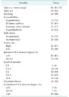
One hundred fifty-nine patients that underwent CEA from August 2004 to December 2013 were retrospectively reviewed. All CEA were done under general anesthesia. Intraoperative TCD was routinely used to detect cerebral ischemia. Of these 159 patients, 27 patients were excluded for a poor transtemporal isonation window and 30 because additional cerebral monitoring systems, such as, EEG or SSEP, were used. Accordingly, 102 patients constituted the study cohort. Patients with monocular blindness or that had experienced a transient ischemic attack or ischemic stroke within the previous 6 months were regarded as symptomatic. Patient and lesion characteristics are summarized in Table 1. Patient mean age was 66 years (43–78 years), 92% were male, 75% were symptomatic, and 81% had severe (>70%) internal carotid artery (ICA) stenosis. Nine patients had contralateral ICA stenosis exceeding 50%.
Carotid endarterectomy
All CEAs were performed under general anesthesia with the use of nitrous oxide and halothane or isoflurane. Before carotid clamping, heparin was administered intravenously; protamine reversal was not used. The eversion technique was used in 56 patients (55%) and conventional CEA in 46 patients (45%, 36 primary closures and 10 patch closures).
TCD monitoring
Intraoperative TCD monitoring was performed using a TC8080 (Pioneer TC 8080, Nicolet Vascular, Madison, WI, USA) (Fig. 1A). Mean flow velocity (MFV) of the ipsilateral middle cerebral artery (MCA) was monitored before and after carotid clamping (Fig. 1B, C). And the mean blood pressure (MBP) was also checked at the same time. The criterion for selective shunting was a MFV reduction >50% after carotid clamping versus baseline.
RESULTS
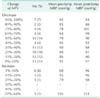
Mean preclamp ipsilateral MCA MFV and MBP were 50 cm/sec (21–123 cm/sec) and 88 mmHg (47–130 mmHg), and mean postclamp ipsilateral MFV and MBP were 32 cm/sec (0–106 cm/sec) and 91 (60–144 mmHg). Table 2 shows MFV change after carotid clamping. The mean decrease in ipsilateral MFV was 33% (-56%–100%). Of the 102 patients, 31 (30%) showed an ipsilateral MFV decrease >50% after carotid clamping, and carotid shunting was performed in all these patients. Accordingly, the shunt rate was 30%.
Regarding early postoperative outcomes, perioperative stroke occurred in 2 patients (2%). One minor ischemic stroke caused by embolism occurred in a patient without a carotid shunt. The other was due to intracerebral hemorrhage (ICH) caused by hyperperfusion syndrome. One perioperative death developed in the patient with hyperperfusion syndrome.
DISCUSSION
Type of anesthesia (general vs . locoregional) remains a controversial issued for CEA. A large randomized controlled trial failed to identify a definite difference between general and local anesthesia with respect to the incidences of stroke, myocardial infarction, or death [13]. One of the theoretical benefits of locoregional anesthesia is a lower carotid shunt rate. Reported shunt rates from the awake test range from 4.4% to 14% [891011], and patients with contralateral ICA occlusion required carotid shunting more frequently than those with a unilateral lesion [914]. To reduce shunt rates during general anesthesia, several intraoperative monitoring modalities have been introduced. TCD is one of these modalities and concerns have been expressed regarding its safety and efficacy.
TCD cannot determine the presence of cerebral ischemia precisely during carotid clamping. Reported values for the sensitivity and specificity of TCD for predicting the need for carotid shunting range from 75% to 83% and from 75% to 96%, respectively, although these values depend on the cutoff used [1516]. Moritz et al. [17] investigated the accuracies of monitoring tools with respect to the detection of cerebral ischemia during CEA. In this previous study, the cutoff value for 100% sensitivity for TCD was determined to be a 48% reduction in ipsilateral MCA MFV versus baseline, the cutoff for 100% specificity was 70%. And the best fit cutoff value was 48%, which provided a sensitivity of 100% and a specificity of 86%, because safety is most important in carotid surgery. In the present study, our shunt criterion was similar to their result. No patient that did not undergo carotid shunting experienced cerebral ischemia during carotid clamping. Therefore, it would appear that a reduction in ipsilateral MCA MFV of >50% as determined by TCD is a safe criterion for shunt placement to prevent cerebral ischemia during carotid clamping. However, the overall shunt rate was 30%, which is greater than previously reported shunt rates from the awake test. This elevated rate was presumably due to false-positives. If the cutoff value is lowered, unnecessary shunting would be avoided. However, it will increase a risk of missing cerebral ischemia. Therefore, to determine an optimal TCD criterion that has lowest shunt rate and do not make cerebral ischemia during carotid clamp, additional large-scale clinical studies are necessary.
Delayed shunt occlusion is a complication of carotid shunting [181920]. Kink of shunt or adhesion of the shunt stump to the arterial wall could cause shunt malfunction. Furthermore, carotid shunt occlusion would not be recognized in the absence of cerebral ischemia monitoring. If these anomalies are not corrected immediately, cerebral ischemia may result. On the other hand, continuous TCD monitoring can confirm shunt patency and represents an additional benefit of TCD.
TCD monitoring not only provides information on hemodynamic changes but also enables the detection of cerebral microembolism. Ackerstaff et al. [21] reported the outcomes of 1,058 patients that underwent CEA with TCD monitoring. They found 31 patients with ischemic stroke and 8 patients with hemorrhagic stroke. TCD-detected microembolism during dissection and wound closure, ≥90% MCA velocity decrease at carotid clamping and ≥100% increase of pulsatile index (PI) were associated with perioperative stroke or stroke-related death. It was suggested that information on microembolization can caution surgeons regarding they need for more careful dissection or early carotid clamping and TCD monitoring is a sensitive tool for the prediction of postoperative hyperperfusion syndrome. In our series, the low sample size prevented our analyzing the association between microembolism and stroke. Regarding hyperperfusion syndrome, the patient that died of ICH showed an increase in PI of 92% after CEA.
Summarizing, TCD monitoring was found to be a safe modality for indicating the need for selective shunting during CEA. For patients that exhibited am ipsilateral MCA MFV reduction of ≤50%, carotid clamping could be done without a carotid shunt. Although the technical limitations of TCD were apparent as 10%–15% of patients did not have an adequate temporal window [22], its use during general anesthesia can reduce shunt and shunt-related complication rates.

 XML Download