

 PDF
PDF ePub
ePub Citation
 Print
Print


INTRODUCTION
Differentiated thyroid carcinoma (DTC) is a malignancy with a favorable prognosis, and DTC patients, who were confirmed to be disease-free during the follow-up period, can expect a normal lifespan [1]. According to the Korea National Cancer Incidence Database, crude and age-standardized cancer incidence rates during 2011 were 81.0 per 100,000 (27.9 in males, 134.1 in females) and 58.3 per 100,000 (20.2 in males, 96.8 in females), respectively [2]. The incidence of thyroid cancer was increased by 23.3% per year in both sexes, and since 2009 it has been the most common cancer in women in Korea [3]. The increased prevalence of thyroid cancer in Korea leads to increased thyroid cancer mortality [4]. The long-term follow-up is designed to monitor recurrence of the disease and to confirm that patients remain disease-free after total thyroidectomy and RAI remnant ablation [5].
The guidelines of the American Thyroid Association (ATA) are widely used for long-term management of DTC [5]. ATA guidelines recommend measuring stimulated Tg 12 months after the completion of thyroidectomy and RAI ablation, using thyroid hormone withdrawal (THW) or recombinant human thyroid-stimulating hormone (rhTSH) stimulation in patients at a low risk of recurrence (with negative findings on neck ultrasonography [US] and undetectable stimulated Tg levels in the first year after treatment) [5]. For patients at intermediate or high risk of persistent disease, ATA guidelines recommend measuring T4-off Tg at 6–12 months, along with a diagnostic whole body scan (DxWBS) [6]. The Korean Thyroid Association recommendation also proposes performing the T4-off Tg measurement 12 months after thyroidectomy [7]. The diagnostic cutoff for T4-off Tg at the 12-month follow-up is 1 ng/mL [8].
The definition of disease-free status comprises the following: (1) no clinical evidence of tumor, (2) no imaging evidence of tumor by radioiodine imaging (no uptake outside the thyroid bed on the initial posttreatment whole body scan [WBS] if performed, or, if uptake outside the thyroid bed had been present, no imaging evidence of tumor on a recent diagnostic or posttherapy WBS) and neck US, (3) low Tg levels during thyroid-stimulating hormone suppression (Tg < 0.2 ng/mL) or after stimulation (Tg < 1.0 ng/mL) in the absence of interfering antibodies [6].
Although it has an important role in long-term follow-up, THW during T4-off Tg measurement could be troublesome for patients. THW induces several discomforts such as cognitive dysfunction, physical and emotional discomfort, and impaired quality of life [9]. Since a repeated measurement of T4-off Tg is of limited value in patients who exhibit undetectable stimulated Tg at least once [10], it is essential to stratify the risk of detectable T4-off Tg after initial treatment (total thyroidectomy and RAI remnant ablation). So the aim of this study was to find clinical and pathologic factors that predict detectable T4-off Tg during follow-up after initial therapy.
METHODS
Patients and study design
Three hundred fifty-five patients of papillary thyroid carcinoma (PTC) underwent total thyroidectomy and prophylactic ipsilateral central compartment node dissection then RAI ablation from October 2008 to August 2012. Of those, 26 were positive for serum Tg antibody and were excluded from the study. A total of 329 patients were included in the retrospective evaluation. A majority of the patients had undergone low-dose RAI ablation, with a mean dose of 66.7 mCi (Table 1). Stimulated Tg levels immediately prior to ablation (ablative Tg) were measured to predict the status of T4-off Tg during follow-up. Patients were followed up by neck US and serum Tg measurement at 4 months after RAI ablation (6 months postoperatively), and by serum Tg measurement alone at 10 months after RAI ablation (12 months postoperatively).
Twelve months after RAI ablation, 319 patients underwent THW therapy, and 10 patients were administered rhTSH. Serum T4-off Tg levels were then measured in all patients. The patients were assigned to high (>1.0 ng/mL, n = 53) and low (≤1.0 ng/mL, n = 276) groups, based on T4-off Tg level measured 12 months postoperatively. Demographic, clinicopathological, and laboratory characteristics at diagnosis and during follow-up were compared between the 2 groups.
Laboratory measurements
Serum Tg was assayed using immunoradiometric assay (IRMAZENco Tg-S, Zentech s.a., Belgium), which is based on coated-tubes with monoclonal antibodies directed against distinct epitopes of the molecule of Tg. Analytical sensitivity was calculated based upon the calibration curve and expressed as the minimal dose showing a significant difference from the Zero Calibrator (mean value + (mean ± 2 standard deviation). This dose is 0.3 ng/mL. The functional assay sensitivity is the lowest value which is measured with a precision maximum of 20% interassay variance. This value is lower than 0.1 ng/mL. Serum anti-Tg antibody was measured with an immunoradiometric assay (anti-Tg RIA; BRAHMS), and anti-Tg antibody values >60 U/mL were considered positive. The sensitivity and specificity of various Tg assays vary in different laboratories, even with the use of an international standard (Certified Reference Material 457 [CRM-457]). Thus, National Comprehensive Cancer Network guidelines recommend monitoring Tg levels via the same Tg assay performed in the same laboratory [11]. Tg assays with function sensitivity of ≤0.1 ng/mL is a highly sensitive and reliable method for detecting persistent or recurrent disease. Meta-analysis published by Giovanella et al. [12] showed that highly sensitive Tg assay has a very high negative predictive value (NPV) for monitoring DTC patients. Since functional sensitivity of our Tg assays was ≤0.1 ng/mL, Tg assay in our study was adequate and reliable in monitoring disease status.
Assessment by ATA 3-level stratification
We stratified risk factors associated with high and low T4-off Tg value into 3 groups in terms of risk of recurrence (low, intermediate, and high risk) according to the ATA guidelines (Tables 1 and 2) [6]. The low-risk group was defined as follows: No local or distant metastases, all macroscopic tumors were resected, no tumor invasion of locoregional tissues or structures, the tumor showed no aggressive histology (e.g., tall-cell, hobnail variant, or columnar-cell carcinoma) or vascular invasion, and no 131I uptake outside the thyroid bed on the first posttreatment WBS. The intermediate-risk group had tumors with aggressive histology or vascular invasion and either microscopic invasion of the tumor into the perithyroidal soft tissues at initial operation, cervical lymph node metastases, or 131I uptake outside the thyroid bed on the WBS performed after 131I remnant ablation. The high-risk group had macroscopic tumor invasion, incomplete tumor resection, distant metastases, and thyroglobulinemia out of proportion to what was seen on the post-treatment scan. However, cases of incomplete tumor resection, distant metastases, and American Joint Committee on Cancer (AJCC) stage IV were excluded to evaluate Tg level as a prognostic factor more independently.
Response to initial therapy according to ATA restratification
The initial ATA risk estimates can be refined based on the assessment of the response to the initial therapy (dynamic restratification) [6] (Table 1). This new concept was applied to this study. A total of 329 patients were reclassified into the 3 groups based on the response to the initial therapy: Excellent response (suppressed and stimulated Tg < 1 ng/mL, neck US without evidence of disease, negative WBS), acceptable response (suppressed Tg < 1 ng/mL, stimulated Tg between 1 and 10 ng/mL, neck US with nonspecific change or WBS with nonspecific change), and incomplete response (suppressed Tg ≥ 1 ng/mL or stimulated Tg ≥ 10 ng/mL, rising Tg value, persistent or newly identified disease).
Statistical analysis
The statistical significance of differences between qualitative variables was determined using Fisher exact test. While normally distributed quantitative variables were compared using Student t-test, abnormally distributed quantitative variables were compared using the Mann-Whitney U-test. Univariate analysis was performed to assess the predictive value of the variables, followed by multivariate logistic regression analysis, which included all variables found to be important in the univariate analyses. All statistical analyses were performed using SPSS v. 12.0 (SPSS Inc., Chicago, IL, USA) with statistical significance set at P < 0.05. Means and standard deviations are reported in the text and tables, where appropriate.
RESULTS
Baseline characteristics of the study population
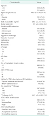
The baseline clinical characteristics of the patients are shown in Table 1. The mean ages of the patients were 48.9 ± 10.9 years (range, 23–76 years). Of the 329 subjects, 251 patients were female (76.3%) and 78 patients were male (23.7%). The mean tumor size was 0.9 ± 0.5 cm (range, 0.2–5.2 cm). About 46% and 11% of the patients had microscopic and macroscopic extrathyroidal extension, respectively. About 42% of the patients had cervical lymph node metastasis. Approximately 60% of subjects were AJCC TNM stage I. According to ATA 3-level stratification, low-, intermediate- and high-risk groups were 32%, 52%, and 15% of the study population, respectively. According to ATA restratification, 46%, 17%, and 36% of patients showed excellent, acceptable, and incomplete response, respectively.
The parameters predicting high 12-month T4-off Tg level
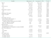
Fifty-three patients (16.1%) had 12-month T4-off Tg levels >1 ng/mL and 276 (83.9%) had levels ≤1.0 ng/mL (Table 2). Predictive factors such as tumor size, preoperative Tg, ablative Tg, cervical lymph node metastasis, thyroglobulinemia out of proportion to results of DxWBS, ATA 3-level stratification and ATA restratification were significantly associated with a 12-month T4-off Tg level >1 ng/mL (Table 2). Since some of the factors were continuous variables, additional analysis using a second model was performed, in which patients were divided into groups by age (≥45 years vs. <45 years), preoperative Tg level (≥9.0 ng/mL vs. <9.0 ng/mL) [13], ablative Tg level (1.0≥ ng/mL vs. 1.0< ng/mL) [6], and tumor size (>1.0 cm vs. ≤1.0 cm). The AJCC TNM staging system categorizes tumor size into 4 groups: group T1a ≤ 1 cm, and groups T1b–T3 > 1 cm [14]. Therefore, a tumor size of 1 cm is a reasonable cutoff value. Based on ATA guideline, it is reasonable to set a cutoff number of cervical lymph node metastasis as 5. The modified 2009 risk stratification system classifies patients with small volume lymph node metastases (clinical N0 or ≤5 pathologic N1 micrometastases) as low-risk [6].
Among histopathological parameters, tumor size >1 cm, preoperative Tg > 9.0 ng/mL ablative Tg > 1.0 ng/mL, more than 5 cervical lymph node metastasis, thyroglobulinemia out of proportion to results of DxWBS, ATA based high-risk group and incomplete response group were significantly associated with high T4-off Tg in univariate analysis (P < 0.05) (Table 3).
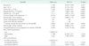
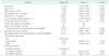
In multivariate analysis, ablative Tg > 1.0 ng/mL, more than 5 cervical lymph node metastasis, and thyroglobulinemia out of proportion to results of DxWBS exhibited T4-off Tg > 1 ng/mL 12 months postoperatively (P < 0.05) (Table 4).
DISCUSSION
T4-off Tg measurement combined with cervical US is widely used for detecting persistent or recurrent disease with patients who underwent total thyroidectomy and subsequent RAI ablation. Since T4-off Tg measurement has high sensitivity and NPV, it aids to determine not only persistent or recurrent disease but also intensity and frequency of cancer surveillance. Newly revised ATA guidelines do not recommend stimulated Tg assay in low-risk patients with no clinical evidence of tumor in cervical US [6]. Since the recent development of highly sensitive basal Tg assay, of which the functional sensitivity is around 0.1 ng/mL, the role of T4-off Tg assay is questioned especially in low-risk patients.
So the aim of the present study was to determine the risk factors associated with a T4-off Tg level >1 ng/mL at the 12-month follow-up, in order to differentiate between high-risk patients who should undergo a 12-month T4-off Tg test and low-risk patients who do not need the test. Risk factors associated with high T4-off Tg were large tumor size, high preoperative Tg, high ablative Tg, cervical lymph node metastasis, thyroglobulinemia out of proportion to results of DxWBS, high-risk group based on ATA 3-level stratification and incomplete response group based on ATA restratification. Results of multivariate analyses indicated ablative Tg > 1.0 ng/mL, more than 5 cervical lymph node metastasis, and thyroglobulinemia out of proportion to results of DxWBS were significant independent risk factors.
Cervical lymph node metastasis is observed in 30%–80% of patients with PTC on postoperative biopsy, which has shown to be an independent predictor of tumor recurrence in addition to age, tumor size, and surgical completeness [1516]. We found that more than 5 cervical lymph node metastases were present in 30.2% of patients in the high T4-off Tg group, compared to 4.7% in the low T4-off Tg group (Table 2), and lymph node metastasis was a strong indicator of risk (Tables 3, 4). This result is in agreement with previous findings. Leboulleux et al. [17] found that higher numbers of metastasized and extracapsular extension lymph nodes were associated with a higher likelihood of a 12-month T4-off Tg level >1 ng/mL and tumor recurrence after treatment. Another study found that the only 2 risk factors that were independent predictors of disease progression and prognosis were lymph node metastasis and 12-month T4-off Tg >1 ng/mL [18]. Therefore, serum T4-off Tg measurement in patients without these risk factors is recommended with reluctance.
Age is the most powerful single prognostic factor used in most PTC staging systems [1920]. For example, the AJCC TNM system stages patients <45 and ≥45 years differently [14], and total thyroidectomy is usually recommended for patients aged ≥45 years [521]. The old age group shows a poor prognosis regardless of other clinicopathological factors [22]. In this study, older age was not a statistically significant risk factor in both the univariate and multivariate analyses (Tables 3, 4). However, the mean age of all 329 subjects was 49 years, and the mean ages of the high and low groups were 47 and 49 years, respectively. Thus, our sample did not accurately reflect differences associated with the standard cutoff age of 45 years.
Previously, Tg concentration measured at the time of diagnosis of thyroid cancer, was considered to be of little importance as a prognostic marker for PTC [23]. Our study did not show preoperative Tg concentration was a significant risk factor for high 12-month T4-off Tg level. In other literature, when the patients were divided into 2 groups, based on a different cutoff value of Tg concentration of 9 ng/mL [13], univariate analysis no longer showed preoperative Tg concentration to be a significant risk factor. We conclude that variability in preoperative Tg concentration is too great to provide a reliable indication of risk for high 12-month T4-off Tg level. Furthermore, Tg concentration has been shown to increase during the course of most thyroid diseases, and the sensitivity and specificity of the serum Tg test are low. This result suggests that this parameter needs further validation as a role for predicting high 12-month T4-off Tg level or recurrence.
Several studies have demonstrated the excellent NPV of postoperative stimulated Tg at the time of 131I remnant ablation [2425]. Kim et al. [26] showed that when a cutoff serum Tg level of 2 ng/mL after THW at the time of 131I remnant ablation was used, the NPV was 98.4% when excluding the recurrence of disease. According to Lee's study, when the cutoff value of postoperative stimulated Tg was reduced to 1 ng/mL, the NPV was 97.8%, which increased to 100% when the cutoff value was combined with negative neck US findings [27]. Based on these studies, it is reasonable to set a cutoff value of ablative Tg at 1.0 ng/mL.
Lee at al. [27] also conducted a study evaluating the usefulness of T4-off Tg and the result was consistent with ours. Postoperative-stimulated Tg < 2.0 ng/mL had a NPV of 94.9%, which increased to 97.7% when low Tg was combined with negative neck US findings. They reported that the stimulated Tg measurement and DxWBS, which are usually performed 6–12 months after 131I ablation therapy, may be skipped, at least in low- and intermediate-risk patients with postoperative stimulated Tg of < 2.0 ng/mL and negative neck US findings.
Padovani et al. [28] described the Tg changes over time after initial therapy with total thyroidectomy and RAI ablation. A slow decline in Tg over several years after initial therapy was observed in patients with suppressed serum Tg values in the 1–5 ng/mL range with a negative neck US findings evaluated 6 months after RAI ablation. In addition, cautious observation would be recommended rather than the additional RAI ablation for patients with stable, persistently low serum Tg levels in the absence of a structurally identifiable disease. Careful monitoring of this group of patients with low-level Tg values is therefore recommended rather than excessive evaluations including repeated measurement of Tg.
In our study, rhTSH was administered to 10 patients (Table 1). The ATA guidelines mentioned that in patients with DTC with or without extensive lymph node involvement, in whom RAI ablation is planned, preparation with rhTSH stimulation is an acceptable alternative to THW. According to the ATA guidelines, rhTSH stimulation shows similar outcomes for achieving remnant ablation, based on evidence of superior short-term quality of life, noninferiority of remnant ablation efficacy, and multiple consistent observations suggesting no significant difference in long-term outcomes compared to THW [6]. Emmanouilidis et al. [29] also showed that there was no statistically significant difference in mean Tg levels directly before the first RAI ablation and on the last follow-up between patients treated with rhTSH stimulation and THW.
The present study had several limitations. First, our study was not prospective. However, our study population included the same pathology (PTC) and followed the same treatment protocol. Another limitation is that the relatively small number of patients was clustered in the same age range, making it difficult to determine the significance of age as a predictive factor. Finally, low-risk patients were minimally managed and lacked long-term follow-up. Future studies should follow long-term outcomes in low-risk patients, with small tumors and without metastasis or invasion, to determine the likelihood of recurrence and assess prognostic factors for long-term survival. Despite these limitations, our study demonstrated whether the measurement of 12-month T4-off Tg level in patients with PTC is necessary. Furthermore, these data are of interest and may help others in planning postoperative evaluations.
In conclusion, ablative Tg > 1.0 ng/mL, more than 5 cervical lymph node metastasis, and thyroglobulinemia out of proportion to results of DxWBS were independent factors for T4-off Tg > 1 ng/mL 12 months postoperative, in patients with PTC following total thyroidectomy and RAI ablation. In low-risk patients without these clinical risk factors, the possible omission of stimulated thyroglobulin measurements could be considered during follow-up period.
 XML Download