

 PDF
PDF ePub
ePub Citation
 Print
Print


INTRODUCTION
Laparoscopic appendectomy (LA) has been widely accepted as a treatment of choice for acute appendicitis for pediatric surgeons because of its advantages of lower complication rate, shorter length of hospital stay, reduced pain, and improved cosmetic outcomes compared to open appendectomy [12]. In the 1990s, laparoscopic-assisted appendectomy employing an umbilical incision as a single-port access was implemented by Pelosi and Pelosi [3]. This technique, termed single incision laparoscopic surgery (SILS), has attracted attention from surgeons in anticipation of less invasiveness and better cosmetic results than conventional multiport procedures. However, recent randomized controlled studies did not find that SILS was more effective in terms of operative time and postoperative pain [45].
Our group has performed transumbilical laparoscopic-assisted appendectomy (TULA), approaching through a single umbilical incision using a multichannel separable glove port to exteriorize the appendix and perform the appendectomy extra-abdominal side. This simple, quick technique has the advantages of both laparoscopic and open methods. Recently, several studies have reported favorable outcomes with single incision techniques similar to ours compared with conventional laparoscopy in terms of length of hospital stay, postoperative pain and complications [67]. However, these studies are different from ours in technique, since they describe use of a rigid single channel trocar for exploration of the appendix, and also include cases of complicated appendicitis or a small sample size, potentially leading to bias in the results. The aim of this study was to compare the efficacy of TULA and LA focusing on perioperative outcomes for pediatric patients with acute uncomplicated appendicitis.
METHODS
Patients
We retrospectively reviewed the records of all pediatric patients under the age of 15 who were diagnosed with acute appendicitis and underwent LA at Korea University-affiliated Hospitals between January 2010 and January 2014. A total of 499 patients underwent laparoscopic surgery with acute appendicitis during this period. Among them, 196 children who were diagnosed with complicated appendicitis were excluded. Of the remaining 303 patients, 85 underwent TULA and 218 underwent conventional 3-port LA. All operations were performed by attending surgeons who had practiced laparoscopic surgery for more than 5 years.
Diagnosis of acute appendicitis
Suspicion of appendicitis was based on either patient history and physical examination such as abdominal pain in the right lower quadrant (RLQ) or migration of pain from the periumbilical area to the RLQ, or laboratory findings indicating elevation of inflammatory markers. Abdominal ultrasonography was used as the first diagnostic tool for these cases. Children underwent surgery based on diagnosis using ultrasonography or CT scan, and a confirmatory diagnosis was made based on pathologic reports.
Exclusion criteria
Children who had a complicated overall status with an abscess in the abdominal cavity or diffuse peritonitis confirmed by imaging reports were not included. Cases identified as complicated appendicitis on the surgical record or pathologic report were also excluded.
Surgical procedures
Laparoscopic appendectomy
Each patient was placed in the supine position under general anesthesia and mechanical ventilation. Laparoscopic access into the abdominal cavity was carried out using a vertical umbilical incision. After inserting a 10-mm laparoscopic trocar into the umbilicus, capnoperitoneum was achieved by carbon dioxide (CO2) gas. Two additional 5-mm trocars were introduced in the right lower abdomen. A 30° 10-mm laparoscope was inserted for abdominal examination. A grasper was used to identify the mesoappendix and to dissect adhesions. After ligating vessels using clips, the appendix was ligated using a laparoscopic endoloop and then resected. The appendix was exteriorized through the umbilicus using a laparoscopic endo-bag. The fascial defect on the 10-mm port site was closed with absorbable sutures.
Transumbilical laparoscopic-assisted appendectomy
Patient preparations were the same as for conventional LA. An approximately 10-mm-sized single vertical umbilical incision was performed. A multichannel separable glove port was used for single-port access. This port can be used as a wound protector after separation during surgical procedures for an exteriorized appendix through the umbilicus. After the establishment of capnoperitoneum, a 30° 5-mm laparoscope was introduced for explorative laparoscopy. The tip of the appendix was grasped with a Babcock clamp and exteriorized through the umbilical incision, which was easily done in most cases. For the few cases with difficulty in exteriorization of the appendix, further dissection was performed to free the lateral peritoneal attachment of the cecum to facilitate exteriorization. The appendix was dissected and resected outside the abdominal cavity as in an open procedure (Fig. 1).
Data
Information related to age, sex, initial body temperature, WBC count, levels of CRP, operative time, length of hospital stay, day of first oral intake after surgery, and postoperative complications was collected.
Statistical analysis
Differences between the 2 groups were tested by SAS ver. 9.3 (SAS Institute Inc., Cary, NC, USA). Continuous variables were compared using the Student t-test. Discrete variables were analyzed with the chi-square test or Fisher exact test. All hypothesis tests were 2-sided. Multiple logistic regression analysis was performed to measure the adjusted odds ratios (ORs) of characteristics for complications. A P-value of less than 0.05 was considered statistically significant.
RESULTS
Table 1 shows the demographic and clinical characteristics of study patients. The age and sex ratios of these 2 groups were not statistically different. The clinical characteristics, including initial temperature, preoperative WBC count, or CRP levels, showed no significant differences between the LA and TULA groups.
In terms of perioperative outcomes, the mean operative time of the TULA group was 30.39 ± 13.12 minutes, which was significantly shorter than that of the LA group (47.83 ± 16.59 minutes) (P < 0.001). The time to first oral intake was shorter for the TULA group compared to the LA group (1.05 ± 0.43 days vs. 1.32 ± 0.52 days) (P < 0.001), and the TULA group also had a shorter average length of hospital stay (2.54 ± 0.72 days vs. 3.22 ± 1.01 days) (P < 0.001) (Table 2).
Postoperative complications were experienced in 19 of 218 patients (8.7%) in the LA group versus 1 of 85 (1.25%) in the TULA group (P = 0.018) (Table 2). Complications were frequently associated with superficial surgical site infection (SSI), accounting for 17 patients in the LA group and 1 patient in the TULA group. The LA group included 1 case of deep SSI and 1 case of intra-abdominal abscess. There were no intra-abdominal abscess cases in the TULA group.
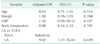
In order to identify variables independently associated with postoperative complications, a multiple logistic regression was performed. LA was significantly associated with a higher risk of postoperative complications compared to TULA (adjusted OR [95% confidence interval], 9.82 (1.27-76.04), P = 0.029), while age, weight, CRP levels, and initial body temperature were not shown to be contributable factors to postoperative complications (Table 3).
DISCUSSION
In recent years, several innovative procedures have been introduced to improve cosmetic outcomes and optimize surgical outcomes of minimally invasive surgery such as natural orifice transluminal endoscopic surgery (NOTES) [89] and SILS [10]. However, NOTES has yet to be clinically applied in humans and requires specialized instruments and training [11]. Moreover, a prospective randomized study comparing SILS and LA in children argued that SILS resulted in significantly longer mean operative times without the advantages related to reductions in complications [5]. Since SILS approaches the abdominal cavity via a single incision site through the umbilicus, the intricate nature of the intracorporeal dissection and resection of the appendix may have led to prolonged operative times [12].
TULA is similar to SILS in that it uses a single port during the surgery. However, TULA uses a combined laparoscopic and open technique, including intra-abdominal laparoscopic mobilization and extracorporeal removal of the appendix [6]. TULA is more cost-effective since it requires smaller numbers of trocars and surgical supplies compared to conventional LA [1314]. In children, where the distance between the appendix and umbilicus is shorter and the abdominal wall is more flexible than in adults, it is easier to exteriorize the appendix through the umbilicus than in adults [6].
We found that the TULA group had significantly shorter operation times than the LA group (P < 0.001). However, Bergholz et al. [7] reported from a prospective study that TULA showed a propensity to take longer than LA, although the difference was not statistically significant. The discrepancy between studies is likely due to differences in study patients and surgical instruments. Bergholz et al. [7] included cases of complicated appendicitis and the instruments used for intra-abdominal dissection were rigid 12-mm ports and all-in-one laparoscopic instruments that involved a rigid side-arm viewing laparoscope and a long single grasper. These instruments might result in interference and collision between surgical instruments. In contrast, our study only included children with simple acute appendicitis. In addition, the separable multichannel glove port used in our study was more flexible, making it easier to mobilize the appendix.
The TULA group started oral intake earlier (P < 0.001), and stayed in the hospital for less time than the LA group (P < 0.001). These results stem from the straightforward process and minimal invasiveness of the TULA procedure, which revisits the concept of "fast-track surgery". TULA is broadly in accordance with the concept of fast-tracking, a comprehensive program for optimization of perioperative care in elective surgery by alleviating stress and discomfort, and where maximization of advantages includes better postoperative outcomes [15].
The rate of postoperative complications was significantly lower in the TULA group than the LA group (1.2% vs. 8.7%; P = 0.018). In support of this finding, multiple logistic regression analysis revealed that undergoing LA compared to TULA (adjusted OR [95% confidence interval], 9.82 (1.27–76.04), P = 0.029) was significantly associated with postoperative complications. Superficial SSI was the most common complication in the LA group in our study. The shorter operation time of TULA and reduced surgical trauma to the incisional site with extracorporeal management of the appendix could be attributed to the lower SSI rate. TULA offers additional advantages of a more favorable cosmetic result compared to 3-port LA. Furthermore, TULA is simple and easy, rescuing surgeons from ergonomically difficult and prolonged single incision techniques. Accordingly, if a patient is diagnosed with an uncomplicated appendicitis, TULA appears to be the preferred option to LA in pediatric patients.
This study has several limitations. First, the retrospective design may have included selection biases, although multiple logistic regression analysis was performed to control for confounders. Second, the procedures were not performed in the same time period. TULA was more frequently performed in more recent surgeries, which may also have led to a bias in patient selection.
In conclusion, TULA was a more effective surgical option compared to conventional LA in patients with acute uncomplicated appendicitis, offering reduced operative time, early initiation of oral nutrition, shorter length of hospital stay, and less postoperative complications.


 XML Download