Journal List > Ann Surg Treat Res > v.91(1) > 1093526

Choi and Han: Ongoing higher infection rate in ABO-incompatible kidney transplant recipient: is it a serious problem? A single-center experience

Abstract

Purpose

Additional clinical experience and knowledge regarding the barrier to transplantation of ABO blood type incompatibility could reduce the higher rate of infectious complications in ABO-incompatible kidney transplantation.

Methods

A total of 79 ABO-incompatible kidney transplantation (ABOiKT) patients were compared with 260 ABO-compatible kidney transplantation (ABOcKT) patients for basic clinical characteristics, infectious complications, rejection episodes, and graft survival.

Results

There were no significant differences in baseline characteristics, rejection rates, or graft survival between the ABOiKT and ABOcKT patients. No significant difference in the infection rate was shown for cytomegalovirus (26.6% vs. 30.0%; P = 0.672), BK virus (19.0% vs. 21.5%; P = 0.752), herpes disease (10.1% vs. 5.0%; P = 0.082), pneumonia (5.3% vs. 3.8%; P = 0.746), or urinary tract infection (8.9% vs. 10.0%; P > 0.999). Female sex (hazard ratio [HR], 2.20; P = 0.003), advanced age (≥60 years) (HR, 2.5; P = 0.019), history of rejection episodes (HR, 2.28; P = 0.016), and history of surgical complications (HR, 4.64; P = 0.018) were significant risk factors for infection. ABO incompatibility demonstrated a tendency toward higher infection risk without statistical significance (HR, 1.74; P = 0.056).

Conclusion

In spite of immunosuppressant protocol modification, the rate of infectious complications following ABOiKT is still higher than with ABOcKT when a modified desensitization protocol is used. However, this was not sufficient to avoid ABOiKT.

INTRODUCTION

The gap between supply and demand for donor kidneys is greater in Asian countries due to the shortage of deceased donors with similar cultural backgrounds; therefore, living donors are a major source of kidneys for transplantation. ABO-incompatible kidney transplantation (ABOiKT) is an alternative option to ABO-compatible kidney transplantation (ABOcKT) in Korea and other Asian countries. However, ABO incompatibility increases postoperative infection rates due to preoperative conditioning, including the use of anti-CD20 antibody (rituximab) and plasmapheresis [1].
Following successful ABOiKT, use of rituximab without splenectomy reduced postoperative complications. However, several studies illustrated the higher risk of postoperative infection following desensitization in ABOiKT patients [234]. Recently, reduced rituximab was reported as a successful strategy for decreasing the incidence of postoperative infections [56]. However, there is still no consensus protocol for reducing infectious complications following ABOiKT. In the interim, modifying the ABOiKT protocol according to ongoing clinical experience may help reduce postoperative complications.
The purpose of this study was to compare infectious complications in ABOiKT recipients with those in ABOcKT recipients. In addition, the risk factors for infectious complications and those associated with ABO incompatibility were assessed.

METHODS

Study design

This retrospective, observational study assessed data from the electronic medical records at our hospital. The examined baseline characteristics included patient age, sex, relationship to the donor (e.g., relative or unrelated), number of human leukocyte antigen mismatches, prior transplant history, history of receiving immunosuppressants, cause of end-stage renal disease (ESRD), and cold ischemic time. To determine the incidence of infectious complications, cytomegalovirus (CMV) and BK virus infection, pneumonia, and urinary tract infection (UTI) were also considered.
The study protocol was approved by Asan Medical Center, Institutional Review Board (IRB No, S2012-1025-0007).

Study population/inclusion and exclusion criteria

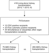
A total of 418 living-donor KTs were performed for ESRD at our institution between January 2012 and June 2013. To reduce compounding influences, we attempted to remove all factors that affect infectious complications, except for the ABO status. Exclusion criteria were the following: other organ transplantations, such as the liver or pancreas; sensitized recipients who needed preoperative conditioning (e.g., complement-dependent cytotoxicity [CDC]-positive, T- or B-flow cytometry cross-match positive); and pediatric patients. After exclusion, 79 ABOiKT and 260 ABOcKT recipients were included in the final cohort (Fig. 1).

Definitions of infectious complications

CMV disease was defined according to evidence for localized CMV infection (e.g., CMV inclusion cells) in biopsies or other appropriate specimens (e.g., bronchoalveolar lavage, relevant symptoms or signs of organ dysfunction that were most likely not due to other causes). The definition for viral syndrome was consistent with the current recommendations of the American Society of Transplantation for use in clinical trials, and > 1 of the following criteria were required: body temperature ≥ 38℃; new or significantly increased malaise; leukopenia (WBC count < 3,500/L); atypical lymphocytosis (i.e., ≥5%); or thrombocytopenia (platelet count < 100,000/L). Clinically significant CMV antigenemia which was needed antiviral therapy was defined as ≥50 antigen-positive cells/200,000 leukocytes at our medical center with or without symptom. BK nephropathy was defined according to evidence of BK disease on graft kidney biopsies [78].
In kidney transplant, there are no clear recommendations about whether or not to treat asymptomatic bacteriuria [9]. However, frequent episodes of asymptomatic bacteriuria have been found to be associated with development of acute allograft pyelonephritis and acute rejection [10]. For this reason, we treated all bacteriuria with or without symptom. Therefor in this paper, the UTI was defined as all bacteriuia.

Monitoring of CMV and BK virus infection

CMV antigenemia was assessed using the Complete CMV pp65 antigenemia IFA kit (Argene, Varihes, France). The CMV antigenemia tests were performed at 1, 2, 3, 4, 6, 8, 12, 16, 20, and 24 weeks postoperatively, and annually thereafter. The BK viral load in the blood was determined using a quantitative polymerase chain reaction (PCR). Blood PCR was performed again at 2, 12, 24, 36, and 48 weeks postoperatively, and annually thereafter.
In Korea, >95% of kidney donors and recipients are positive for CMV IgG antibodies [11]. At our medical center, universal CMV prophylaxis is not performed, and ganciclovir is only administered to CMV IgG antibody-positive donors and CMV IgG-negative recipients. UTI and pneumonia were defined as the need to prescribe antibiotics for accompanying symptoms. All patients who developed ≥ 1 infection were registered.

Immunosuppressant protocols

Conventional immunosuppressive therapy administered to ABOcKT recipients at our institution consists of induction with an interleukin (IL)-2 receptor antagonist (20 mg basiliximab) and a calcineurin inhibitor (CNI; cyclosporine or tacrolimus), an antimetabolite (mycophenolate mofetil [MMF] or cyclophosphamide), and a steroid (methylprednisolone) (Fig. 2). Steroids were administered at a high dose during the first week after KT and serially reduced to 4 mg twice daily until the second week. Twenty-four recipients who wanted their steroids withdrawn received rabbit antithymocyte antibody (thymoglobulin) as the induction therapy, rather than an IL-2 receptor antagonist followed by a CNI and antimetabolite, and the steroid was withdrawn over the first 7 days and then stopped. Thirty-one recipients did not receive induction therapy, and the other immunosuppressant protocols were the same as those administered to the IL-2 receptor antagonist group.
Two weeks before the transplant, all ABOi recipients received anti-CD20 antibody (200-mg rituximab). The patients were admitted 1 week before transplant and received plasmapheresis to reduce the anti-ABO antibody. Most recipients received plasmapheresis 4 to 5 times (range, 2–9 times) until the isoagglutinin anti-ABO antibody ratio was ≤1:4. A CNI (i.e., selective cyclosporine, if the patient was ≥55 years old), antimetabolite, and steroids were started with plasmapheresis. All ABOi recipients received an IL-2 receptor antagonist as part of the induction therapy. The MMF dose was reduced in all ABOi recipients from 1.5 g/day to 1 g/day at one week after transplant. We did not administer intravenous immunoglobulin for desensitization to any patient.
The target trough level for tacrolimus was 5–8 ng/mL during the early postoperative period, but this was decreased to 3–4 ng/mL after one year. The target trough level for cyclosporine was 100–150 µg/L, and this was gradually decreased after 3 months in the ABOc and ABOi recipients. Episode biopsies were obtained when rejection was suspected due to serum creatinine elevation or proteinuria. Acute cellular rejection and acute antibody-mediated rejection were analyzed according to the 2013 Banff criteria [12].
High dose acyclovir was used for prophylaxis of herpes infections regardless of ABO compatibility (Fig. 2). Trimethoprim-sulfamethoxazole was used for prophylaxis for pneumocystis jiroveci pneumonia in all cases.

Statistical analysis

The results were expressed as the mean ± standard deviation. Student t-test was used to compare the results of the quantitative assays, and the chi-square test was used to compare categorical variables. Parameters independently associated with infectious episodes were identified using univariate and multivariate binary logistic regression analysis. Variables that were statistically significant at a 10% level according to the univariate analysis were included in the multivariate model. Differences were considered to be significant for P-values < 0.05. Statistical calculations were performed using IBM SPSS Statistics ver. 20.0 (IBM Co., Armonk, NY, USA).

RESULTS

Among the 418 living-donor KT recipients who were treated between January 2012 and June 2013 at our hospital, 79 ABOi and 260 ABOc recipients were analyzed after excluding sensitized patients and patients who had previously received other organ transplants (liver or pancreas) (Fig. 1). The demographic and baseline characteristics of both groups at the time of transplant surgery are compared in Table 1. There were no significant differences found in terms of age, sex ratio, causes of ESRD, dialysis period, number of transplants, or cold ischemic time between groups and immunosuppressant trough level. The number of HLA antigen mismatches was somewhat less in the ABOc group because there were more unrelated donors in the ABOi group and most of the unrelated donors to the ABOi recipients were spouses.
Regarding the use of immunosuppressant agents, the CNI ratio (tacrolimus vs. cyclosporine) was similar between groups, although MMF was higher in ABOi recipients than ABOc recipients (98.8% vs. 83.1%, respectively). In most donors and recipients, the presence of CMV IgG antibodies was preoperatively noted (99.7% of donors and 98.9% of recipients). The mean follow-up period was 19.5 months (range, 12–29 months) in the ABOi group vs. 20.5 months (range, 12–29 months) in the ABOc group. Regarding the postoperative clinical outcomes, there were 2 graft losses in ABOcKT patients. Acute cellular- and antibody-mediated rejections totaled nine ABOi patients (11.4%) and 38 ABOc patients (14.6%) (P = 0.578). Intervention-requiring surgical complications, e.g., hematoma, lymphocele, wound infection, and wound dehiscence, developed in four ABOi (5.1%) and nine ABOc recipients (3.5%) (P = 0.510) (Table 2). Serum creatinine levels were similar, demonstrating mean values of 1.07 and 1.25 mg/dL between groups during the postoperative period, and ABOiKT was not inferior to ABOcKT in terms of graft function.
We also compared infectious complications between groups. UTI developed in 7 (8.9%) and 26 ABOi and ABOc patients (10.0%), respectively (P > 0.999). Pneumonia developed in four (5.0%) and 10 patients (3.8%), respectively (P = 0.752). Herpes-related diseases, such as herpes zoster and chickenpox, developed in 8 (10.1%) and 13 patients (5.0%), respectively (P = 0.082). CMV infection was similar. Antigenemia positivity was noted in 21 (26.6%) and 78 ABOi and ABOc patients (30.0%), respectively (P = 0.672). Clinically significant CMV antigenemia (defined as ≥ 50 antigen-positive cells/200,000 leukocytes at our medical center) developed in 4 (5.0%) and 12 patients (4.6%; P = 0.771), respectively. All the patients of clinically significant CMV antigenemia were treated by ganciclovir for at least 2 weeks. CMV disease or syndrome developed in 2 patients (2.5%) in the ABOi group and 5 patients (1.9%) in the ABOc group (P = 0.667). Positive BK blood PCR results were noted in 15 ABOi patients (19.0%) and 56 ABOc patients (21.5%) (P = 0.752). The incidence of a viral load ≥ 4 logs, which is regarded to be within the threshold for clinical significance, did not differ between groups (6.3% vs. 8.1%; P = 0.810). BK nephropathy developed in 1 patient (1.3%) in the ABOi group and 6 patients (2.3%) in the ABOc group (P > 0.999). The earlier detection of CMV antigenemia and BK viremia was noted in ABOiKT recipients, but this finding was not statistically significant.
In a previous study, we examined the effect of immunosuppressant protocol modification [1]. Before modification of the protocol, we used a higher dose of rituximab, a CNI trough level, and a high dose of MMF (Fig. 3). After modifying the immunosuppressant protocol, the incidence of infectious complications (e.g., UTI, pneumonia, herpes-related disease, CMV, and BK) did not differ between ABOiKT and ABOcKT patients, although overall infectious complications still occurred more often in ABOiKT recipients (39.2% vs. 18.5%; P = 0.04) (Table 2). Based on the rate of delayed discharge after kidney transplantation or readmission due to infection, severe infections did not occur more often in ABOiKT recipients.
Univariate analysis was performed to detect the factors that affect infectious complications in all KT recipients, and ABO-incompatible status, female sex, advanced age (≥60 years), >3 HLA antigen mismatches, rejection episodes, treatment-requiring surgical complications, and a long, cold ischemic time (<60 minutes) were found to be positive predictive factors (Table 3). In addition, female sex, advanced age, rejection episode, and surgical complications were significant factors according to multivariate analysis (P < 0.05). ABO-incompatible status tended (hazard ratio [HR], 1.742) to result in infectious complications (P = 0.056). Surgical complications were the greatest risk factor for postoperative infection (HR, 4.635; P = 0.018). Advanced age and rejection episodes were also high-risk factors for infectious complications (HR, 2.503 and 2.281, respectively).

DISCUSSION

ABOiKT is no longer considered challenging by experienced transplant centers. Based on the reported short- and long-term outcomes [613141516], ABOiKT demonstrates similar rejection and graft survival rates as ABOcKT if early postoperative morbidity can be overcome. In our current study, ABOiKT recipients demonstrated surgical complications comparable to those of ABOcKT recipients and similar acute rejection rates and graft survival during the short-term follow-up period. Regarding graft function, creatinine levels were similar at all times during the postoperative period in both ABOi and ABOcKT recipients.
However, postoperative morbidity is the Achilles heel of successful outcomes. In 2011, Habicht et al. [2] reported that ABOiKT demonstrates more infectious complications than ABOcKT. During the initial period, we also noted a higher rate of infectious complications, including mortalities [1]. More recently, Lentine et al. [3] reported an increased risk of early postoperative complications following ABOiKT. They also analyzed national data and concluded that ABOiKT demonstrates a higher risk of infectious and hemorrhagic complications. Even though they analyzed a huge database that provided a high rate of statistical power, the data also had some limitations. Because every transplant center has its own ABOiKT protocol, the characteristics of the national data were heterogeneous. The national data exhibited a general trend, but not center-specific effects, and the authors admitted that the national data might not reflect the results of highly experienced and successful medical centers. Another issue is that the data were collected between 2000 and 2007. During that time, splenectomies were performed during earlier periods, and the rituximab dose during the early phase might have been higher than in later periods. Regarding postoperative infectious complications in ABOcKT recipients, 7.3% of wound infections, 3.8% of pneumonia cases, and 15.3% of UTIs have been reported, respectively [3].
In contrast to the national data, our current data are homogeneous. We excluded other organ transplantations, such as liver or pancreas, sensitized recipients (e.g., CDC-positive, T- or B-flow cytometry cross-match positive), and pediatric patients. We evaluated the cohort that was treated using the same immunosuppressant protocol between January 2012 and June 2013, and analyzed ABOi and ABOcKT, which were performed during the same time period. This is a short period of time to evaluate long-term graft survival, but it is sufficient for evaluating infectious complications.
At our medical center, 184 ABOiKT procedures were performed between February 2009 and June 2013. Despite using rituximab without splenectomy during the initial period (2009–2011), ABOiKT recipients developed more infectious complications, including mortalities, than ABOcKT recipients. Since January 2012, we have used a modified immunosuppressant protocol, i.e., reduced rituximab and MMF doses, selective cyclosporine administration in older recipients (≥55 years), and prophylactic antiviral agents (Fig. 2), thus reducing the infection rate. In our present analyses, ABOiKT recipients were included in the cohort that received the modified protocol [1].
Regarding preoperative preparation, differences between the ABOi and ABOc transplants included rituximab, plasmapheresis, and the early use of immunosuppressants, including MMF, in ABOiKT recipients. There were no differences in the CNI levels between groups. MMF is reportedly associated with CMV and BK infection [171819]. Following our initial experience with postoperative infectious complications, we reduced the MMF dose and used acyclovir to prevent infections, as administering rituximab for the induction is considered a risk factor for herpes-related diseases in KT recipients [20].
After modifying the immunosuppressant protocol, the incidence of infectious complications, such as UTI, pneumonia, CMV, and BK, did not differ between ABOiKT and ABOcKT patients, although overall infectious complications still occurred more often in ABOiKT recipients. One of the most important description factors was herpes virus infection. Even though statistical differences were not found, herpes virus infections occurred more often in ABOiKT recipients. The postoperative rate of infectious complications including herpes virus infection could also have improved due to other modifications, such as reducing the rituximab dose or use of acyclovir. However, the complete removal of effects that result from preoperative conditioning, including rituximab and plasmapheresis, cannot be easily achieved, but only minimized.
Univariate analysis was performed to determine the risk factors that affect infectious complications in all KT recipients, and ABO-incompatibility status, female sex, advanced age (≥60 years), >3 HLA antigen mismatches, rejection episode, treatment-requiring surgical complications, and a long, cold ischemic time (≤60 minutes) were considered predictive factors by multivariate analysis. According to multivariate analysis, female sex, advanced age, rejection episode, and surgical complications were also significant factors. As previously reported, female KT recipients demonstrate a greater UTI prevalence than males [21]. Advanced age is not only a risk factor for infection, but also a risk factor for mortality in KT recipients [22]. The treatments for rejection, such as high-dose steroids or biologic agents, as well as plasmapheresis, are also well-known risk factors for infection. In our current study, the most powerful factor that affected infectious complications was intervention-requiring surgical complications, including surgery and radiologic modalities. ABO incompatibility demonstrated a low HR (1.742) among all evaluated risk factors, with borderline significance (P = 0.056). ABO incompatibility was still a risk factor, even though the risk was decreased. However, according to readmission or delayed discharge rate reports (Table 2), ABO incompatibility might not be a risk factor for infectious complications sufficiently serious to avoid kidney transplantation.
This study had several limitations. First, this was a retrospective study. The immunosuppressant protocol for ABOcKT was not the same for all recipients, especially for antimetabolites. Moreover, the number of transplants was not large due to the one year to one and half year recruitment period data available from our center. Studies on larger cohorts with longer follow-up periods are warranted in the future.
In conclusion, ABOiKT still demonstrates a higher rate of infectious complications in comparison with ABOcKT. However, it was not a major hurdle to performance of ABO-incompatible kidney transplantation.

Figures and Tables

Fig. 1

Selection of the study cohort. CDC, complement-dependent cytotoxicity.

astr-91-37-g001
Fig. 2

Immunosuppressant protocols for ABOi and ABOc kidney transplant recipients (after January 2012). MMF, mycophenolate mofetil; PD, prednisolone; LDKT, living donor kidney transplantation; PP, plasmapheresis.

astr-91-37-g002
Fig. 3

Initial immunosuppressant protocols for ABOi kidney transplant recipients (February 2009-December 2012). KT, kidney transplant; FK, FK 506; MMF, mycophenolate mofetil; PD, prednisolone; LDKT, living donor kidney transplantation; PP, plasmapheresis.

astr-91-37-g003
Table 1

Demographic characteristics of ABOi and ABOc kidney transplant recipients

astr-91-37-i001

Values are presented as mean ± standard deviation or number (%) unless otherwise indicated.

ABOi, ABO-incompatible; ABOc, ABO-compatible; ESRD, end-stage renal disease; GN, glomerulonephritis; PCKD, polycystic kidney disease; HLA, human leukocyte antigen; CNI, calcineurin inhibitor; CMV, cytomegalovirus.

a)The donor was positive for CMV IgG, and the recipient was IgG-positive. b)The donor was negative for CMV IgG, and the recipient was IgG-positive. c)The donor was positive for CMV IgG, and the recipient was IgG-negative.

Table 2

Postoperative clinical outcomes of ABOi and ABOc kidney transplant recipients

astr-91-37-i002

Values are presented as number (%) or mean ± standard deviation.

ABOi, ABO-incompatible; ABOc, ABO-compatible; ACR, acute cellular rejection; AMR, antibody mediated rejection; UTI, urinary tract infection; CMV, cytomegalovirus; PCR, polymerase chain reaction.

a)Procedure-requiring complications, e.g., wound infection, lymphocele, hematoma, etc. b)Summary of the development of UTI, pneumonia, clinically significant CMV infection or BK infection, and herpes-related disease.

Table 3

Binary logistic regression analysis of pooled infectious complicationsa)

astr-91-37-i003

CI, confidence interval; HR, hazard ratio; HLA, human leukocyte antigen; CNI, calcineurin inhibitor; ESRD, end-stage renal disease.

a)All variables are categorical variables (yes/no).

Notes

CONFLICTS OF INTEREST No potential conflict of interest relevant to this article was reported.

References

1. Choi BH, Cho HK, Jung JH, Choi JY, Shin S, Kim YH, et al. How to reduce lethal infectious complications in ABO-incompatible kidney transplantation. Transplant Proc. 2015; 47:653–659.
2. Habicht A, Broker V, Blume C, Lorenzen J, Schiffer M, Richter N, et al. Increase of infectious complications in ABO-incompatible kidney transplant recipients: a single centre experience. Nephrol Dial Transplant. 2011; 26:4124–4131.
3. Lentine KL, Axelrod D, Klein C, Simpkins C, Xiao H, Schnitzler MA, et al. Early clinical complications after ABO-incompatible live-donor kidney transplantation: a national study of Medicare-insured recipients. Transplantation. 2014; 98:54–65.
4. Hwang JK, Kim YK, Kim JM, Chung BH, Choi BS, Yang CW, et al. Comparative analysis of ABO-incompatible living donor kidney transplantation with ABO-compatible grafts: a single-center experience in Korea. Transplant Proc. 2013; 45:2931–2936.
5. Shirakawa H, Ishida H, Shimizu T, Omoto K, Iida S, Toki D, et al. The low dose of rituximab in ABO-incompatible kidney transplantation without a splenectomy: a single-center experience. Clin Transplant. 2011; 25:878–884.
6. Hatakeyama S, Fujita T, Murakami R, Suzuki Y, Sugiyama N, Yamamoto H, et al. Outcome comparison of ABO-incompatible kidney transplantation with low-dose rituximab and ABO-compatible kidney transplantation: a single-center experience. Transplant Proc. 2014; 46:445–448.
7. Asberg A, Humar A, Rollag H, Jardine AG, Mouas H, Pescovitz MD, et al. Oral valganciclovir is noninferior to intravenous ganciclovir for the treatment of cytomegalovirus disease in solid organ transplant recipients. Am J Transplant. 2007; 7:2106–2113.
8. Bohl DL, Brennan DC. BK virus nephropathy and kidney transplantation. Clin J Am Soc Nephrol. 2007; 2:Suppl 1. S36–S46.
9. Singh R, Geerlings SE, Bemelman FJ. Asymptomatic bacteriuria and urinary tract infections among renal allograft recipients. Curr Opin Infect Dis. 2015; 28:112–116.
10. Fiorante S, Lopez-Medrano F, Lizasoain M, Lalueza A, Juan RS, Andres A, et al. Systematic screening and treatment of asymptomatic bacteriuria in renal transplant recipients. Kidney Int. 2010; 78:774–781.
11. Lee YM, Kim YH, Han DJ, Park SK, Park JS, Sung H, et al. Cytomegalovirus infection after acute rejection therapy in seropositive kidney transplant recipients. Transpl Infect Dis. 2014; 16:397–402.
12. Haas M, Sis B, Racusen LC, Solez K, Glotz D, Colvin RB, et al. Banff 2013 meeting report: inclusion of c4d-negative antibody-mediated rejection and antibody-associated arterial lesions. Am J Transplant. 2014; 14:272–283.
13. Fuchinoue S, Ishii Y, Sawada T, Murakami T, Iwadoh K, Sannomiya A, et al. The 5-year outcome of ABO-incompatible kidney transplantation with rituximab induction. Transplantation. 2011; 91:853–857.
14. Muramatsu M, Gonzalez HD, Cacciola R, Aikawa A, Yaqoob MM, Puliatti C. ABO incompatible renal transplants: Good or bad? World J Transplant. 2014; 4:18–29.
15. Opelz G, Morath C, Susal C, Tran TH, Zeier M, Dohler B. Three-year outcomes following 1,420 ABO-incompatible living-donor kidney transplants performed after ABO antibody reduction: results from 101 centers. Transplantation. 2015; 99:400–404.
16. de Weerd AE, van Agteren M, Leebeek FW, Ijzermans JN, Weimar W, Betjes MG. ABO-incompatible kidney transplant recipients have a higher bleeding risk after antigen-specific immunoadsorption. Transpl Int. 2015; 28:25–33.
17. Asberg A, Jardine AG, Bignamini AA, Rollag H, Pescovitz MD, Gahlemann CC, et al. Effects of the intensity of immunosuppressive therapy on outcome of treatment for CMV disease in organ transplant recipients. Am J Transplant. 2010; 10:1881–1888.
18. Bataille S, Moal V, Gaudart J, Indreies M, Purgus R, Dussol B, et al. Cytomegalovirus risk factors in renal transplantation with modern immunosuppression. Transpl Infect Dis. 2010; 12:480–488.
19. Brennan DC, Legendre C, Patel D, Mange K, Wiland A, McCague K, et al. Cytomegalovirus incidence between everolimus versus mycophenolate in de novo renal transplants: pooled analysis of three clinical trials. Am J Transplant. 2011; 11:2453–2462.
20. Ko GB, Kim T, Kim SH, Choi SH, Kim YS, Woo JH, et al. Increased incidence of herpes zoster in the setting of cytomegalovirus preemptive therapy after kidney transplantation. Transpl Infect Dis. 2013; 15:416–423.
21. Lee JR, Bang H, Dadhania D, Hartono C, Aull MJ, Satlin M, et al. Independent risk factors for urinary tract infection and for subsequent bacteremia or acute cellular rejection: a single-center report of 1166 kidney allograft recipients. Transplantation. 2013; 96:732–738.
22. Karim A, Farrugia D, Cheshire J, Mahboob S, Begaj I, Ray D, et al. Recipient age and risk for mortality after kidney transplantation in England. Transplantation. 2014; 97:832–838.
TOOLS
Similar articles