

 PDF
PDF ePub
ePub Citation
 Print
Print


INTRODUCTION
Hyperparathyroidism is the most common and first manifestation of multiple endocrine neoplasia type 1 (MEN1), most of which are due to hyperplasia of multiple parathyroid glands [1]. Treatment of hyperparathyroidism in MEN1 patients consists of surgical removal of the parathyroid glands. However, the rate of persistent or recurrent hyperparathyroidism after primary operation is high, for which one of the reasons is the presence of supernumerary or ectopic parathyroid gland [1]. For complete surgical removal of parathyroid glands, precise localization using an appropriate imaging method is needed. Technetium-99m (Tc-99m) sestamibi scintigraphy is a highly sensitive diagnostic tool for the detection of hyperfunctioning parathyroid glands. Tc-99m sestamibi single-photon emission computed tomography (SPECT) or single-photon emission computed tomography/computed tomography (SPECT/CT) can improve both sensitivity and specificity compared to planar scan [2]. We report here a MEN1 patient with anterior mediastinal parathyroid adenoma who underwent successful surgical excision after localization via SPECT/CT. To our knowledge, this is the first case report in the literature of Tc-99m sestamibi SPECT/CT image of mediastinal parathyroid adenoma in MEN 1 patient.
CASE REPORT
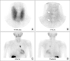
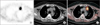
A 51-year-old female reported to the Endocrinology Department for genetic counseling. She was the eldest of the 3 sisters, of whom had been diagnosed with MEN1. The middle sister (46 years of age) had hyperparathyroidism and a pancreatic neuroendocrine tumor. The youngest sister (42 years of age) had hyperparathyroidism, pituitary microadenoma, and a pancreas neuroendocrine tumor. Upon examination of the MEN gene (11q13), mutations c.669+3A>G (intron 3), which results in a splicing defect, and missense variation c.1636G>A (exon 10) were confirmed. In a screening test for MEN1, blood sampling and imaging studies were performed. Intact parathyroid hormone (iPTH) (731.59 pg/mL) and serum calcium (12.5 mg/dL) levels were elevated, and 25-hydroxy vitamin D (15.21 ng/dL) level was decreased. Tc-99m sestamibi scintigraphy was performed for diagnosis of hyperfunctioning parathyroid gland (Fig. 1). Four-hour late imaging showed focal uptake in the right upper and lower thyroid beds, suggestive of hyperfunctioning parathyroid glands. To exclude incidental ectopic parathyroid glands, a planar chest image was obtained, and focal hot uptake was seen in the mediastinum. When additional SPECT/CT was performed to localize the mediastinal hot uptake, a lobulated mass with focal sestamibi uptake was noted in the mediastinal left paraaortic region (Fig. 2). There was no other abnormality suggestive of MEN1 in blood tests or imaging studies. Total parathyroidectomy with autotransplantation of a parathyroid gland on the left forearm and video-assisted thoracoscopic surgery on an anterior mediastinal mass were performed. Final pathology revealed hyperplasia of bilateral superior and inferior parathyroid glands and a parathyroid adenoma of anterior mediastinal mass. The size of the hyperplasia involving the four parathyroid glands was about 1.0 cm × 1.0 cm, and the size of the parathyroid adenoma was 3.5 cm × 2.2 cm. After operation, the patient's iPTH and serum calcium level were restored to normal.
DISCUSSION
MEN1 is an autosomal dominant familial tumor syndrome in which tumors can develop in the endocrine system, mostly in the anterior pituitary gland, parathyroid gland and pancreatic islet cells [3]. Hyperparathyroidism resulting from parathyroid adenoma or hyperplasia occurs in most patients and is typically the first manifestation of MEN1 [4]. Hyperparathyroidism manifests in most patients by their third decade of life [5]. However, as in our case patient, there can be a long asymptomatic period even through the biochemical changes of hyperparathyroidism and decreased bone mass [6]. The treatment of choice for hyperparathyroidism in MEN1 is surgical removal of parathyroid glands. Subtotal parathyroidectomy or total parathyroidectomy with autotransplantation of parathyroid tissue is usually recommended [7]. After surgical excision of parathyroid glands, persistent or recurrent hyperparathyroidism can result from supernumerary or ectopic parathyroid glands, regrowth of remnant parathyroid glands or hyperfunctioning autograft tissue [1]. The major cause of persistent or recurrent hyperparathyroidism is ectopic or supernumerary parathyroid gland. Therefore, precise localization of parathyroid glands is essential for complete surgical removal.
Parathyroid glands originate from the third and fourth branchial pouches and usually are located posterior to the thyroid. Superior parathyroid glands arise from the fourth branchial pouch and migrate with the thyroid gland. Inferior parathyroid glands arise from the third branchial pouch and migrate with the thymus, a relatively longer distance than the movement of the superior parathyroid glands. Therefore, the locations of inferior parathyroid glands are more variable [8]. Ectopic inferior parathyroid glands can be located in the cervical portion of the thymus, the posterior aspect of the middle third of the thyroid gland, the anterior mediastinum, the intrathyroidal region, or along the carotid sheath. Among these locations, ectopic parathyroid glands in the anterior mediastinum are rare, representing about 3.9%–5% of incidences [9]. Ectopic or supernumerary parathyroid glands are more common in MEN patients than in sporadic hyperparathyroidism patients [10].
Tc-99m sestamibi scintigraphy is regarded as the best imaging modality for identification of hypefunctioning parathyroid tissue. It shows only hyperfunctioning parathyroid tissue, not normal parathyroid glands. Therefore, it is helpful for discriminating hyperfunctioning parathyroid glands from normal parathyroid glands. Tc-99m sestamibi parathyroid scintigraphy is performed in 2 phases on the anterior neck using a pinhole collimator: early (5–15 minutes) images and late (2–4 hours) images after intravenous injection of Tc-99m sestamibi. A chest image must also be obtained using a parallel collimator in order to detect incidental or unsuspected mediastinal ectopic parathyroid tissue. Obtaining chest planar images is a good method to exclude ectopic parathyroid tissue without additional radiation exposure and cost. Therefore, chest images should be obtained before the parathyroidectomy is scheduled for the patient.
Precise localization of hyperplastic parathyroid glands or parathyroid tumors is also important to improve surgical outcome. It is better to use SPECT/CT for precise localization of parathyroid glands. Large-sized adenomas or hyperplasia can be detected or localized easily by CT or ultrasonography; however, it is difficult to identify small parathyroid tissue. Tc-99m sestamibi SPECT or SPECT/CT show high sensitivity and specificity for diagnosis of hyperfunctioning parathyroid glands even of small size [8].
In conclusion, we report a case of a MEN1 patient with anterior mediastinal parathyroid adenoma that was successfully removed following localization via Tc-99m sestamibi scintigraphy and SPECT/CT.
 XML Download