

 PDF
PDF ePub
ePub Citation
 Print
Print


INTRODUCTION
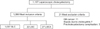
The standard management of cholecystitis is laparoscopic cholecystectomy (LC). Standard laparoscopic cholecystectomy (SLC) requires the dissection of Calot triangle and exposure of the cystic duct (CD). Then, the CD is divided and the entire gallbladder (GB) is dissected from the liver (Fig. 1A). Sometimes, dissecting Calot triangle, dividing the CD, and dissecting the entire GB from the liver are very difficult. SLC has considerable risks of bile duct injury, bleeding from the liver bed in cases of severe inflammation or fibrosis [1], Mirizzi syndrome [2], and biliary anomalies [3]. Laparoscopic subtotal cholecystectomy (LSC) can be an alternative procedure in these situations. In LSC, the GB is divided at the level of the Hartman pouch instead of at the CD, and both the anterior and posterior wall of the GB are removed from the liver bed (Fig. 1B). However, removing the entire GB is still difficult and time-consuming when the GB is tightly adhered to the liver. This can result in considerable bleeding from the liver bed. Instead of removing the whole gall bladder, LSC removing only the anterior wall of the GB (LSCA) is done in order to avoid liver injury and bleeding from the liver (Fig. 1C). The primary concerns of LSCA are bile leak from the GB stump and the severe inflammation of the remaining posterior wall. The purpose of this study was to evaluate the clinical outcomes of SLC such as bile duct injury, surgical site infection, mortality.
METHODS
Patients enrolled
LSC and LSCA were defined as leaving the CD regardless of whether the duct was left closed or open. The inclusion criteria for patients were age ≥18 years and laparoscopic cholecystectomies that were completed without conversion to open surgery. The exclusion criteria were GB cancer, sepsis due to cholecystitis and precholecystectomy complications such as small bowel injury or abdominal wall bleeding due to trocar insertion. Preoperative diagnosis was made with medical history, physical examination, ultrasound or abdominal CT. The diagnosis of cholecystitis was made when patients have typical pain at right upper quadrant accompanied by leukocytosis and radiologic findings of GB wall thickening, pericholecystic fluid collection. Final diagnosis was made by pathologic findings.
Operative technique
For LSC, a dissection was made through the anterior and posterior side of the neck or body of the GB. If the GB wall was not too thick, the transection was made primarily with a laparoscopic stapler. When the stapler could not be used because of GB wall thickening, the transection was made with electrocautery. The whole GB was dissected from the liver bed with electrocautery. When we had to perform LSC, we made a small hole on the anterior wall of the GB, removed the GB contents with suction, and collected the gall stone into a laparoscopic plastic bag so that we could minimize intraabdominal spillage before transecting the GB. For LSCA, only the anterior wall of the GB was transected with electrocautery or ultrasonic scissors (Fig. 1). The mucosa of the remaining posterior wall of the GB was cauterized with electrocautery to eliminate any functioning mucosa. If the inside of the CD was identified, it was closed with continuous running suture. If it was not identified, we tried to close the wall of the GB stump. In case suturing of the CD or the GB wall was impossible because of hardness or being too friable, a fabricated hemostatic agent or fibrin sealant was packed in the GB stump. We defined these patients as open CD group. In all cases, the initial goal was to perform SLC. We changed SLC to LSC or LSCA when dense inflammation or fibrosis might have resulted in a hazardous cholecystectomy which has high risk of major bile duct injury or bleeding. When the GB was adhered tightly to the liver, we performed LSCA instead of LSC. The decision was made by a surgeon once dissection of Calot triangle was performed. Closed suction drainage was placed in Morrison's pouch in all cases of LSC for early detection of bile leakage.
Data collection and analysis
We retrospectively reviewed the medical records of laparoscopic cholecystectomies performed between January 2006 to December 2015 To compare characteristics and clinical outcomes between SLC, LSC, and LSCA groups, analysis of variance for continuous variables or linear-by-linear association test for categorical variables was used. When LSC and LSCA groups were compared, Mann-Whitney test and Fisher exact test were mainly used. All statistical analyses were performed using the IBM SPSS Statistics ver. 23.0 (IBM Co., Armonk, NY, USA).
RESULTS
Patients enrolled
LC was performed on 1,107 patients during the study period. Twenty-one patients were excluded because of the exclusion criteria and 1,086 patients were enrolled. SLC was performed on 1,037 patients, LSC on 22 patients, and LSCA on 27 patients (Fig. 2).
Preoperative characteristics of patients
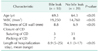
The mean ages of the patients who underwent SLC, LSC, and LSCA were 61.7, 67.3, and 68.3 years respectively (range, 18–91 years). The mean body mass indices of the patients in these groups were 24.7, 24.4, and 24.1 (range 16.1–40.3). The mean lengths of hospitalization before cholecystectomy were 1.4, 5.6, and 6.1 days. Compared to SLC, LSC and LSCA groups had longer preoperative hospitalization (P < 0.05; range 0–12 days). Preoperative white blood cell counts were 8,900, 15,650, and 18,760 (P < 0.05). The mean thicknesses of the GB walls (calculated with ultrasound or CT) were 4.6, 7.4, and 8.1 mm (P < 0.05; range, 3.4–9.2 mm). In the LSC and LSCA groups, the mean preoperative WBC count and thickness of the GB wall were significantly higher than SLC group. The most frequent indications for cholecystectomy in the SLC group were colic due to gall stones, cholecystitis, polyps, and adenomyomatosis in that order. In the LSC and LSCA groups, cholecystitis was the primary preoperative diagnosis for cholecystectomy (Table 1).
Surgical outcomes
The mean operating times of SLC, LSC, and LSCA were 41, 74, and 67 minutes respectively. The duration of SLC was significantly shorter than LSC and LSCA. The duration of LSCA was significantly shorter than LSC. The estimated blood loss for each type of surgery was 5 (0–100), 45 (5–200), and 33 mL (5–150). LSCA had significantly less blood loss compared to LSC. The mean length of postoperative hospitalization was 3.4 days for SLC patients, 6.2 days for LSC patients, and 5.8 days for LSCA patients. Mortality and morbidity occurred in 37 patients in the SLC group (3.3%), 5 patients in the LSC group (9%), and 7 in the LSCA group (3.7%). In the SLC group, 2 patients died of postoperative myocardial infarction and congestive heart failure. Four patients had major bile duct injury (0.38%) requiring hepaticojejunostomy. Seven patients had minor bile duct injury managed with endoscopic procedures such as nasobiliary drainage or sphincterotomy. Five patients had temporary bile leakage from the GB stump (0.48%), which was cured with conservative management. Two patients had intraabdominal abscess, which was cured with percutaneous drainage and conservative management. Eight patients had surgical site infections of the trocar sites. Five patients had postoperative bleeding which was cured conservatively. Two patients had transection of the right hepatic artery without any adverse effect on the liver. In the LSC group, four patients had bile leakage from the GB stump, which was cured conservatively and with endoscopic nasobiliary drainage. One patient had a subhepatic abscess, which was managed conservatively. In the LSCA group, six patients had bile leaks which were cured conservatively and with endoscopy. One patient had a superficial surgical site infection. There was no mortality in the LSC and LSCA groups (Table 2). Bile leak from the GB stump was more common in LSC and LSCA than in SLC but cured nonoperatively.
When we stratified the patients according to the closure of the CD, bile leak occurred in 4 out of 11 patients (36.3%) whose CDs were left open. When the CD was sutured, 6 out of 38 patients had bile leak (15.7%). The rate of bile leak was significantly lower when the CD was closed. All patients who had bile leak were cured conservatively or with endoscopic intervention (Table 3). Patients who had bile leaks were older and had higher WBC counts, thicker GB walls, and longer hospitalizations compared to patients who had no bile leak (Table 4).
DISCUSSION
Two major complications of cholecystectomy are major bile duct injury and bleeding from the liver bed or the vasculature around the GB. Surgeons always have to balance the benefit of removing the whole GB with the major complications of doing so. When the complications of removing the whole GB are considered more significant than the complications of leaving part of the GB, subtotal cholecystectomy (SC) can be performed. There is no universal definition of SC. Initially, SC meant leaving the posterior wall of the GB to prevent massive bleeding from the GB bed. This operative procedure was particularly effective when a patient had portal hypertension due to liver cirrhosis or chronic pancreatitis. Leaving the posterior wall of the GB in a SC was regarded as a simple, safe option for patients in whom standard cholecystectomy could be hazardous [4]. Sometimes, we can leave the CD as well to avoid bile duct injury. Strasberg classified the SC as either the fenestrating SC or the reconstituting SC, according to the surgical technique used for the lowest part of the GB. In a fenestrating SC, Hartmann pouch is opened and the CD is closed from inside with a pursestring suture. The CD can be left open. In a reconstituting SC, the lowest part of the GB is closed by suture or stapler [5]. In the past, SC was usually used for open cholecystectomies. It was adapted to laparoscopic surgery and became a surgical option for difficult LCs [6]. Because of the rarity of GB disease with portal hypertension, leaving the posterior wall of the GB after transecting the CD seemed to lose popularity. Instead, it became more prevalent to leave the CD and transect at the level of the GB neck or body to avoid major bile duct injury. Usually, this surgical option is applied when the dissection of Calot triangle has significant risk of bile duct injury because of severe inflammation or fibrosis. Some authors have called this scenario a frozen Calot triangle. The purpose of leaving the CD and transecting the GB at the level of the body or Hartmann pouch is to avoid major bile duct injury. The incidence of major bile duct injury requiring biliary reconstruction in SC is very low (0%–0.08%) compared to SLC (0.3%) [7]. LSC can effectively prevent major bile duct injury. However, there are several major concerns with LSC, including bile leak, spillage of GB contents, and infection.
Most of the literature has revealed that LSC has a higher incidence of bile leak from the GB stump. Laparoscopic suturing of the interior of the CD is difficult. Closure of the CD is recommended to decrease the incidence of bile leak. However, leaving the CD open can also be attempted. Michalowski et al. [8] reported that bile leak occurred in only one LSC patient out of fourteen in whom the CD was left open. Sinha et al. [9] reported that, among 28 LSC patients without CD ligation, two temporary bile leaks resolved spontaneously and three patients required endoscopic retrograde cholangiopancreatography. Philips et al. [10] and Shingu et al. [11] had similar results. Most studies of LSC have revealed that bile leak is a trivial complication. There was no mortality caused by bile leak, even when the CD was left open. Bile leak resolved spontaneously or with endoscopic intervention. Thickened GB wall and CD caused by severe inflammation seem to decrease the patency of the CD and prevent the bile leak, though it is unclear how. If suturing the inside of the CD or the GB stump wall is not easy, one can try packing with a fabricated hemostatic agent or fibrin sealant. In our study, eight out of 15 patients did not have bile leak. If the surgeon is not accustomed to laparoscopic suturing or it is technically impossible, simple packing of the GB neck or the CD can be an alternative procedure. Because of its infrequency, we could not compare the statistical significance between suturing and packing the CD.
Spillage of the GB contents is unavoidable when LSC (especially LSCA) is performed. Surgeons should try to minimize the spillage of GB contents. However, it is not GB perforation but bacteriobilia that is the significant risk factor for surgical site infections [12]. It is helpful to know of the presence of bacteriobilia in patients with acute cholecystitis [13]. Percutaneous cholecystostomy is an effective means of diagnosing bacteriobilia. However, preoperative laboratory tests are also available. The known risk factors of bacteriobilia are age >55 years, alkaline phosphatase >100 IU/L, and total bilirubin >1.2 mg/dL [14]. If patients meet these criteria, it may be useful to penetrate the GB and aspirate the GB contents with a suction tip in order to minimize the risk of surgical site infection. In our study, many medical records omitted the presence of spillage of the GB contents, and so we could not analyze the relationship between the spillage of GB contents and surgical site infections.
The other major concern of LSCA is leaving the posterior wall of the GB, which often has severe inflammation. LSCA is usually carried out to avoid bleeding from the liver bed. Severe inflammation, a sunken GB, or fibrosis can hinder surgeons from dissecting the GB from the liver. Difficult dissection can cause liver injury and troublesome bleeding. Compared to SLC, LSC and LSCA had significantly less blood loss in difficult cholecystectomies [15161718]. With the use of ultrasonic scissors, it becomes easier and faster to transect the anterior wall of the GB without bleeding. This is especially true when patients have had portal hypertension or Mirrizzi syndrome [418]. LSCA would be expected to have a higher incidence of infection because a part of the severely inflamed GB is left behind. Contrary to our expectations, however, the frequency of subhepatic abscesses and surgical site infections was not influenced by the remaining posterior wall of the GB. According to one large-scale systemic meta-analysis, the rates of subhepatic collections and wound infections were not different [6]. The most common cause of conversion to open cholecystectomy from LC is bleeding. LSCA seems to reduce the conversion rate, especially when the patient has had portal hypertension or severe inflammation. The rate of wound infection in our study was lower than in previous studies. When LSCA is performed, we irrigate the port site meticulously with over 1,000 mL of saline.
Preoperative cholecystostomy is useful procedure when the patient's condition is poor or the age is too old [19]. Although we excluded septic patient with cholecystitis, it is not uncommon clinical scenario. In these situations, cholecystectomy has considerable risk. Preoperative cholecystostomy is safe and effective alternative procedure for the initial management [20].
In conclusion, LSC and LSCA is a clinically feasible and safe operative procedure. It is superior to SLC in terms of major bile duct injury and bleeding. LSC and LSCA have similar clinical outcomes when compared to SLC, in terms of surgical site infections and development of intraabdominal abscesses. LSC and LSCA have a higher risk of bile leak than SLC, but it can usually be managed with endoscopic intervention. LSCA has shorter operation times and less bleeding than LSC. LSC and LSCA can be tried if SLC has a considerable risk of major bile injury or bleeding, such as in the cases of severe inflammation, fibrosis, Mirizzi syndrome, or portal hypertension. LSCA is an easier but effective alternative to LSC.

 XML Download